






Abstract
Background
Knowledge about the clinical features of Darier disease, an orphan autosomal-dominant genetic disorder, is sparse and has been evaluated only in few studies.
Objectives
To investigate the clinical features of a large group of patients with Darier disease, and to explore for associations between disease characteristics and severity of the disease.
Methods
Seventy-six individuals with Darier disease were evaluated utilizing a structured questionnaire-based interview, a physical examination, and a retrospective assessment of their medical records.
Results
The most frequent locations of lesions were hands (99%) and fingernails (93%). Wart-like lesions on the hands were more visible after soaking them in water for 5 minutes, we therefore named this phenomenon the “wet hand sign”. Oral involvement was found in 43% of patients, while 48% of women and 16% of men showed genital lesions. Patients with severe Darier disease had a tenfold greater risk of developing genital lesions than those with mild disease (P = .01). Most patients (88%) in our study exhibited a combination of the four types of the disease patterns of distribution (flexural, seborrheic, nevoid, and acral).
Conclusions
Documentation of disease on the hands and fingernails provides a highly sensitive means to aid in the diagnosis of Darier disease. It is important to evaluate mucosal lesions including genital and oral mucosa.
Introduction
Darier-White disease (DD) was initially described independently in 1889 by the two legendary dermatologists; Professor Jean Darier (1856-1938), and Professor James C White (1833-1916). 1,2
DD is an autosomal-dominant disorder characterized by loss of adhesion between epidermal cells (acantholysis) and abnormal keratinization (dyskeratosis, “corps ronds,” and “grains”). 3 -6 Exact figures of prevalence are unknown but there are estimates of around 1 in 55,000. Penetrance is complete and delayed, onset is usually before the third decade, and expressivity varies. 7
The gene ATP2A2 on chromosome 12q23-24.1 was recognized as the causative gene for DD by Sakuntabhai et al. in 1999. 8 ATP2A2 encodes the sarco/endoplasmic reticulum Ca2 +ATPase isoform 2 (SERCA2), which plays a central role in intracellular calcium signaling. 9
Clinical features include brown, keratotic papules, often associated with itching and malodor. 10 Acral involvement is very common and includes punctate depressions on the palms and soles (palmoplantar pits) and wart-like lesions on back of hands and feet. 7 Nail involvement is highly common and include red or white longitudinal bands, V-shaped notches at the free margin, longitudinal ridging, and subungual hyperkeratotic debris. 7
The disease is historically classified according to patterns of distribution (flexural, seborrheic, nevoid, and acral). 11
Few clinical studies have been conducted to define the various aspects of DD. 7,12 -15
The most comprehensive one, carried out in England in 1992 on 163 patients from 107 unrelated families, described various clinical manifestations. 11
In this descriptive study, we aimed to comprehensively characterize the various clinical manifestations of DD in skin and mucous membranes, to explore for associations between disease characteristics and severity of the disease and to learn from patients’ knowledge and experience on the disease by studying in-depth a large sample of patients with DD.
Materials and Methods
Study Population
Seventy-six patients with DD from 34 families were recruited for the study from dermatology department at Emek Medical Center and other hospitals along with community dermatology clinics in Israel (Table 1). Inclusion criteria were age above 18 years, biopsy-proven diagnosis of DD and at least one of the classical lesions of DD (keratotic papules, acral pits, and acral wart-like lesions). In order to apply these inclusion criteria, each participant was assessed by an experienced dermatologist (the primary investigator (PI), R.P.D-G) prior to enrollment in the study. Exclusion Criteria: Age under 18 years and pregnancy.
Clinical and Demographic Characteristics of 76 Patients With Darier Disease (DD).
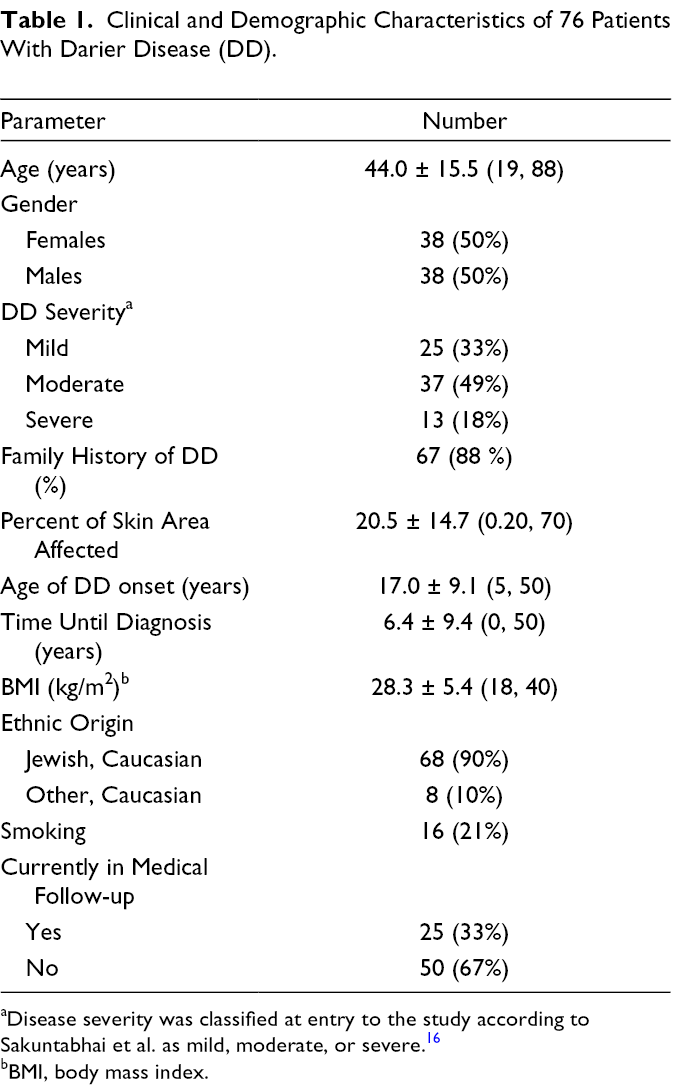
aDisease severity was classified at entry to the study according to Sakuntabhai et al. as mild, moderate, or severe. 16
bBMI, body mass index.
Study Design
Participants were invited individually to a meeting with the study supervisor, during which inclusion criteria were verified, the study was explained, and signed informed consent was obtained. An interview was conducted based on a structured questionnaire (Supplemental Appendix 1), and a physical examination (Supplemental Appendix 2) was performed, which included being photographed. Medical information was also gathered by retrospective assessment of patient’s medical records.
The study protocol was approved by the institutional ethics committee at Emek Medical Center.
Instruments
Questionnaire
The highly comprehensive questionnaire (27-pages with over 300 items) designed for the study, gathered the following information: personal and family demography and medical history, family tree, initial manifestations of DD, its course, triggering factors, medical follow-up and hospitalizations, disease symptoms, location of the lesions, and involvement of nail, oral, and genital regions (Supplemental Appendix 1). The questionnaire was completed by the study investigator during a medical interview, which took approximately 60 minutes per participant.
Physical examination
A thorough cutaneous examination of the skin, hair, nails, oral mucus membranes, and external genitalia was performed by the primary investigator (R.P.D-G) to determine the skin findings, areas involved, disease severity, and classification of the disease. 11,16 Height and weight were also recorded. Disease severity was classified as mild, moderate, or severe according to the criteria of Sakuntabhai et al. 16 Mild—keratotic papules scattered sparsely over the trunk or flexures, or disease limited to one or two areas. Moderate—more extensive papular lesions or localized verrucous plaques. Severe—coalescent verrucous plaques involving most of the trunk or grossly hypertrophic flexured disease. 16
The examination was conducted according to a form designed for the study (Supplemental Appendix 2), and took approximately 25 minutes to complete.
Photography
Skin and mucous membranes were photographed by a professional medical photographer.
Statistical Analysis
Data were analyzed with SAS statistical software, version 9.4 (SAS Institute, Cary, NC, USA); significance was set at P < .05. Demographic and clinical data are presented as frequencies and percentages for categorical variables and by mean ± SD for continuous variables. Wilcoxon’s two sample test was applied to compare patients with severe and mild disease by age of onset. Spearman’s rank correlation coefficient was used to determine the relationship between body surface area (BSA) (as continuous variable) and age of onset of the disease. Logistic regression model was used to explore the association between severity of disease or BSA (as categorical variable) and several categorical variables.
Results
Disease Characteristics
Initial onset
Average age of onset of the disease was 17 ± 9 years (5-50); and time from onset of cutaneous symptoms to diagnosis was 6 ± 9 years (0, 50).
The disease appeared in the summer in 53 patients (83%), in the spring in 8 patients (13%), in the winter in 2 patients (3%), and in the fall in 1 patient (1%). Twelve patients could not recall the season in which the disease first appeared.
Only 6 patients (8%) stated that the disease was triggered by a sunburn.
Exacerbating factors
Exacerbation of the disease was reported by 67 patients (88%) during the summer and while sweating, by 56 patients (74%) following exposure to sunlight, by 45 (59%) at times of mental distress, by 33 (43%) following contact with wool, and by 23 (30%) during febrile illness.
Exacerbation of the disease was reported by 21 patients (28%) following surgical procedures and by 26 patients (34%) following skin injury. Twenty patients (26%) reported being hospitalized due to disease exacerbation.
Of the 38 females in the study, 18 (47%) reported disease onset prior to menarche, 15 (39%) reported change in disease course during pregnancy, 9 (60%) of whom described amelioration of symptoms. Twenty-five (67%) reported no effect of oral contraceptive pills on the disease.
Clinical Features
Distribution
Seventy-five of the 76 patients agreed to a physical exam. The most frequently involved areas were the palms (99%) and nails (93%). Other areas involved were the chest (77%), neck (71%), ears (69%), feet (69%), and body folds (67%). The most severely involved areas were the chest (28%), neck (24%), and body folds (24%). The areas most lightly involved were the upper extremities (18%), abdomen (15%), lower extremities (15%), and back (12%). Most of the patients (88%) exhibited a combination of the patterns suggested by Burge et al with no clear dominance of a specific location. 11 One patient exhibited a linear pattern of lesions on the trunk.
Morphology
The most frequent primary lesion was a keratotic papule, found in 95% of patients (Figure 1a), mostly on the trunk. The most frequent lesions on the hands were wart-like papules and pits (Figure 1b and 1c), and on the fingernails were red and white lines, ridging, V-shaped notches, and subungual hyperkeratosis (Figure 1d). The wart-like lesions on the hands were more visible after soaking in water for 5 minutes. This phenomenon was noted in 58 of 59 patients in the physical examination and could be termed the “wet hand sign.” (Figure 2)

Classical lesions of Darier disease in the skin and nails. (
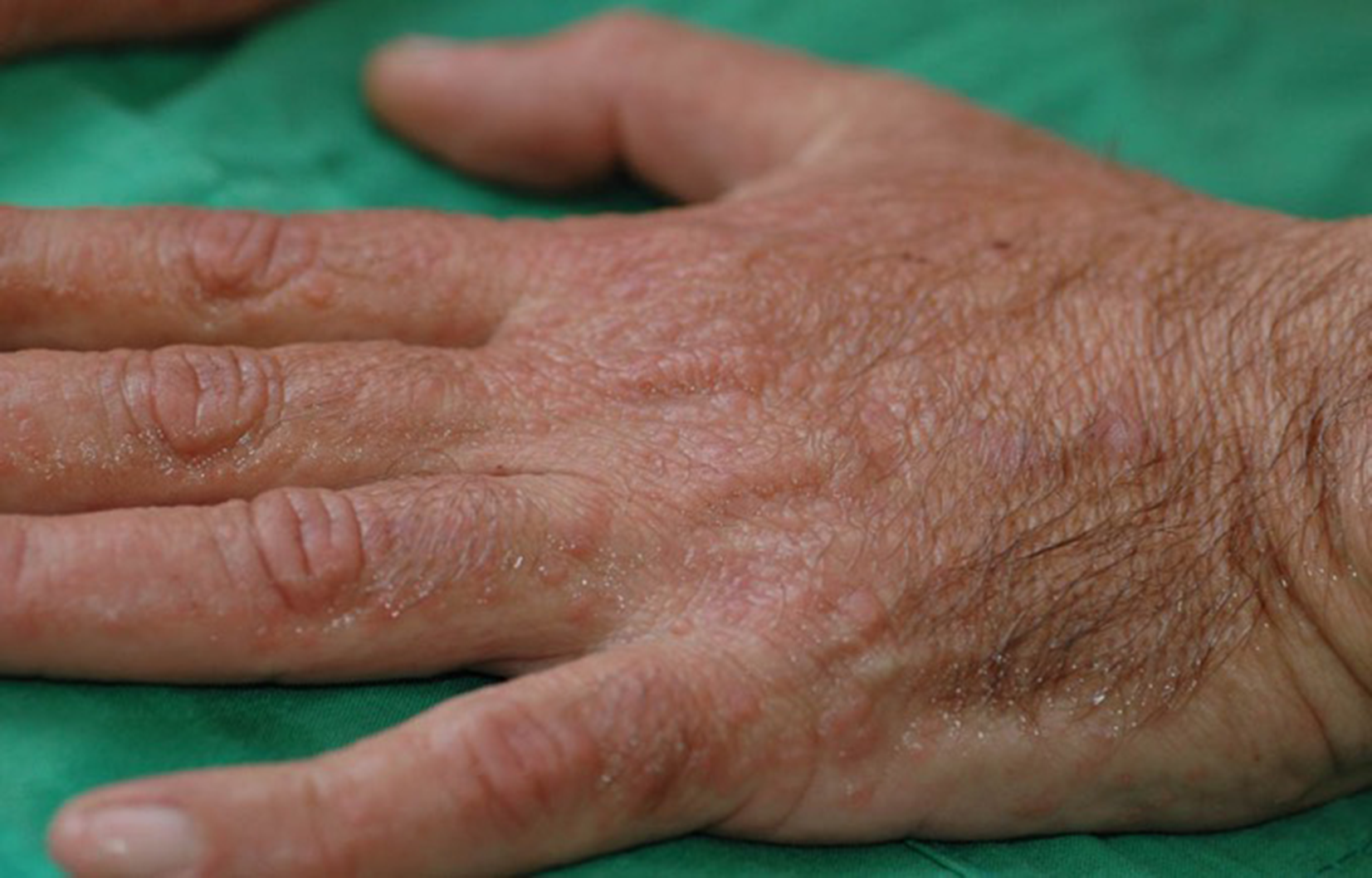
The “wet hand” sign. Heightened wart-like lesions on the hands following soaking in water for 5 minutes.
Rare skin findings included palmar keratoderma in six patients (8%) from three families, plantar keratoderma in 11 patients (15%) from six families (Figure 3a), leukodermic macules in five patients (7%) from four families (Figure 3b), numerous giant comedones in one patient (1%) (Figure 3c), keloid-like vegetations in body folds in six patients (8%) from four families (Figure 3d), and blisters on plantar area and small black hemorrhagic macules with jagged borders on the palms in five patients (7%) from two families (Figure 3e and f).

Non-classical lesions of Darier disease (variants of DD) (
The patient with giant comedones and two patients with hemorrhagic bullae also had acral keratoderma.
Mucous membranes and nails
Oral involvement was found in 30/69 (43%) of patients. Examination of the genitalia in 50 patients (25 men and 25 women) revealed lesions in 12 women (48%) and 4 men (16%). Nail involvement was found in 67/72 patients (93%). The objective and subjective findings are presented in Table 2.
Oral, Genital, and Nail Findings in Patients With Darier Disease.

N/A not available.

Manifestations of Darier disease in oral and genital mucous membranes. (a) White papules on hard palate (arrow). (b) Pink papules, a few with central erosions on labia minor and clitoris (arrow).
Correlation to Disease Severity
There was a statically significant difference between severity of disease and age of onset. Patients with severe disease were younger at onset (average 13.7 ± 11.4 years) than patients with mild disease. (average 18.6 ± 10.0 years, P = .01).
Disease severity influenced the probability of having genital and oral lesions. Patients with severe DD had 10 times greater chance of having genital lesions than patients with mild disease (95% CI 1.61-60.24, P = .01). Female patients with severe DD had 22 times greater chance of having genital lesions than those with mild disease (95% CI 1.54-314.27, P = .03). There was a clear trend toward the association of disease severity and having oral lesions: patients with severe DD had a 4.5 greater chance of having oral involvement than patients with mild disease (95% CI 0.85-24.11, P = .07).
Not surprisingly, disease severity was correlated with the probability of being under dermatological medical follow-up and having been hospitalized for treatment of DD. Patients with severe DD had nine times greater odds of being supervised under follow-up care than patients with mild disease (95% CI 1.78-43.49, P = .01), and 38 times greater chances of being hospitalized than patients with mild disease (95% CI 3.88-379.69, P = .002)
Discussion
Darier disease is an orphan disease, which accounts for the meager number of studies describing the clinical manifestations of the disease on large cohorts of patients. 15,17 -19 The largest cohort on DD was studied by Burge et al. and published in 1992. 11 In accordance with the results of this study, we found the hands to be the most common location of the disease. Indeed, 100% of our patients had cutaneous and/or nail involvement of hands. Thus, assessment of the skin on the hands for wart-like lesions, pits, and even keratoderma, and of the fingernails for red/white lines, ridging, V-shape notches and subungual hyperkeratosis in a patient with suspected DD offers a sensitive diagnostic approach.
The “wet hand sign” found in our study can also assist in diagnosis (Figure 2). This discovery was made thanks to the patients who reported that the wart-like papules on their hands become prominent after soaking in water for several minutes.
We found high percentage of oral involvement (43%) (Figure 4a) as previously documented in studies and case reports. 20,21
We also found high percentage of genital lesions: pink-red papules with central erosion. (Figure 4b) These lesions, reported previously only in rare case reports, were a common finding in our study, especially in female patients (48% of female patients and 16% of male patients). 22,23
Genital lesions were significantly more common in patients with a severe disease. Interestingly, patients with a severe disease also experienced younger age of onset of their disease.
Burge et al. suggested classifying the disease into four cutaneous categories according to the location of the lesions: seborrheic (predominantly scalp, face, and trunk with mild flexural involvement), flexural (predominantly flexural with mild involvement elsewhere), nevoid (unilateral zosteriform involvement), or hands only. 11 We found it problematic to apply this classification to our patients, since most of them (88%) exhibited a combination of the patterns suggested by Burge et al. 11 with no clear dominance of a specific location.
We found that the cutaneous manifestations of DD can be easily divided into two types of lesions; classical and non-classical. The classical lesions—keratotic papules, acral pits and acral wart-like lesions—are common and characterize the disease (Figure 1), whereas the non-classical lesions—acral keratoderma, leukodermic macules, giant comedones, kelloid-like vegetations, and acral hemorrhagic blisters—are rare and appear only in patients with DD variants (Figure 3).
In our study, all patients (100%) had at least one of the classical lesions and 33% of patients had non-classical lesions of DD.
Limitation of the study is collection of some of the data by interview, a technique which is subject to recall bias. There may be bias due to non-response when data are missing. Differential recall bias is also possible as patients diagnosed with DD know the reported exacerbation risk factors and may be more likely to report them (eg, onset in summer in 80% may be real vs may be suspected by the patient as they know UVR leads to disease exacerbation). Differential exposure ascertainment was minimized since questionnaire and physical examination were performed by same PI.
Darier disease is a debilitating and chronic condition, with both cutaneous and systemic manifestations, which can have a severe impact on patients’ general health and quality of life. Therefore, prompt and accurate diagnosis and treatment are crucial. This study, which follows our previous research on DD, 9,24 -26 sheds new light on the clinical aspects of DD; a novel skin manifestation (the “wet hand” sign), prominent genital involvement found to be more frequent in patients with severe disease, and combination of previously suggested disease patterns (flexural, seborrheic, nevoid, and acral) in most of our patients (88%), indicating that there may be a need to re-evaluate the classification of DD.
Further studies on this rare disease should be conducted to better characterize its full magnitude and develop improved treatment modalities.
Supplemental Material
Supplementary Material 1 - Supplemental material for An Update on the Cutaneous Manifestations of Darier Disease
Supplemental material, Supplementary Material 1, for An Update on the Cutaneous Manifestations of Darier Disease by Algit Yeshurun, Michael Ziv, Eran Cohen-Barak, Shiraz Vered, Dganit Rozenman, Muhammad Sah, Morad Khayat, Olga Polyakov, Boaz Amichai, Abraham Zlotogorski, Stavit Shalev and Roni P. Dodiuk-Gad in Journal of Cutaneous Medicine and Surgery
Supplemental Material
Supplementary Material 2 - Supplemental material for An Update on the Cutaneous Manifestations of Darier Disease
Supplemental material, Supplementary Material 2, for An Update on the Cutaneous Manifestations of Darier Disease by Algit Yeshurun, Michael Ziv, Eran Cohen-Barak, Shiraz Vered, Dganit Rozenman, Muhammad Sah, Morad Khayat, Olga Polyakov, Boaz Amichai, Abraham Zlotogorski, Stavit Shalev and Roni P. Dodiuk-Gad in Journal of Cutaneous Medicine and Surgery
Footnotes
Acknowledgments
We thank the patients for their participation in this study; Hadas Baumgarten, the study coordinator, and the dermatologists and other health professionals for their assistance in recruiting participants; Inbar Bizinski, Efrat Yaskil, and Rita Yuval for assistance in designing the study; Sandra Trumper and Yael Meshulam for photography of patients; and Dr Naama Schwartz and Paula Herer for assistance with the statistics. We wish to express our deepest gratitude to Dr Orna Blondheim, the CEO of Emek Medical Center, for her full support in our study in all of its stages. We are indebted to the Harold Grinspoon Foundation for funding the study; their support played no role in the design, methods, data collection, analysis or interpretation of the results, and its publication was not contingent on their approval.
Declaration of Conflicting Interests
The author(s) declared no potential conflicts of interest with respect to the research, authorship, and/or publication of this article.
Funding
The author(s) disclosed receipt of the following financial support for the research, authorship, and/or publication of this article: This work was supported by a grant from the Harold Grinspoon Foundation.

Supplemental Material
Supplemental material for this article is available online.
References
Supplementary Material
Please find the following supplemental material available below.
For Open Access articles published under a Creative Commons License, all supplemental material carries the same license as the article it is associated with.
For non-Open Access articles published, all supplemental material carries a non-exclusive license, and permission requests for re-use of supplemental material or any part of supplemental material shall be sent directly to the copyright owner as specified in the copyright notice associated with the article.
