
Abstract
Magnetic resonance imaging (MRI) has had a profound impact on both research and clinical management of multiple sclerosis (MS), but signal changes reflect underlying neuropathology only indirectly and often non-specifically. Positron emission tomography (PET) offers the potential to complement MRI with quantitative measures of molecularly specific markers of cellular and metabolic processes. PET radiotracers already available promise new insights into the dynamics of the innate immune response, neuronal function, neurodegeneration and remyelination. Because PET is an exquisitely sensitive technique (able to image even picomolar concentrations), only microdoses of radioligand (<10 µg) are needed for imaging. This facilitates rapid implementation of novel radioligands because extensive toxicology data is not required. In the future, molecular imaging could assist clinical decision-making with patient stratification for optimization of treatment selection.
Introduction
Potential applications of positron emission tomography (PET) in molecular imaging-based research for MS
Principles of molecular imaging
PET imaging relies on the design and manufacture of radiolabelled ligands which can bind selectively to a target of interest with minimal non-specific binding. These ligands are labelled with positron emitting radioisotopes that decay with a short half-life (approximately 20 and 110 min for 11C and 18F, respectively), reducing long-term health risks associated with the ionizing radiation. Following intravenous administration of the radioligand, positrons will collide with nearby electrons to produce pairs of photons that travel at 180° to each other and can be detected as coincident events by γ-detectors surrounding the subject. The relative position of detection of coincident events and their precise timing enable localization of the annihilation events, and the spatial distribution of the radiolabelled ligand can be reconstructed. The additional correction for degrading factors such as attenuation, scatter, random detection events, detector efficiencies and dead time allows for a quantitative image of the radio-labelled ligand’s concentration to be generated. Spatial resolution is limited (approximately 4 mm), but the PET data can be co-registered with structural data from computed tomography (CT) or MRI images to provide additional anatomical context to the functional PET data.
Development of new, molecularly specific PET radioligands is challenging. The binding affinity of a tracer for the target must be high enough to produce sufficient signal for detection, but it must dissociate from the target quickly enough to allow binding equilibrium to be reached within the timeframe of the scan (typically 1–2 h). A tracer with poor lipid solubility will not cross the blood–brain barrier, but if it is too lipid soluble, it will interact non-specifically with cell membranes and produce a high background signal. 4 Even if these hurdles are overcome, a tracer candidate can still be rendered unusable if it is metabolized rapidly. This can yield a mix of radiolabelled molecules that produce signals too complex to interpret. Finally, the tracer must be able to be prepared to pharmaceutical standards within the short time frame of approximately a single radioactive half-life of the isotope.
PET data can be acquired and analysed in different ways. Signal-to-background contrast obtained from a single 3D image is used clinically, for example to localize tumours with 18F-fluorodeoxyglucose (FDG) PET. The standardized uptake value is a simple means of signal quantification. It is calculated by normalizing the signal measured by the PET camera within the region of interest to the injected dose and body weight. 5 However, this analysis does not distinguish between radioligand specifically bound to the target, non-specifically bound to other structures, or free in the blood or tissue compartments. More complex analyses may be applied to 4D data acquired to provide kinetic information on the changing tracer time course in regions of interest. The application of tracer kinetic biomathematical models to these data allows for the determination of quantitative measures of specific binding, which is aided by the presence of a reference region devoid of specific binding. Such ‘reference regions’ facilitate modelling because signal localizing here cannot represent specific binding. Where targets are expressed throughout the brain and therefore do not have a reference region, more complex approaches are needed to estimate non-specific binding. 6
Current applications of molecular imaging relevant to MS
Imaging activated macrophages and microglia
Microglia are monocytes-derived, brain-resident macrophages contributing to the innate immune response. 7 They are rapidly activated by a wide range of stimuli, and upon activation undergo a stereotyped response which includes the adoption of an amoeboid shape, 8 proliferation and the expression of inflammatory mediators. 9 Microglial activation is an early consequence of brain insults of various types, 8 including MS, 10 although the nature and degree of activation varies depending on the stimulus. 11 In MS, haematogenous monocytes also are recruited to sites of focal acute inflammation, where they differentiate into macrophages. 12 There is therefore great interest in developing methods to detect and quantify activation or recruitment of monocyte-derived cells to allow dynamic assessment of the evolution and resolution of the innate immune response in vivo.
The 18-kDa Translocator Protein: a lead target
The 18-kDa Translocator Protein (TSPO), previously known as the peripheral benzodiazepine receptor, is the most widely explored PET marker of monocyte-derived cells. The TSPO has a well-defined role in steroid synthesis, translocating cholesterol from the outer to the inner mitochondrial membrane prior to conversion to pregnenolone. 13 Although the TSPO is expressed to some degree in many cell types, baseline expression in the healthy human brain is low. 14 The activation of resident microglia and recruitment of haematogenous monocytes seen in MS lesions is associated with a substantial increase in TSPO expression, making it a plausible molecular marker of the innate immune response. 15 It should be noted, however, that TSPO is not specific to microglia, and in certain animal models astrocytes’ TSPO expression is significant. 16,17 The TSPO is also implicated in various pathways including immunomodulation, porphyrin transport and apoptosis, but the mechanisms are currently unclear. 15
[11C]PK11195 is a radioligand with nanomolar affinity for the TSPO. 18 It has been applied to studying disease processes which involve microglial activation or the recruitment of macrophages such as MS, as well as ischaemic stroke, herpes encephalitis, Parkinson’s disease and Alzheimer’s disease. 19 – 23 In animal inflammatory models, increased expression precedes and correlates with subsequent demyelination. 24 In MS, increased [11C]PK11195 uptake co-localizes with areas of focal pathology identified by MRI as gadolinium-enhancing lesions. 23 However, uptake also is detected in areas of the brain defined by MRI as normal appearing white matter (NAWM), with higher uptake by NAWM reported in patients with greater disability. 25 [11C]PK11195 uptake in NAWM appears to correlate with brain atrophy. 26
MS has traditionally been regarded as a disease of white matter; however, over the last decade substantial grey matter (GM) involvement has been recognized. Cognitive deficits such as memory impairment and attention deficits are common in patients with MS,
27
and these deficits are not well explained by focal demyelination, which is restricted to white matter.
28
Neuropathological evidence of both subcortical
29
and cortical
30
–
32
GM disease in MS is emerging from post-mortem studies, and cortical lesions have been detected by MRI.
33,34
However, imaging GM lesions with MRI is challenging because there is poor MRI contrast between lesions and normal GM using conventional sequences. Techniques aimed at improving GM lesion detection include multisequence imaging protocols
35
and use of high-field MRI.
36
These approaches are problematic due to limited sensitivity, availability and safety of high-field magnets, as well as susceptibility artefacts in high-field imaging. However, GM lesions are characterized neuropathologically by pronounced macrophage and microglial activation.
37
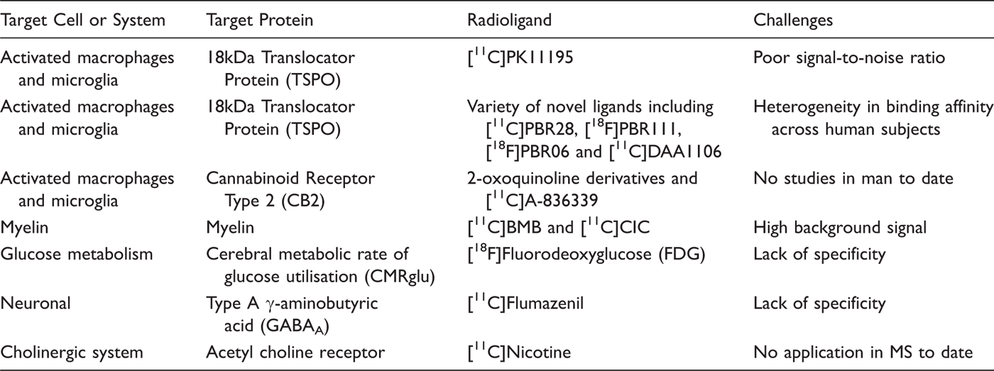
The sensitivity of PET combined with the low TSPO expression in normal GM make TSPO PET promising as an improved method for GM lesion imaging. In a recent study, substantial increases (around 120%) in [11C]PK11195-specific binding were detected in cortical GM of patients with secondary progressive MS relative to healthy controls (Figure 1).
38
The magnitude of the signal correlates with both expanded disability status scale (EDSS) and multiple sclerosis impact scale (MSIS-29), suggesting that TSPO–PET can provide an estimate of the contribution from microglia to disability in MS.
Summed [11C]PK11195 PET image at the cortical level of a patient with secondary progressive MS. The image is coregistered and fused with a 1.5 Tesla MRI image. This study demonstrates increased microglial activation in vivo in the cortical GM (arrow) for the first time. Image courtesy of M. Politis and P. Piccini, Imperial College London.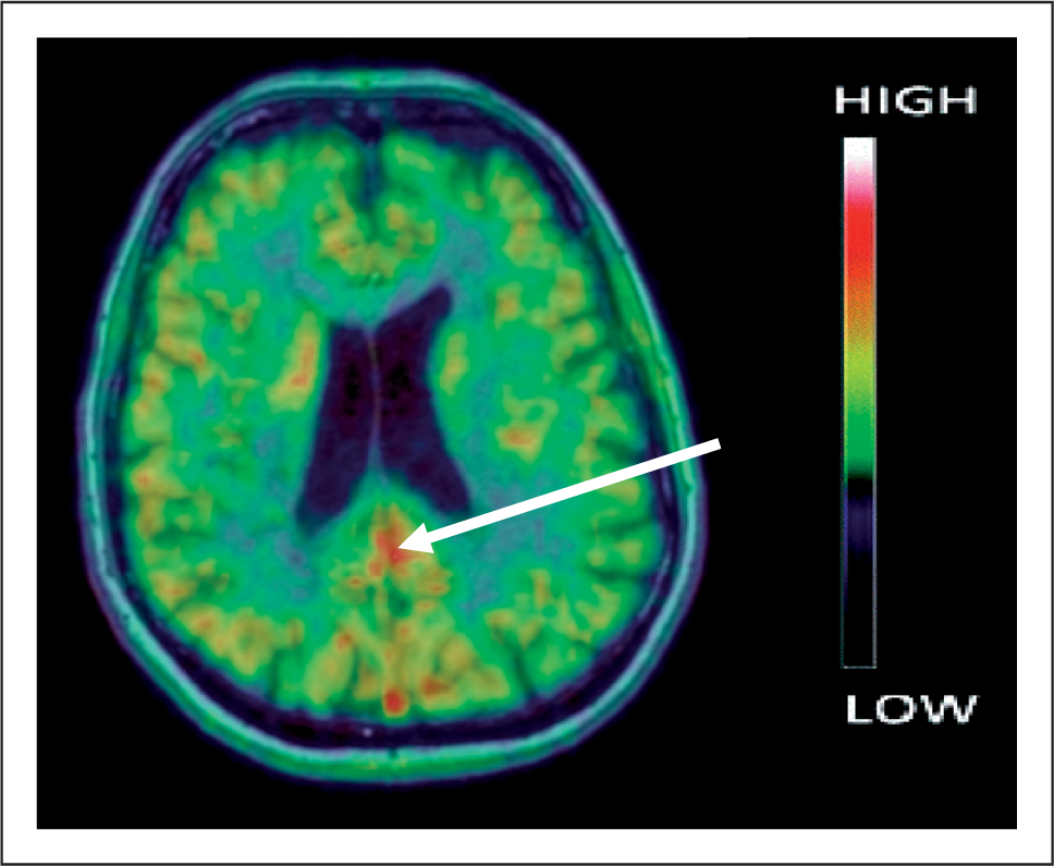
However, despite the insight that the radioligand has provided, it is well recognized that in vivo PET studies using [11C]PK11195 are limited by relatively poor signal-to-noise ratio. 23 This is a result of a combination of factors, the most significant of which is that [11C]PK11195 binds non-specifically to sites in addition to TSPO. The high non-specific binding increases the complexity of modelling and limits accurate quantification. This precludes confident use of [11C]PK11195 in monitoring treatment responses for clinical or drug development applications. There has therefore been considerable interest in developing TSPO tracers with improved signal-to-noise ratio relative to [11C]PK11195. Approximately 40 alternative radioligand candidates have been evaluated preclinically. 39 However, only a handful including [18F]PBR06, 40 [18F]FEPPA, 41 [11C]DAA1106, 42 [11C]DPA-713, 43 [18F]PBR111, 44,45 and [11C]PBR28 46 have been evaluated in humans.
Recent work has shown how exquisitely specific these radiotracers can be, but highlighted an additional consideration in their use. A pilot clinical PET study with [11C]PBR28 suggested that a minority of subjects lack a specific TSPO signal.
47
In recent work, we have clarified that this arises not because they lack TSPO, but because in approximately 20% of the population the affinity of the radioligand for the target is markedly reduced (low-affinity binders, LABs).
48
Furthermore, among patients who show a measurable PET signal with [11C]PBR28, two groups can be defined: high-affinity binders (HABs) who express a single class of high-affinity binding sites, and mixed-affinity binders (MABs) who behave as if they express the two different binding sites found in HABs and LABs (Figure 2). Other recently reported TSPO ligands (including DPA713, DAA1106, PBR111 and PBR06) all behave in an analogous fashion (Owen et al, Journal of Nuclear Medicine, in press).
49
Competition assays with [3H]PK11195 and unlabelled TSPO ligands in human brain tissue in vitro. Each data point represents the mean value of at least four subjects. Error bars represent SEM. The data show the variation in binding affinity in human subjects with PBR28 (right hand panel), with three non-overlapping groups evident: High affinity binders, Low affinity binders and Mixed affinity binders. Novel TSPO ligands including PBR111, DAA106, PBR06 and DPA713 bind analogously to PBR28, but the traditional TSPO ligand, PK11195, does not distinguish the three groups (left hand panel). Modified with permission from Owen et al.
48
Prior knowledge of differences in binding affinity will allow differences in PET signal between subjects to be interpreted directly in terms of receptor density, provided that binding affinity remains constant over time (which has yet to be confirmed directly) and that the binding affinity in the brain can be predicted from binding affinity in peripheral blood cells, which could therefore be used as a screening test. Early evidence suggests that binding affinity is constant over time and across tissues, 50 but it remains important to demonstrate this directly for the three binding affinity groups.
We anticipate that these challenges to accurate quantification will be met. PET studies with TSPO ligands therefore offer the opportunity to serially measure macrophage and microglial activation. While TSPO expression does not distinguish between activated resident microglia and recruited macrophages, future applications could couple TSPO–PET with MRI enhanced with ultrasmall superparamagnetic iron oxide particle (USPIO) contrast, as USPIOs are avidly endocytosed by circulating monocytes and therefore offer the possibility of specifically imaging newly recruited macrophages. 51 – 53
Cannabinoid Receptor Type 2 (CB2): an alternative target
Alternative approaches to microglial imaging are possible. Integration of information from use of multiple molecular markers should provide a more complete profile of the inflammatory response. The cannabinoid receptors belong to a class of G protein-coupled receptors with seven transmembrane spanning domains.
54
Two subtypes of cannabinoid receptor have been identified to date: CB1 receptors are distributed predominantly in the brain and are responsible for the psychoactive effects of the exogenous cannabinoids extracted from the marijuana plant;
55
CB2 receptors, in contrast, are expressed in very low levels in healthy brain, but are abundant in immune cells
56
including microglia.
57
As microglia become activated, CB2 expression increases by approximately 10-fold;
58
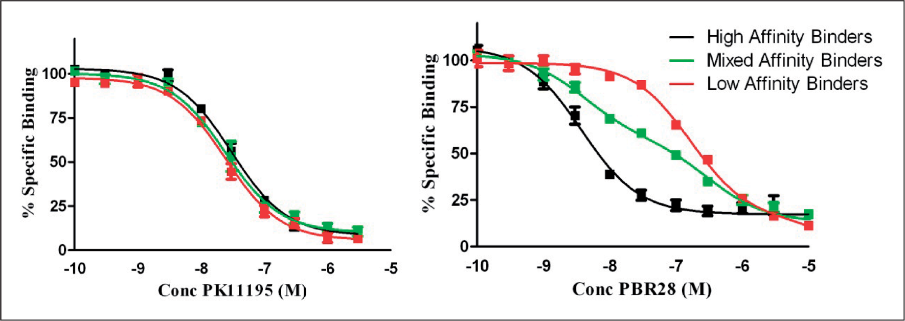
post-mortem human brain tissue from patients with MS shows substantial CB2 expression, co-localizing with microglial markers (Figure 3).
59
CB2 therefore has been proposed as a possible PET target to identify, quantify and monitor microglial activation. CB2 PET ligands have been synthesized, and early evaluation in the mouse confirms they cross the blood–brain barrier and demonstrate high specificity for the target.
60,61
Studies in man are awaited.
Co-localization of CB2 with microglia, monocytes, astrocytes and T cells. Double-immunofluorescence assays of CB2-positive cells (left column) and phenotypic markers (middle columns). 
Molecular imaging of myelin
The limited correlation between acute inflammatory demyelination and persistent neurological deficit can be partly ascribed to spontaneous myelin repair, which occurs to a variable degree. When acute lesions are remyelinated structural integrity is partially restored, resulting in functional recovery of saltatory conduction. Reducing demyelination and promoting remyelination in MS patients are therefore potential therapeutic objectives. 62 An imaging marker capable of quantifying myelin in vivo could act as a useful endpoint with which to assess drugs in development, and might also have clinical application as a means to monitor response to treatment. Currently, there are no well-validated, direct methods for assessing myelin. MRI magnetization transfer 63 or diffusion-tensor 64 methods are in widespread research use, but suffer from lack of specificity and limits in absolute quantification of the myelin changes. 65 PET potentially offers greater specificity in the detection of myelin and the ability to quantify myelination. Radiotracers which bind with high affinity to components of the myelin sheath have hence been recently developed for this purpose illustrating this potential, although few reports have yet been published.
1,4-bis(paminostyryl)-2-methoxy benzene (BMB) is a distilbene derivative, structurally similar to the amyloid imaging probe AV45, which binds white matter tracts in mouse brain sections ex vivo.
66
Binding to white matter is reduced in areas of demyelination both in a rodent model (Figure 4), and in post-mortem brain samples from patients with MS.
66
Studies in vivo confirm that BMB crosses the blood–brain barrier following intraperitoneal injection, and PET data demonstrate that the higher binding potentials of white relative to cortical GM could be distinguished.
66
However, in the baboon BMB also binds non-specifically, which increases the background signal. This and lack of a clearly saturable specific binding site are likely to limit accurate quantitation, but relative changes in myelin in individuals over time may still potentially be assessed. The use of BMB in humans has not yet been reported.
[11C]-Case imaging compound (CIC) binding to myelin in a rat model of demyelination. Axial views of corpus callosum of control and demyelinated rat model. Lysolethicin was injected into the right hemisphere of the corpus callosum (arrow) to induce demyelination. The reduction in the demyelination (panel D) relative to the control (panel B) is evident from the lower [11C]-CIC binding in the PET images (red represents high signal, blue represents low signal). The PET images are fused with MRI (rapid-acquisition relaxation-enhanced acquisition, response time/echo time = 2000/40 ms, 4 echoes; field of view = 45 mm × 45 mm, matrix = 256 × 256). 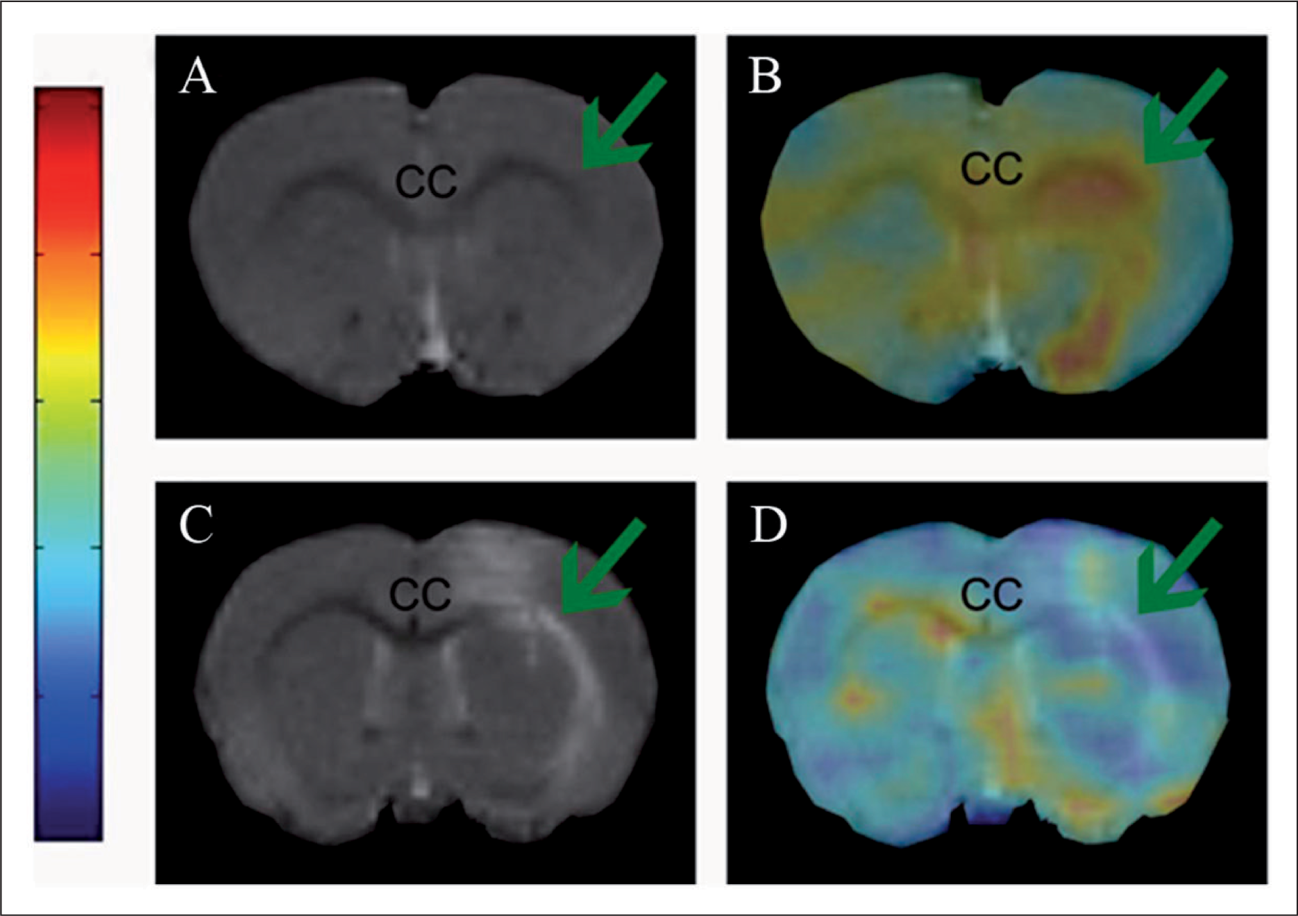
[11C]-Case imaging compound (CIC) is a similar molecule to BMB. Ex vivo fluorescent staining of mouse brain tissue sections indicates that CIC selectively stains white matter lesions such as the corpus callosum. 67 An in vivo PET study confirmed that [11C]CIC readily crosses the blood–brain barrier following intravenous injection, showed a correspondence between brain regions binding in vivo and ex vivo and, in a rat model of focal demyelination, localized demyelinating lesions. 67 However, the usefulness of CIC, an analogue of BMB, also is likely to be limited by high background signal.
The potential advantages offered by PET over MRI in myelin imaging (improved specificity and signal quantification) cannot be realized until suitable radiotracers, with specific, saturable and high-affinity binding to components of the myelin sheath and low background signal are developed and implemented. Given that the challenges of developing a useful radiotracer for myelin are considerable, an alternative approach would be to develop molecules that target receptors expressed specifically on demyelinated axons.
Assessing neuronal function and neurodegeneration
Measurement of brain metabolic function with 18F-fluorodeoxyglucose
Glucose is the major energy source for the brain, and because activity-related glucose metabolism is predominantly presynaptic, glucose uptake reflects the integrity and functional state of the synapse. 68 In the absence of an active inflammatory response, which increases regional glucose uptake because of the high level of anaerobic metabolism of lymphocytes and macrophages, 69,70 the measurement of glucose consumption can be used as an index of neuronal function. 71 Quantitative metabolic measurements are possible with FDG, a glucose analogue. Once transported intracellularly, FDG is phosphorylated by hexokinase, the first enzyme in the glycolytic pathway. The phosphorylation prevents egress of the labelled molecule from the cell, while the fluorination prevents further glycolytic metabolism.
FDG PET therefore provides a means of measuring the cerebral metabolic rate of glucose utilization (CMRglu) and for independently estimating glucose transport rate and hexokinase flux, which provides a bioenergetic surrogate for neuronal function. In diseases associated with a loss of synapses, CMRglu decreases. A global reduction in GM CMRglu in patients with MS compared with healthy controls has been reported 72,73 that showed regional variation from 3–18% and correlated with total lesion volume on MRI. 74 A longitudinal study in MS has demonstrated a decrease in CMRglu over a 2-year period, 75 consistent with the anticipated progression of neuropathology. Other studies have demonstrated associations between CMRglu and clinical symptoms. Global CMRglu correlates with cognitive function (r = 0.58), 74 and regional reductions have been associated with fatigue 73 or memory loss. 76
Markers of specific neurotransmitter systems
Central benzodiazepine receptor
[11C]-flumazenil binds the benzodiazepine site on the γ-aminobutyric acidA (GABAA) receptor, which is abundant on cortical neurones, and has therefore been used to measure cortical loss or dysfunction in various diseases including ischaemic stroke, 77 amyotrophic lateral sclerosis 78 and Alzheimer’s disease. 79 The potential for molecular imaging to use this as a tool to discriminate between changes in synaptic function and neurodegeneration is illustrated by a case report of a patient with a demyelinating lesion in the optic radiation. 80 In this patient, the synapse loss in the occipital cortex, evidenced by a marked reduction in CMRglu, was accompanied by only a modest decrease in neuronal density, measured by [11C]flumazenil binding.
The cholinergic system
Targeting the cholinergic system to improve memory and cognitive function in MS has been attempted on the basis of the success (albeit modest) in Alzheimer’s disease. 81 Cerebrospinal fluid markers show a reduction in cholinergic activity in patients with MS. There have been a few small pilot trials with anticholinesterases in MS, of which two have suggested modest increases in cognitive function. 82,83 Imaging studies with PET tracers that have been developed to probe cholinergic activity within the brain may have utility for defining which patients could respond, and in understanding mechanisms for any response in MS. For example, they could be used to investigate to what degree the cholinergic pathway contributes to cortical dysfunction in MS, and whether there exists a subgroup of patients who will derive clinically significant benefit from anticholinesterase use. Such tracers are already in use for other applications, most commonly to investigate Alzheimer’s disease, 84 but have yet to be used in MS.
Conclusion
Molecular imaging provides a powerful tool for dynamic, in vivo neuropathology. While MRI-based molecular imaging approaches have been applied preclinically for a range of applications, 85 the relatively high plasma concentrations needed for MRI contrast agents increase toxicity risks, complicating their rapid translation to human studies. The high selectivity of PET allows microdosing and, in favourable situations, radioisotopes can be incorporated directly into the chemical structures of drugs already evaluated for human use. Already, PET radioligands suitable for application to the problems posed by therapeutic strategies for MS, such as the control of innate immune response, are available. Moreover, molecular imaging can be used to test novel therapeutics to ascertain whether they reach the target and have the desired pharmacodynamic action in early phase studies, thereby helping decision-making in the selection of candidate molecules for phase III trials. 86 In the clinic, molecular imaging also offers the possibility of allowing specific individual patient stratification for selecting or changing treatments. PET already is widely used for diagnosis, stratification and treatment monitoring in oncology. 87 However, if the full potential of PET in applications to MS is to be realized, development and validation of a wider range of relevant probes is required. This is a complicated and costly process, dependent upon interdisciplinary partnerships between centres with the necessary academic expertise, financial resources and access to patients, but one that the MS research community should begin to champion.
Footnotes
Acknowledgements
All co-authors thank Dr Paolo Muraro and Professor Roger Gunn for their thoughtful review of the draft manuscript. PP thanks the MRC for funding.
Funding
DRJO has been funded by the Wellcome Trust-Imperial College-GSK Translational Medicine Training Programme.
Conflict of interest statement
PMM is a full time employee of GlaxoSmithKline. The authors declare that they have no other conflicts of interest.