






Abstract
Background:
Memory disturbance is a common symptom of multiple sclerosis (MS), but little is known about autobiographical memory deficits in the long-term course of different MS subtypes. Inflammatory activity and demyelination is pronounced in relapsing–remitting multiple sclerosis (RRMS) whereas, similar to Alzheimer’s disease, neurodegeneration affecting autobiographical memory-associated areas is seen in secondary progressive multiple sclerosis (SPMS).
Objective:
In light of distinct disease mechanisms, we evaluated autobiographical memory in different MS subtypes and hypothesized similarities between elderly patients with SPMS and Alzheimer’s disease.
Methods:
We used the Autobiographical Memory Interview to assess episodic and semantic autobiographical memory in 112 education- and gender-matched participants, including healthy controls and patients with RRMS, SPMS, amnesic mild cognitive impairment (aMCI) and early Alzheimer’s dementia (AD).
Results:
Patients with SPMS, AD, and aMCI, but not with RRMS, exhibited a pattern of episodic autobiographical memory impairment that followed Ribot’s Law; older memories were better preserved than more recent memories. In contrast to aMCI and AD, neither SPMS nor RRMS was associated with semantic autobiographical memory impairment.
Conclusion:
Our neuropsychological findings suggest that episodic autobiographical memory is affected in long-term patients with SPMS, possibly due to neurodegenerative processes in functional relevant brain regions.
Keywords
Introduction
Autobiographical memory (AM) is a mental representation of personal events and data that allows for the retrieval of personal semantic ‘facts’ (e.g. date and place of wedding) and the recollection of specific events from our past (episodic memory; e.g. the situation of one’s wedding ceremony).
The neuroanatomical substrates of AM include the posterior cingulate cortex; the medial temporal lobe (including the hippocampus); the medial prefrontal cortex; and the inferior parietal lobe. The role of the medial temporal lobe and hippocampus on AM encoding and retrieval is still under debate, even with respect to the recollection of recent and remote memories. 1
The standard model of consolidation 2 suggests a temporary dependency on medial temporal lobe and hippocampal structures for formation and consolidation of declarative knowledge. After the consolidation process, remote memories are stored in the neocortex and the hippocampus is no longer required for the retrieval of these memories. By contrast, the Multiple Trace Theory 3 proposes that the hippocampal formation encodes all information and forms memory traces that include both hippocampal and neocortical neurons. In this model, each time a memory is retrieved, a new hippocampally mediated trace is created. Thus, frequently repeated remote memories are represented by more and stronger hippocampal–neocortical traces than recent memories, making them less susceptible to disruption by brain damage. 3
Several studies have examined AM in amnesic syndromes 4 and degenerative dementias. 5 AM impairment is one of the most apparent symptoms in Alzheimer’s disease, 6 where there is a pattern of retrograde amnesia that follows Ribot’s Law 7 such that older memories are better preserved than more recent ones. Even patients with amnesic mild cognitive impairment (aMCI) exhibit deterioration of AM, including personal incident memory and personal semantic data, that follows a temporal gradient. 8 As the presence of aMCI is a risk factor for Alzheimer’s dementia (AD), 9 AM impairment in MCI may signal the onset of hippocampal dysfunction associated with neurodegenerative (Alzheimer’s disease-related) pathology. 8
Multiple sclerosis (MS) is characterized by the appearance of widespread lesions and plaques in the brain and spinal cord. These lesions and plaques affect the myelin sheath, thus impairing axonal propagation of the action potential. 10 While inflammatory demyelination has traditionally been seen as the main disease process in MS, axonal damage or loss is receiving increasing attention. 11 Grey matter damage may be the pathological correlate of the cognitive dysfunction that arises in 40–70% of patients with MS. 12
The topography of grey matter atrophy in MS differs among the MS subtypes: relapsing–remitting MS (RRMS), secondary progressive MS (SPMS), and primary progressive MS (PPMS). 13 Whereas demyelination and inflammatory activity is pronounced and seems to be the dominant process in RRMS, 14 slowly expanding demyelinating lesions and neurodegenerative events predominate in SPMS and PPMS. 14 -16 Axonal loss and neurodegeneration occur in cortical areas involved in cognitive processing 13,15 and seem to increase cognitive deterioration in long-term patients with progressive MS subtypes. 17,18
Various aspects of cognitive functioning are affected in MS, 19 but only a few studies have examined MS-related AM dysfunction. 20,21 Kenealy et al. 20 found poorer performance on the autobiographical incident schedule of the Autobiographical Memory Interview (AMI) 22 than in the retrieval of personal semantic information in elderly patients with MS. They also found a temporal gradient in autobiographical episodic memory, with better preservation of memory for remote than for recent incidents. By contrast, Paul et al. 21 found an impairment of memory for personal semantic information but not autobiographical incident memory as assessed with the AMI 22 in a sample of MS patients with an average disease duration of 11 years. They did not find a temporal gradient in the retrieval of either personal semantic information or autobiographical incidents.
Cortical thinning varies regionally in patients with SPMS and is most prominent in areas of the brain that have extensive cortico-cortical connections. 13,15,16 These areas play an important role in AM storage and retrieval. 1 Similarly, brain regions involved in AM seem to be affected by the process of grey matter atrophy in patients with AD. 23
In the present study we compared AM retrieval in an education- and gender-matched sample of healthy controls (HC) and patients with aMCI, early AD, RRMS, or SPMS, using the AMI. 22 As neurodegeneration occurs in similar cortical areas in (pre)dementia and progressive MS, we hypothesized that SPMS patients would exhibit a graded loss of AM akin to that seen in patients with early AD or aMCI. We predicted that patients with RRMS, in which inflammatory activity and demyelination dominate 14 and primarily affect speed of information processing, 24 would exhibit normal AM similar to that of HC participants.
Methods
Participants
There were 112 participants in this study, including 67 females and 45 males, with a mean age of 65.9 ± 9.4 years. All groups had an n of 20 except SPMS, which comprised 32 patients. All participants had normal or corrected-to-normal visual acuity and sufficient hearing ability. None of the participants had a physical handicap that affected his or her ability to perform the required tasks, nor any indication of neurological or psychiatric disorders unrelated to his or her diagnosis. The local ethical committee of the University Hospital of Tübingen approved the study. All participants signed an informed consent form after receiving a detailed explanation of the study.
Patients with aMCI or AD
Patients with aMCI or AD were recruited from the Memory Clinic of the Department of Psychiatry and Psychotherapy of the University Hospital of Tübingen. They underwent physical, neurological, neuropsychological, and psychiatric examinations, as well as brain imaging. Routine laboratory tests included Lues (syphilis) serology as well as analysis of vitamin B12, folic acid, and thyroid-stimulating hormone levels.
The diagnosis of aMCI was defined by the Mayo criteria, 25 which include the presence of a memory complaint (corroborated by an informant), objectively impaired memory function, preserved general cognitive function, intact activities of daily living, and the absence of dementia. All patients with AD met diagnostic criteria of probable AD according to the Diagnostic and Statistical Manual of Mental Disorders, fourth edition. 26 All patients with AD had a score of four on the Global Deterioration Scale. 27
Patients with RRMS or SPMS
In total, 52 elderly patients who were under regular follow-up care at the University Hospital Tübingen, Center of Neurology, and who had definite MS according to the McDonald criteria, 28 participated in this study and were classified as RRMS or SPMS. All underwent a detailed comprehensive neurological examination and were scored accordingly on the Expanded Disability Status Scale (EDSS). 29 The EDSS is a method to quantify disability in patients with MS. Eight functional systems (pyramidal, cerebellar, brainstem, sensory, bowel and bladder, visual, cerebral, other) are scored on an ordinal clinical rating scale ranging from 0 (normal neurological examination) to 10 (death due to MS) in half-point increments. 29
Healthy control group
HC individuals did not have a history of neurological or psychiatric disease or any sign of cognitive decline, as confirmed by a clinical interview.
Neuropsychological assessment
All participants underwent neuropsychological assessment of global cognition (Mini-Mental State Examination (MMSE)), 30 executive functions (Trail Making Test Part B) 31 and verbal learning and memory (Consortium to Establish a Registry for Alzheimer’s Disease (CERAD) word list immediate and delayed recall; word list recognition). 32 Episodic memory was assessed by verbal learning of 10 words over three trials as well as recall and recognition of the 10-word list. Trail Making Test Part B includes numbers (1–13) and letters (A–L) which must be connected in an ascending alternating pattern (i.e. 1–A–2–B–3–C, etc.) as fast as possible. As the subjects have to switch between mental sets the Trail Making Test B is used to assess cognitive flexibility and reflects executive functions. 33
All participants took part in the AMI. 22 The AMI is a semi-structured interview consisting of two parts that independently test recall for the two components of AM, autobiographical incidents and personal semantic information. The personal semantic schedule requests facts from childhood, early adult life, and recent years (i.e. the last 5 years before test administration). For each time period, a maximum of 21 points can be achieved. The autobiographical incident questionnaire requests personal experiences of the same three time periods. For each time period, the maximum score is nine.
Data analysis
The SPSS-16 statistical package for Windows was used for data analysis. For all tests, the level of significance was set to p<0.05. Levene’s test was used to assess homogeneity of variance. Differences in age, education, global cognition, anterograde memory (verbal learning, recall and recognition) and frontal/executive functions were assessed using a one-way analysis of variance (ANOVA) followed by a post hoc Tukey test. Independent samples t-test was used to detect differences among the MS subgroups in years since diagnosis. We applied the Pearson chi-square test to detect group differences in gender distribution and the nonparametric Mann–Whitney U test to detect group differences among the MS subgroups in the EDSS scores. We used two-way ANOVAs with group and time period as factors to examine semantic and episodic autobiographical memory recall. We examined between-group differences within each time period using a one-way ANOVA followed by a post hoc Scheffé test.
To test for a temporal gradient (i.e. better preservation of remote than more recent memories) paired samples t-tests (childhood vs. recent years; childhood vs. early adulthood; early adulthood vs. recent years) within each group were calculated. Results were corrected for multiple comparisons using Bonferroni correction (i.e. comparisons were performed at the p<0.017 level of significance).
To test for a substantial influence of age on memory recall, we used two-way analyses of covariance with group and time period as factors and age as a covariate to examine semantic and episodic memory recall.
Results
Clinical and demographic characteristics of the participants
Table 1 presents the clinical and demographic characteristics of the participants. There were no significant group differences in gender distribution (χ2 [4]=1.194; p=0.879) or years of education (F[4,107]=1.058; p=0.381). There was a significant group difference in age (F[4,107]=50.297; p<0.001). Participants in the RRMS and SPMS groups were younger than those in the HC, aMCI, and AD groups.
The RRMS group scored lower on the EDSS than did the SPMS group. These two groups did not differ in years since diagnosis.
Clinical and demographic characteristics of the participants.

Note: Values are expressed as mean (standard deviation).
p<0.001
HC: healthy controls; RRMS: relapsing-remitting multiple sclerosis; SPMS: secondary progressive multiple sclerosis; aMCI: amnesic mild cognitive impairment; AD: early Alzheimer’s dementia; M/F = male/female; EDSS: Expanded Disability Status Scale; n/a: not applicable; n.s.: not statistically significant.
Neuropsychological performance on global cognition, anterograde memory and frontal/executive functions
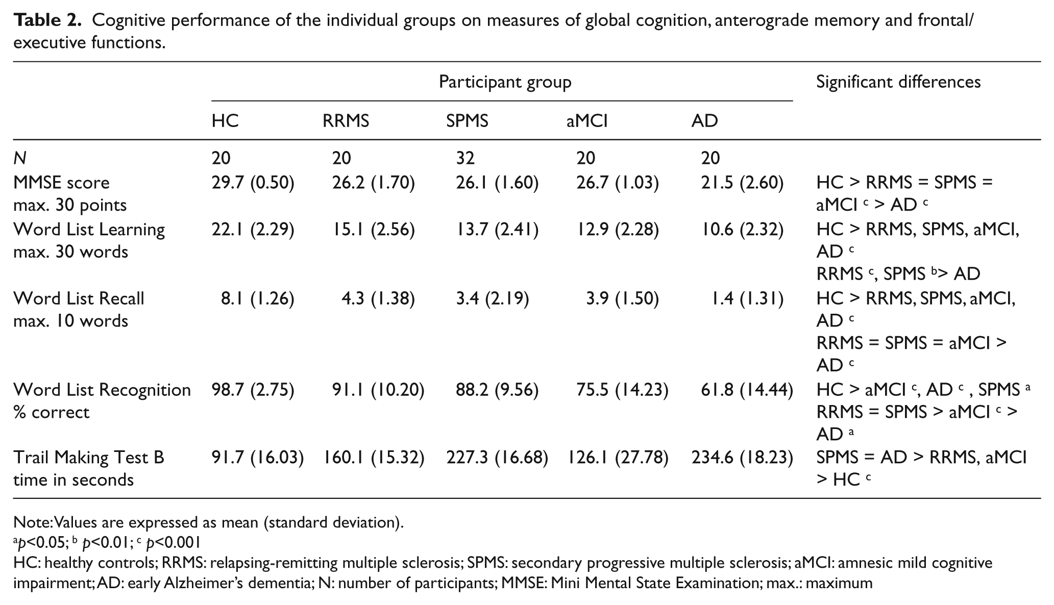
Table 2 presents the neuropsychological performance of the participants on measures of global cognition, anterograde memory (verbal learning, recall and recognition) and frontal/executive functions. There were significant group differences in the MMSE scores (F[4,107]=64.561; p<0.001), word list learning (F[4,107] = 51.951; p<0.001), word list recall (F[4,107] = 44.715; p<0.001), word list recognition (F[4,107] = 35.629; p<0.001), and Trail Making Test part B (F[4,107] = 38.860; p<0.001).
Cognitive performance of the individual groups on measures of global cognition, anterograde memory and frontal/executive functions.
Note: Values are expressed as mean (standard deviation).
p<0.05; b p<0.01; c p<0.001
HC: healthy controls; RRMS: relapsing-remitting multiple sclerosis; SPMS: secondary progressive multiple sclerosis; aMCI: amnesic mild cognitive impairment; AD: early Alzheimer’s dementia; N: number of participants; MMSE: Mini Mental State Examination; max.: maximum
MMSE scores in the AD group were lower than those in the RRMS, SPMS, and aMCI groups, which did not differ. Verbal learning was worst in patients with AD compared with the RRMS/SPMS groups but did not differ from that of patients with aMCI. Word list recall was mostly impaired in patients with AD compared with patients with RRMS, SPMS, and aMCI where no differences could be detected. Word list recognition was similar in patients with RRMS and SPMS and better compared with patients with aMCI and AD that were in turn worse than patients with aMCI. Time to complete Trail Making Test B was longest (i.e. indicating greater impairment) in patients with AD and SPMS compared with patients with aMCI and RRMS. In all tests the HC group performed better than the patient groups.
Autobiographical episodic memory
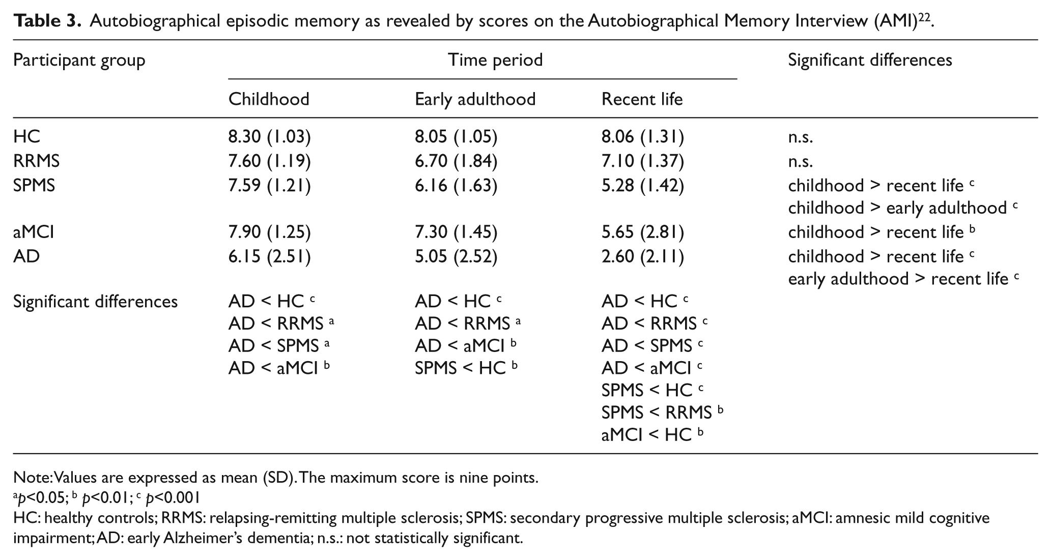
Table 3 presents the autobiographical episodic memory AMI scores in all groups. We found a significant main effect of time period (F[2,214]=43.070; p<0.001) and group (F[4,107] = 21.444; p<0.001). Overall, the AD group performed more poorly than did the other groups. The HC and RRMS groups performed similarly to one another, as did the SPMS and aMCI groups. The HC/RRMS scores were higher than the SPMS/aMCI scores.
Autobiographical episodic memory as revealed by scores on the Autobiographical Memory Interview (AMI) 22 .
Note: Values are expressed as mean (SD). The maximum score is nine points.
p<0.05; b p<0.01; c p<0.001
HC: healthy controls; RRMS: relapsing-remitting multiple sclerosis; SPMS: secondary progressive multiple sclerosis; aMCI: amnesic mild cognitive impairment; AD: early Alzheimer’s dementia; n.s.: not statistically significant.
Whereas recall of autobiographical incident memories for childhood, early adulthood and recent life was similar in HC and patients with RRMS, patients with SPMS, aMCI and AD were affected dependent on time period (significant group × time period interaction: F[8,214]=6.021; p<0.01). Recall of autobiographical incident memories from childhood was poorer in the AD group than in all other groups, which did not differ from one another. Recall of autobiographical incident memories from early adulthood was poorer in the AD group than in the HC, RRMS, and aMCI groups, which did not differ from one another, and poorer in the SPMS group than in the HC group. There were no other significant group differences. Recall of autobiographical incident memories from recent life was poorer in the AD group than in all other groups; poorer in the aMCI group than in the HC group; and poorer in the SPMS group than in the RRMS and HC groups. There were no other significant group differences.
Paired t-tests showed that temporal gradients (i.e. better preservation of remote than more recent memories) could be found in patients with AD, aMCI, and SPMS. In the aMCI/AD group, recall of autobiographical incident memories from childhood and early adulthood was better than from recent life. In the SPMS group, recall of autobiographical incident memories from childhood was better than from early adulthood and recent life. In contrast, in patients with RRMS and the HC group, recall of autobiographical incident memories from childhood, early adulthood, and recent life was similar. Figure 1 illustrates the episodic autobiographical memory performance of the individual groups.

Recall of personal incident memories across life periods in healthy controls (HC), patients with relapsing–remitting multiple sclerosis (RRMS), secondary progressive multiple sclerosis (SPMS), amnesic mild cognitive impairment (aMCI), and early Alzheimer’s dementia (AD) patients. Means and standard errors of means are given; max. = maximum. ** p<0.01; *** p<0.001.
Within each time period there was no main effect of the covariate factor age: childhood (F[1,35]=1.185; p=0.324); early adulthood (F[1,35]=0.745; p=0.797); recent life (F[1,35]=1.519; p=0.128).
Autobiographical semantic memory
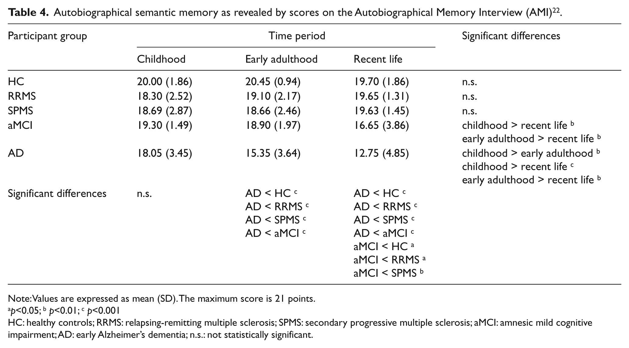
Table 4 presents the autobiographical semantic memory AMI scores in all groups. We found a significant main effect of time period (F[2,214]=10.453; p<0.001) and group (F[4,107] = 15.532; p<0.001). Overall, the AD group performed more poorly than did the other groups, which did not differ from one another.
Autobiographical semantic memory as revealed by scores on the Autobiographical Memory Interview (AMI) 22 .
Note: Values are expressed as mean (SD). The maximum score is 21 points.
p<0.05; b p<0.01; c p<0.001
HC: healthy controls; RRMS: relapsing-remitting multiple sclerosis; SPMS: secondary progressive multiple sclerosis; aMCI: amnesic mild cognitive impairment; AD: early Alzheimer’s dementia; n.s.: not statistically significant.
Whereas recall of semantic memories for childhood, early adulthood and recent life was similar in HC and the RRMS/SPMS groups, patients with aMCI and AD were affected dependent on time period (significant group × time period interaction: F[8,214]=10.156; p<0.001). Recall of semantic memories from early adulthood and recent life was poorer in the AD group than in all other groups. Recall of semantic memories from recent life was poorer in the aMCI group than in the RRMS, SPMS, and HC groups, which did not differ from one another.
Paired t-tests showed that temporal gradients (i.e. better preservation of remote than more recent memories) could be found in patients with AD and aMCI but, in contrast to autobiographical episodic memory not in patients with SPMS. In the AD group, recall of semantic memories from childhood was better than from early adulthood, which in turn was better than from recent life. In the aMCI group, recall of semantic memories from childhood and early adulthood was better than from recent life. In the RRMS, SPMS, and HC groups recall of semantic memories from childhood, early adulthood, and recent life was similar. Figure 2 illustrates semantic autobiographical memory performance of the individual groups.
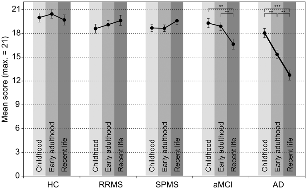
Recall of personal semantic memories across life periods in healthy controls (HC), patients with relapsing–remitting multiple sclerosis (RRMS), secondary progressive multiple sclerosis (SPMS), amnesic mild cognitive impairment (aMCI), and early Alzheimer’s dementia (AD) patients. Means and standard errors of means are given; max. = maximum. ** p<0.01; *** p<0.001.
Within each time period there was no main effect of the covariate factor age: childhood (F[1,35]=6.906; p=0.588); early adulthood (F[1,35]=1.190; p=0.320); recent life (F[1,35]=1.347; p=210).
Discussion
We investigated AM retrieval among elderly patients with RRMS and SPMS versus an education- and gender-matched sample of aMCI patients, early AD patients, and healthy controls. Patients with SPMS but not RRMS exhibited graded loss of personal incident memory akin to that seen in patients with early AD or aMCI. However, there were no differences in personal semantic data retrieval in patients with SPMS, patients with RRMS, or HC participants. These neuropsychological results point at distinct disease mechanisms in different MS subtypes. In patients with long-term SPMS, AM impairment might be due to neurodegeneration in brain areas that are functionally relevant for AM encoding and retrieval.
Patients with SPMS had difficulty recalling personal incidents from early adulthood and recent life as compared with HC participants. This pattern of autobiographical incident memory decline resembles qualitatively that seen in patients with aMCI or AD, where structures of the medial temporal lobe, including the hippocampus, are affected in the early stages of the disease. 34 Prior investigations on AM retrieval in patients with aMCI or AD indicate that AM relies on hippocampal integrity. 8 AM retrieval deficits appear to coincide with consolidation disturbances related to functional impairment in the medial temporal lobe and hippocampus. 3 Similar to aMCI and AD patients, in SPMS patients these brain regions seem to be affected by the grey matter atrophy. 13,15,16 These neurodegenerative processes become progressively more severe in long-term patients with progressive MS subtypes. 17,18
According to the standard model of memory consolidation, 2 formation and consolidation of declarative knowledge depends on neocortical regions including medial temporal lobe and the hippocampus, but only for a limited time period. Observations of a temporal gradient in memory performance resulting from hippocampal damage or atrophy have led to the hypothesis that the hippocampus participates only in memory consolidation processes, 2 and at the completion of these processes, memories are stored in the neocortex where they become resistant to hippocampal disruption. Hippocampal lesions are seen in SPMS patients, and the extent of hippocampal damage depends on disease progression. 35 Disturbed consolidation of autobiographical information should not affect remote memories. This accounts for the observed temporal gradient in our patients.
Consistent with our hypothesis, we did not observe substantial memory loss for autobiographical incidents or facts in the RRMS group. RRMS involves primarily inflammatory activity and is characterized by focal pathology of white matter. 14 Grey matter loss is seen mainly in the right precentral and postcentral gyri, 15 putamen and cingulate sulcus; 36 thus, regions involved in AM are relatively spared. Hippocampal degeneration is observable in RRMS, but differs from that seen in SPMS. RRMS is characterized by selective loss of volume in the cornu ammonis 1 region of the hippocampus, whereas SPMS is characterized by loss of volume in cornu ammonis 1 and other cornu ammonis regions. 37 Deficits in memory encoding and retrieval accompany loss of volume in hippocampal subregions; 35 hence, our findings might be explained in terms of increased involvement of regions that are functionally relevant for memory storage and retrieval in SPMS as compared with RRMS. 15 In addition, increased neural recruitment during episodic memory retrieval as seen in fMRI studies might account for the better performance in the RRMS group versus the SPMS group. 38
Unlike aMCI and early AD patients, patients with SPMS exhibited no memory deficits for personal semantic data. Deficits in memory for personal facts associated with Alzheimer’s disease might be due to early disturbance of the lateral temporal cortex due to neurodegenerative processes. 39 In SPMS this brain region seems to be less affected. 15
In conclusion, this study suggests that distinct disease mechanisms associated with MS subtypes lead to differences in AM. We observed graded AM loss in patients with SPMS but not RRMS. The temporal gradient in patients with SPMS resembles that of aMCI and early AD patients. Our findings could indicate that in long-term SPMS patients AM is affected by neurodegeneration of functionally relevant brain regions.
Footnotes
Funding
This research received no specific grant from any funding agency in the public, commercial, or not-for-profit sectors.
Conflict of interest
The authors declare that they have no conflicts of interest.