






Abstract
Background:
auto-antibodies against the potassium channel inward rectifying potassium channel 4.1 (Kir4.1) have previously been identified in 46% of patients with multiple sclerosis (MS).
Objectives:
to confirm these findings.
Methods:
we evaluated the presence of anti-Kir4.1 antibodies by enzyme-linked immunosorbent assay (ELISA) and immunofluorescence in 268 MS patients, 46 patients with other neurological diseases (OND) and 45 healthy controls.
Results:
anti-Kir4.1 antibodies were found in 7.5% of MS patients, 4.3% of OND patients and 4.4% of healthy controls. Immunofluorescence analysis did not identify any specific staining.
Conclusions:
we confirmed the presence of anti-Kir4.1 antibodies in MS patients, but at a much lower prevalence than previously reported.
Introduction
Demyelination in multiple sclerosis (MS) is thought to be mainly mediated by autoimmune T cells. However, antibodies and complement are found in some MS lesions, suggesting the involvement of auto-antibodies. The identification of a target auto-antigen remains thus far largely uncertain. 10 years ago, Berger et al. 1 suggested that the presence of auto-antibodies against myelin oligodendrocyte glycoprotein and myelin basic protein in patients with a clinically isolated syndrome (CIS) strongly predicted the occurrence of a definite MS. Interestingly, this result was not confirmed. Thus, such antibodies may reflect a response to damage of the central nervous system (CNS) and may not be an indication of causality. Recently, the inward rectifying potassium channel 4.1 (Kir4.1) was identified as a specific target of auto-antibodies in 46% of MS patients and 57% of children with acquired demyelinating disease.2,3 We here aimed to confirm this finding by testing the reliability of anti-Kir4.1 antibody as a marker for MS, using the methods previously described.
Patients and methods
We prospectively included MS patients fulfilling 2010 McDonald criteria, 4 patients with other neurological diseases (OND) and age-matched healthy donors.
We developed an enzyme-linked immunosorbent assay (ELISA) using Kir4.183-120 peptide (JPT peptide Technologies, Berlin, Germany) as described previously. 2 N-terminal biotinylated Kir4.183-120 peptide was diluted in phosphate-buffered saline pH 7.4 (PBS) to 6 μg/ml and coated overnight at +4°C onto Nunc immobilizer streptavidin pre-coated and pre-blocked ELISA plates (Thermo scientific, Waltham, USA). Anti-human Kir4.1 antibodies (MyBioSource, San Diego, USA) were diluted in PBS containing 0.05% Tween-20 (PBS-T), 1% bovine serum albumin (BSA, Euromedex, Strasbourg, France) or healthy control serum and incubated for 2 hours at room temperature. Patient samples were diluted 200 times in PBS-T containing 1% BSA and incubated 2 hours at room temperature. Horseradish peroxidase (HRP)-conjugated protein G (Sigma-Aldrich, St Louis, USA) was used for detection. Optical density (OD) measurements were carried out at 450 nm on a microplate reader (Thermo scientific, Waltham, USA).
Primate brain and cerebellum sections (Instrumentation Laboratory, Barcelona, Spain) were used for immunofluorescence analysis with diluted sera or anti-human Kir4.1 antibodies (1:50) according to the manufacturer’s instructions. The secondary antibodies used were AlexaFluor 488-conjugated anti-rabbit IgG antibodies (Life Technologies, Carlsbad, USA) and FITC-conjugated anti-human IgG antibodies (Instrumentation Laboratory, Barcelona, Spain). Clinical data was collected independently. Written informed consent was obtained from patients. The Nîmes University ethics committee approved the study.
Results
We developed an ELISA, with the peptide Kir4.183-120 corresponding to the first extracellular loop and adjacent intramembrane regions of the Kir4.1 protein since most MS serum activity is directed against this epitope. 2 We demonstrated the ability of our ELISA to detect anti-Kir4.1 antibodies using serial dilutions of commercial polyclonal anti-Kir4.1 antibodies generated using the full-length human Kir4.1 protein as an antigen (linear dilution range: 2.2, 1.1, 0.55, 0.28, 0.14 and 0.07 µg/ml, Spearman rank correlation, r = -0.9429, p = 0.0167).
From October 2012 to April 2013, 268 MS patients, 46 patients with OND and 45 healthy donors were included (Table 1). Anti-Kir4.1 antibodies were detected in 20/268 (7.5%) MS patients, 2/46 (4.3%) OND patients and 2/45 (4.4%) in the control group. There was no significant statistical difference between groups (p = 0.65) (Figure 1(a)). Clinical data was identical in patients with and without anti-Kir4.1 antibodies except for the difference in the type of MS. Of the 248 anti-Kir4.1 antibody-negative MS patients, 65.3% were relapsing remitting (RR) and 22.6% secondary progressive (SP) compared to 35% RR and 50% SP in antibody-positive MS patients (Table 1). Antibody-positive patients were more frequently treated with immunosuppressive drugs than antibody-negative patients. The two anti-Kir4.1 antibody-positive OND patients suffered from a mitochondrial disease and a nemaline rod myopathy. In our ELISA experiments, anti-Kir4.1 antibodies exhibit a sensitivity of 7.5% in MS, a positive likelihood ratio (LR) of 1.70 and a negative LR of 0.97.
Clinical data of healthy controls, OND and MS patients according to the anti-Kir4.1 antibody status.
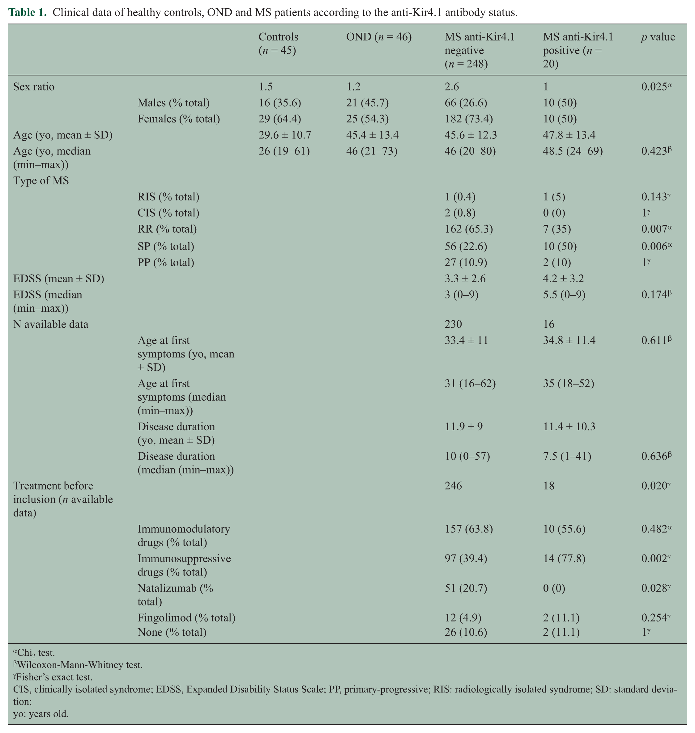
Chi2 test.
Wilcoxon-Mann-Whitney test.
Fisher’s exact test.
CIS, clinically isolated syndrome; EDSS, Expanded Disability Status Scale; PP, primary-progressive; RIS: radiologically isolated syndrome; SD: standard deviation;
yo: years old.
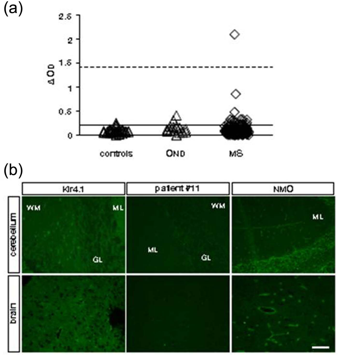
Serum reactivity against Kir4.1. (a) Serum antibody binding to Kir4183-120 peptide was determined by ELISA in healthy controls and in patients with OND or MS. For each sample, OD obtained with peptide-coated wells was corrected for background binding to uncoated wells (ΔOD). The positive threshold (filled line) was defined as 2 standard deviations (SD) above the mean OD of the control group. An anti-Kir4.1-positive serum (patient #11) was used in each experiment as a positive control. The mean ΔOD obtained with this serum is indicated as a dashed line (1.42 ± 0.09, mean ± SD, n = 6). All assays were performed in duplicate and in a blind manner. (b) Indirect immunofluorescence staining was performed on primate brain and cerebellum sections using anti-Kir4.1 antibodies (Kir4.1) and serum from an anti-aquaporin-4-positive patient (NMO) as positive controls, which shows perivascular staining at the blood–brain barrier. Patient #11 data are representative of the microphotographs obtained with sera from anti-Kir4.1 positive patients (n = 10). Scale bar, 100 µm.
We performed indirect immunofluorescence on primate brain and cerebellum sections with serum from patients showing the highest anti-Kir4.1 immunoreactivity by ELISA (n = 10). Although commercial polyclonal anti-Kir4.1 antibodies and serum from an anti-aquaporin-4-positive patient used as positive controls showed a perivascular staining, we did not observe any specific immunolabelling using the serum of MS patients (Figure 1(b)).
Discussion
Our study shows that anti-Kir4.1 antibodies are found in 7.5% of MS patients, as compared to 46.9 % in the study by Srivastava R and colleagues. 2 Furthermore, as demonstrated previously, immunostaining of CNS tissues with MS patient’s serum does not show a specific pattern of IgG binding. 5 The presence of anti-Kir4.1 antibodies in a similar proportion of patients with OND and healthy donors presents strong evidence against a pathogenic role of anti-Kir4.1 antibodies in MS. A progressive course of the disease is more frequent in MS patients with anti-Kir4.1 antibodies than in patients without anti-Kir4.1 antibodies. This finding may be related to a humoral response to chronic damage that occurs in the CNS of patients with MS and other chronic neurological disorders.
The Kir4.1 channel is expressed primarily by CNS glial cells (astrocytes and oligodendroglia) as part of the macromolecular dystrophin-glycoprotein complex which includes the water channel aquaporin-4. 6 Higashimori and Sontheimer 7 established a link between the Kir4.1 channel and growth control of glial cells. Studies of Kir4.1-deficient mice suggested a role of Kir4.1 in oligodendrocyte development and myelination. 8 Thus, anti-Kir4.1 antibodies were an appealing candidate as a suitable biomarker for MS. Unfortunately, we were unable to confirm the previous results reporting a prevalence of anti-Kir4.1 antibodies in MS patients. 2 We followed a methodology similar to that reported previously. 2 Differences among the included patients (most of our patients had RR or SP/primary-progressive (PP) MS, in contrast to the previous study including mainly RR and CIS patients) are slight and might not explain the observed discrepancies. In addition, in our study, the Kir4.1 antibody was mostly found in progressive MS (12/95), with a very low rate in RR MS (7/169). Thus, an anti-Kir4.1 antibody ELISA-based protocol may be an unreliable means to diagnose MS.
Footnotes
Acknowledgements
We are thankful to Dr Melissa Bowerman and Michael Drake for their critical reading of the manuscript.
Conflict of interest
The authors declare that there are no conflicts of interest.
Funding
This work was funded by the institut national de la santé et de la recherche médicale (Inserm).