
Abstract
New DNA sequencing technologies have uncovered non-coding RNA (ncRNA) as a major player in regulating cellular processes and can no longer be dismissed as “junk” or “dark” RNA. Among the ncRNA, microRNA (miRNA) is arguably the most extensively characterized category and a number of studies have implicated them in regulating critical functions that can influence autoimmune demyelination. Of specific interest to multiple sclerosis (MS), miRNA have been implicated in both regulating immune responses and myelination, thus making them an attractive candidate for both pharmacological intervention and as disease biomarkers. In addition, exosomes, small vesicles secreted by most cell types and present in all body fluids, have been also shown to play roles in immune signaling, inflammation and angiogenesis. Therefore, exosomes are also being explored as tools for therapeutic delivery and as biomarkers. This article reviews the recent advances in miRNA and exosome profiling in MS and experimental models.
Introduction
Despite the remarkable progress in the understanding and treatment of multiple sclerosis (MS), the disease course is still highly unpredictable. 1 Therefore, an urgent need exists for the discovery of biomarkers that can track and predict outcomes related to disease progression and response to therapeutic drugs. Currently, no externally validated blood immune marker has adequate sensitivity and specificity to be used for the diagnosis of MS, which probably reflects the heterogeneity of the disease. Owing to the progress in high throughput sequencing technologies and emerging data on the mechanisms of horizontal transfer of material between the cells new opportunities have arisen in this field. In this review, we discuss currently available evidence associating novel intra- and extracellular RNA analysis as putative biomarkers of MS as well as their potential role in autoimmune demyelination development and progress.
microRNA and other non-protein coding RNAs
Progress in molecular biology over the last two decades has unveiled a universe of non-protein coding RNA (ncRNA) present in our cells. It is now apparent that protein coding RNA only represents about 2% of the information encoded in genomic DNA. 2 Furthermore, recent high throughput transcriptome analyses have documented that ncRNA constitutes a major product of our genome. 3 Our current knowledge of the function of ncRNAs is still very limited resulting in the notion that we are just starting to understand the true value of the information encoded in our genomes.
One of the best studied groups of the ncRNA are microRNA (miRNA). miRNA are conserved, single-stranded, non-coding RNAs comprised of approximately 22 nucleotides, that play important roles in gene-regulation by targeting mRNAs for cleavage or translational repression. 4 The master role of miRNAs is regulation of gene transcription at the posttranscriptional level, which is considered as an epigenetic regulatory mechanism, through cleaving or blocking messenger RNAs (mRNAs) from further translation. A single miRNA can directly repress translation of hundreds of genes and each mRNA transcript may be regulated by multiple miRNAs. This many-to-many relationship underscores the complexity of the miRNA regulatory networks that govern cellular programs from inflammation and neurogenesis, to homeostatic maintenance. Several databases and online resources are available to lookup published miRNA sequences and annotations (e.g. miRbase, http://mirbase.org), including specialized software to find associations between given miRNA and its target genes (e.g. miRSystem, http://mirsystem.cgm.ntu.edu.tw).
The role of miRNA in autoimmune demyelination
The last decade has seen great progress in the description of the role miRNA in MS and experimental autoimmune encephalomyelitis (EAE). From unbiased transcriptomic analysis to functional dissection of specific miRNAs, the field has flourished. In one of the earliest studies, miR-155 was identified as a key regulator of these inflammatory responses, as targeted deletion of this gene in mice resulted in a decrease in Th1 and Th17 cellular differentiation both in the central nervous system (CNS) and in peripheral lymphoid organs. Furthermore, both deletion and pharmacological targeting of miR-155 resulted in delayed course and reduced severity of EAE, which prompted authors of the study to propose its therapeutic targeting. 5 In a separate study, a rare form of this same gene (miR-155-3p) was found to drive the development of autoimmune demyelination by regulation of heat shock protein 40. 6 miR-301a was also found to be a critical regulator of myelin-reactive T-helper type 17 cells, further supporting their role as candidates for therapeutic targets for controlling of autoimmune demyelination. 7
In addition to their role in modulating effector inflammatory T cell responses, miRNA also play a role in regulating the regulatory T cell (Treg) population, that normally counterbalances the former. Surprisingly, miR-155 has also been found to influence Treg development and function, as recent studies showed that it is regulated by Foxp3, a transcription factor characteristic of this cell population. 8 The seemingly opposite roles of the same miRNA in different cells highlights the complexity of the regulatory networks at play in the immune system.
While the previous examples highlighted the regulatory role of miRNA in proinflammatory responses during EAE, additional studies also showed their primary role in regulating myelination itself. For example, miR-23a-overexpressing mice have increased myelin thickness, providing in vivo evidence that miR-23a enhances both oligodendrocyte differentiation and myelin synthesis. 9
miRNA as biomarkers of MS
Encouraged by the early discoveries of a handful of miRNA involved in myelination 10 and inflammatory responses, 11 several studies focused on their systematic profiling in serum and other biological fluids as disease biomarkers. In one study including 4 observational cohorts, 5 miRNAs (hsa-miR-484, hsa-miR-140-5p, hsa-miR-320a, hsa-miR-486-5p, and hsa-miR-320c) showed a significant difference between patients with MS and healthy individuals. 12 A follow-up study explored the potential of miRNA profiling to identify subtypes of MS. In this study, miR-92a-1 was differentially expressed in relapsing-remitting MS (RRMS) versus secondary progressive MS (SPMS), and RRMS versus healthy controls. Furthermore, this miRNA showed an association with both Expanded Disability Status Scale (EDSS) and disease duration. 13
In a more recent study, a large group of MS patients (n = 1,088) was stratified based on brain imaging and miRNA profiling was used to identify differences across the different magnetic resonance imaging (MRI)-based phenotypes. 14 Interestingly, each MRI phenotype demonstrated a miRNA signature whose underlying biology implicates blood–brain barrier (BBB) pathology. Specifically, miR-22-3p, miR-361-5p, and miR-345-5p were the most valid differentiators of the MRI phenotypes.
While miRNA profiling from biofluids poses a great promise as a biomarker for MS, it must be noted that conflicting results, heterogeneity and lack of replication are ongoing challenges in the field. For example, a recent review of the literature highlighted that out of the 650 differentially expressed ncRNAs reported in MS patients, only 27.5% were found dysregulated in the same direction in at least two independent studies. If the cutoff criteria is increased to three studies, then only 9% of the reported differences remain. 15 This lack of replication is not foreign to biomarker projects in general and underscore the logistical difficulties behind this kind of studies. While some of these challenges (e.g. small sample size, biospecimen collection methods, profiling techniques, etc.) can be overcome with appropriate planning and funding, some are inherent to the complexity and heterogeneity of the underlying biology. The recent emergence of novel mechanisms of miRNA regulation (e.g. circular RNAs) might provide a better insight and renewed enthusiasm for the development of more robust biomarkers in autoimmune demyelination. 16
Exosomes as cell-to-cell communication system
Exosomes are small extracellular vesicles released from the endocytic compartment of cells, which can be easily detected in serum and other biological fluids. 17 They contain material of cell origin, including proteins, lipids, DNA, mRNA, and non-coding RNA and mediate a wide array of biological functions including the modulation of immune reactions, cell differentiation and apoptosis. 18 Exosomes might in fact provide a mechanism for long distance intercellular communication since they can travel between different compartments of the body and across biological barriers. Due to their small size (30–120 nm) and lipophilic nature, exosomes are taken up by a number of organ tissues and cell populations. 19 Indeed, it was demonstrated that cells transfected with a plasmid coding for the green fluorescent protein (eGFP), when injected into mice, released exosomes carrying eGFP RNA that was found not only in the circulation but in many other cell types proving direct interaction between exosomes and target cells. 20 The production, secretion, and transfer of exosomes arises as a crucial and still largely unexplored system of a cell-to-cell communication. Cancer research in particular provides numerous clues for the significance of exosome-mediated effects. 21 An increasing amount of recently published literature confirms that cancer and cancer-associated cells use exosomes to communicate their message to malignant and non-malignant cells.22–24 This is done through the transfer of functional components, in order to initiate pathways that are necessary for tumor survival and propagation. Additional roles of exosomes include regulation of cell proliferation, induction of programmed cell death, metabolism, differentiation and phenotypic modification. 25 Thus, exosome-mediated effects likely play a role in many pathological conditions and may contribute to every aspect of cellular biology and dysregulation.
Exosomes do not contain a random sampling of components of their parent cells but have a specific composition reflecting the state of cell activation and can be influenced by pathologic processes from the tissue of origin. Indeed, exosome release is considered a cellular adaptation mechanism and its biogenesis, composition and secretion are affected by the microenvironment of cells. Furthermore, extracellular RNA, RNA-binding proteins, and other cellular proteins are differentially expressed in exosomes and non-vesicle compartments. 18 Thanks to the recent progress in high throughput transcriptome techniques, the analysis of the low content material of total exosomes has become possible. 26 The resulting data provide a unique insight into the wealth of the transcripts present in the extracellular vesicles.18,27
RNA species identified as a cargo of the cell culture released exosomes include predominantly different classes of short RNA species including miRNA, small nuclear RNA, small nucleolar RNA, Piwi-interacting RNA, vault RNA, Y-RNA, small conditional RNA, signal recognition particle RNA, 7SK-RNA. 28 Furthermore, a ribosomal RNA, transfer RNA, messenger RNA, long non-coding RNAs, and various intergenic repeats have also been found within the exosomes. So far only few studies have addressed the human biofluid–derived exosomes’ full transcriptome content.27,29–31 All of them demonstrated a wealth of sequences present in the circulating exosomes. Majority of the RNAs were ncRNA, including miRNA. These studies provided evidence that large part of the cellular transcriptome is present in exosomes and therefore could potentially be transferred to other cells. This complexity of the exosomal RNA cargo combined with their seclusion from the circulating RNAses provides an exciting platform for a biomarker discovery research.
Circulating exosomes as a biomarkers
Exosomes provide a means of cell-to-cell communication by transporting their cargo and delivering it to target cells. The content of these microvesicles representing a molecular bioprint of their parental cells. Therefore, exosomes present in blood and other biofluids are potentially noninvasive biomarkers for early diagnosis and prognosis of various types of diseases. 32 Exosome profiling is often called a “liquid biopsy” as it may provide an insight about the content of the hard-to-access organs without the need of their direct sampling. 33 Exosome release is considered a cellular adaptation mechanism and its biogenesis, composition and secretion are affected by the microenvironment of cells.
Currently, clinicaltrials.gov registry enlists 35 active trials aiming at the observation of the exosomes biomarkers analysis. The majority of these trials are dedicated to the cancer and tumor research, and only one study is dedicated to a neurological condition (i.e. dementia). 34 Interestingly, blood circulating exosomes show presence of the CNS markers resulting from the brain origin.35,36 The analysis of these exosomes should provide a unique insight into the status of the CNS bypassing the need for a biopsy. 37 Furthermore, exosomes can provide a source of the long sought CNS antigens trafficking through the BBB. 36 These arguments should prompt for more trials involving exosomes analysis in the CNS conditions including MS.
Exosomes in MS monitoring
Emerging data on the use of miRNA and other molecular cargo carried by circulating exosomes as biomarkers of various conditions have renewed enthusiasm for further exploring the role of these microvesicles as a putative indicator of MS status and for disease monitoring. 38 Current search of the PubMed database, using MeSH terms “exosomes” and “multiple sclerosis,” following an individual curation revealed 16 original publications relating to the biomarker search in MS (Table 1). The majority (9 articles) analyzed serum or plasma-derived exosomes, whereas 3 addressed cerebrospinal fluid (CSF) microvesicles, 3 urine, and 2 described cell cultures. Most frequently analyzed exosomes content was miRNA (9 papers), whereas 3 papers investigated lipids and only 2 proteins. Only 2 publications aimed at global profiling of exosomes cargo, either RNA 27 or protein. 39 These reports document technical feasibility of exosomes extraction and subsequent analysis of MS patients derived material. Promising MS biomarkers (e.g. levels of blood circulating exosomal miR-122-5p) are starting to emerge.27,40 Although the number of publications on the significance of exosomes analysis as biomarkers in MS is still limited, the current results are encouraging and warrant further research.
List of original publication describing analyzing exosomes as biomarkers in MS.
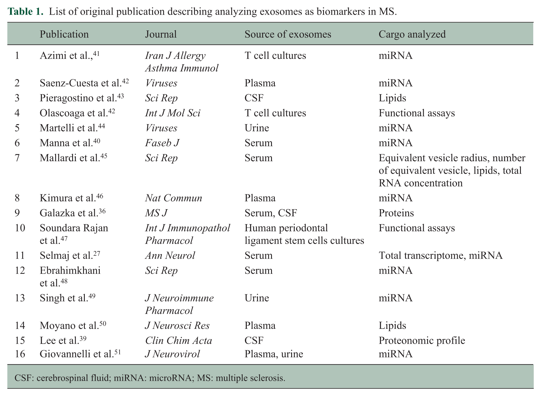
CSF: cerebrospinal fluid; miRNA: microRNA; MS: multiple sclerosis.
Conclusion and further perspectives
Large-scale profiling of circulating miRNA and exosome content in EAE and MS has prompted early enthusiasm for their potential to emerge as the long-sought biomarker that can characterize patient status using a minimally invasive approach, yet with high sensitivity and specificity. However, while promising results have arisen, limited replication and high heterogeneity remain unsolved challenges. Further research is warranted to firmly establish these relatively newly discovered biomolecules as firm contenders in the quest for the MS biomarker.
Footnotes
Declaration of Conflicting Interests
The author(s) declared no potential conflicts of interest with respect to the research, authorship, and/or publication of this article.
Funding
The author(s) disclosed receipt of the following financial support for the research, authorship, and/or publication of this article: This study was supported by National Center of Research Poland grants 2016/23/B/NZ6/02541, 2015/19/B/NZ6/02834, the National Center for Research and Development grant ERA-NET_NEURON/14/2019 and the University of Warmia na Mazury in Olsztyn internal grant to M.P.M.. Additional support was received from NIH/NINDS (R01NS088155 and R01NS099240), NMSS (CA-1072-A-7), and DOD (DOD_W81XWH-15-1-0652) to S.E.B. S.E.B is the Heidrich Family and Friends Endowed Chair in Neurology at UCSF. S.E.B. holds Distinguished Professorship in Neurology I at UCSF.