
Abstract

Keywords
Prolonged venous hypertension is the central pathophysiologic mechanism underlying chronic venous disease (CVD). Sustained elevations in venous pressure promote chronic inflammation, activation of cytokines, chemokines, growth factors, and matrix metalloproteinases, leading to collagen deposition, disruption of elastin architecture, and loss of venous wall compliance. These changes further impair venous return and perpetuate venous insufficiency, ultimately manifesting clinically as varicose veins, edema, skin changes, and venous leg ulceration. 1 Importantly, the downstream consequences of venous hypertension extend beyond the macroscopic venous system, affecting the microcirculation, interstitial compartment, and lymphatic drainage. 2
Duplex ultrasound (DUS) remains the cornerstone of venous disease evaluation, providing essential information on venous anatomy, reflux, obstruction, and flow patterns. Hemodynamically significant reflux is defined by established duration thresholds; however, reflux severity does not consistently correlate with venous hypertension, symptom burden, 3 or clinical progression. 4 This discordance is well recognized in clinical practice: some patients with marked reflux exhibit minimal symptoms, whereas others experience significant edema or skin changes despite limited reflux on DUS. These observations underscore the limitations of relying solely on macrovascular flow assessment to characterize disease burden and highlight the need for complementary physiologic markers that reflect downstream consequences of venous hypertension. 5
Bioimpedance spectroscopy (BIS) offers a noninvasive method to assess tissue electrical properties dependent on both electrical conductivity (e.g., ionic concentration) and volume of tissue, and subsequently infer extracellular fluid accumulation. Because electrical current traverses fluid more readily than solid tissue, increasing extracellular fluid content results in lower tissue impedance. This is reported with a derived index called the L-Dex value, in which lower impedance corresponds to higher index values. This technique is highly sensitive and capable of detecting small changes in extracellular fluid volume, often before overt clinical swelling becomes apparent.6,7 In this context, bioimpedance does not directly measure venous reflux or venous pressure but rather may reflect impaired swelling control and fluid accumulation—key downstream consequences of chronic venous hypertension.
The objective of this study was to characterize leg bioimpedance measurements across the spectrum of CVD and to explore whether bioimpedance trends vary according to Clinical, Etiological, Anatomical, and Pathophysiological (CEAP) clinical class. We hypothesized that bioimpedance would reflect physiologic changes associated with advancing disease that are not captured by DUS alone. To test this hypothesis, we performed a retrospective analysis of 118 patients with CEAP classes ranging from C0 to C6. This study was approved by the Stanford University institutional review board. Bioimpedance measurements were obtained using a validated BIS platform (SOZO®; Impedimed, Lindfield, Australia), with L-Dex values calculated using standard reference comparisons.
Patients with CEAP classes C0 to C2 generally demonstrated L-Dex values within the normal range, suggesting preserved regulation of extracellular fluid. In contrast, patients with C3 and above exhibited progressively higher L-Dex values, consistent with increasing edema and impaired swelling control as disease advanced. Statistically significant differences were observed between early-stage (C0 to C2) and more advanced disease (C3 to C6), as well as between patients with healed ulcers (C5) and those with active ulcers (C6) (Figure 1A). Among patients with venous leg ulcers who achieved healing through a combination of wound care, compression therapy, and venous interventions, bioimpedance values decreased following ulcer resolution (Figure 1B). These findings suggest that improvements in fluid control accompany clinical healing and that bioimpedance may capture physiologic changes associated with transitions between advanced CEAP stages.
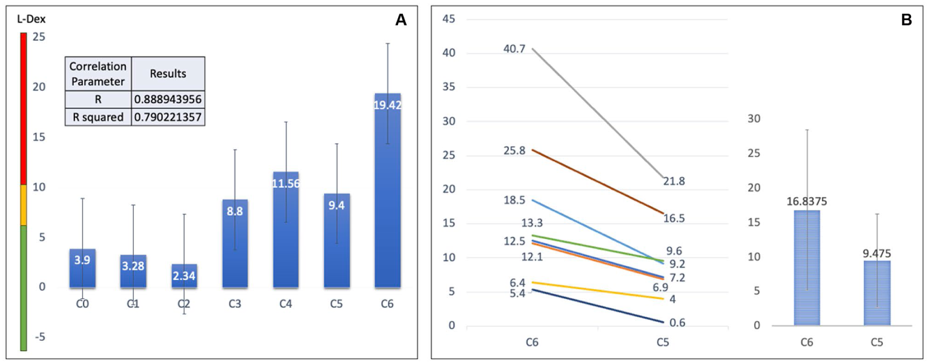
Bioimpedance index (L-Dex) values by CEAP category (
The observed bioimpedance trends are consistent with known mechanisms of CVD progression. Persistent venous hypertension increases interstitial pressures, disrupts endothelial permeability, and impairs lymphatic uptake of filtered fluid, leading to extracellular fluid accumulation. 4 These microcirculatory and interstitial changes are not directly assessed by DUS, which focuses on macroscopic venous flow and reflux. Bioimpedance therefore provides complementary information by providing an indicator of limb fluid accumulation, potentially helping to explain discordance between duplex findings and clinical presentation. For example, bioimpedance may aid in identifying impaired swelling control in patients with minimal reflux on duplex or demonstrate preserved fluid regulation in patients with significant reflux but limited clinical manifestations.
Several limitations warrant consideration. Patients with bilateral disease may introduce variability due to differences in tissue composition, such as electrolyte abnormalities and vascular dynamics when using an alternative reference limb. 8 The retrospective design and lack of validated clinical severity scores or patient-reported outcomes further limit conclusions regarding symptom severity or functional impairment.
Overall, bioimpedance demonstrated limited ability to distinguish early CVD (CEAP ⩽ 2) but appeared more informative in advanced disease (CEAP ⩾ 3), where impaired swelling control is a defining clinical feature. Although this study does not establish bioimpedance as a predictor of clinical outcomes or disease progression, the findings suggest that it may serve as an adjunctive physiologic marker in advanced CVD. By objectively quantifying changes in limb fluid status over time, bioimpedance may complement clinical examination and DUS. Prospective studies incorporating standardized measurement protocols, clinical severity scores, and longitudinal outcomes are needed to define its role more clearly and to determine whether bioimpedance-guided management improves patient care.
Footnotes
Declaration of conflicting interests
The authors declared the following potential conflicts of interest with respect to the research, authorship, and/or publication of this article. Eri Fukaya serves as a consultant for Koya Medical and Boston Scientific. Nicolas Lopez has no conflicting interests.
Funding
This research was funded by the 2020 American Venous Forum-Jobst Clinical Research Grant.