
Abstract
Introduction:
For the majority of patients who experience persistent dyspnea 3 months after an acute pulmonary embolism (PE), the cause of exercise limitation is unknown. We hypothesized that data collected from invasive cardiopulmonary exercise (iCPET) would inform whether pre-load failure (PLF) may be an under-recognized cause of post-PE dyspnea.
Methods:
Between 2015 and 2023, we retrospectively identified patients with prior PE and unexplained dyspnea who underwent iCPET testing. Those with chronic thromboembolic pulmonary hypertension (CTEPH) were excluded.
Results:
Thirty-four patients with PE history and unexplained dyspnea were identified. The mean peak VO2 for the cohort was reduced at 71.0 ± 20.5%. This cardiovascular limit was explained by PLF with a reduced right atrial pressure (RAP) at peak exercise (4.5 [5.0] mmHg). There was no evidence of ventilatory inefficiency (VE/VCO2 slope: 30.8 [10.9]). However, dead space ventilation at peak exercise was increased (0.24 ± 0.10). Owing prior work establishing an association between PLF and small fiber neuropathy (SFN), we evaluated the results of post-PE patients referred for skin biopsies at the providers’ discretion (32.4%, n = 11). Of these, 9 patients (82%) had confirmed SFN.
Conclusion:
This is the first case series to utilize iCPET data to investigate novel causes of post-PE dyspnea. We describe PLF, characterized by a central cardiac limit to exercise and low RAP at peak, in post-PE patients with otherwise unexplained symptoms. We additionally describe an association between PLF and SFN and hypothesize a mechanistic link between an inflammatory PE insult, peripheral neuronal injury, and resultant dyspnea.
Keywords
Introduction
Three months after an acute pulmonary embolism (PE), one-third of survivors experience persistent dyspnea and exercise limitation.1,2 For a small proportion of patients, symptoms are ascribed to chronic thromboembolic disease with or without pulmonary hypertension (CTEPH or CTED).3,4 In the remaining majority, the pathophysiology of post-PE dyspnea remains unknown.
Noninvasive cardiopulmonary exercise testing (CPET) to interrogate persistent dyspnea frequently identifies a reduced peak oxygen consumption (VO2),5,6 although the pathophysiology driving this impairment is not clear and may be heterogenous. 1 Prior studies ascribe this limitation to muscle deconditioning whereas more recent data associate this abnormality with ventilatory inefficiency or elevated dead space ventilation and ventilation/perfusion (V/Q) imaging defects.5–8 These data highlight the prevalence of post-PE exercise limitation but lack of clarity on disease mechanism.
More recently, growing data from invasive CPET (iCPET) identifies reduced right atrial (RA) filling pressures and impaired peak VO2, termed preload failure (PLF), in patients with otherwise unexplained dyspnea.9–11 A reduction in venous return creates a central cardiac exercise limit that drives symptoms; ventilatory inefficiency and muscle deconditioning are also seen.12,13 Data from the myalgic encephalomyelitis/chronic fatigue syndrome (ME/CFS) population further identifies a mechanistic link between PLF and small fiber neuropathy (SFN), a neurologic disorder characterized by excessive firing or degeneration of autonomic peripheral neurons with associated symptoms including exertional intolerance, tachycardia, and hypotension.14,15 However, PLF and SFN remain under-recognized. We hypothesized that post-PE dyspnea may be driven by the presence of PLF physiology, possibly due to SFN.
Methods
From the 2332 iCPETs performed at our institution between 2015 and 2023, we retrospectively identified studies involving patients with unexplained dyspnea and prior PE.11,16 Low-risk PE was defined by normal right ventricular (RV) size and function and normal troponin and NTproBNP values. Intermediate-risk PE was defined by elevated troponin or NTproBNP ± RV dilation or dysfunction. High-risk PE was defined by hypotension and/or syncope as well as RV dysfunction and serum biomarker elevation. Maximum iCPET effort was confirmed by an age-predicted heart rate > 85% or a peak respiratory exchange ratio (RER) > 1.05. Normal peak VO2 was informed by age, sex, and height and defined as > 80% of the predicted value.17,18 Predicted peak cardiac output was calculated from the predicted peak VO2, assuming a normal peak exercise Ca-vO2 of 140 mL/L. 18 PLF as the cause of a cardiovascular limit to exercise was defined by a reduced right atrial pressure (RAP) at peak exercise (< 6.5 mmHg). 9 Exercise pulmonary hypertension was defined as a mean pulmonary artery pressure (mPAP) to cardiac output slope < 3.3,17 Two patients were excluded based on abnormal V/Q scans and an associated diagnosis of CTEPH. Descriptive statistics were used to present normally and nonnormally distributed data as mean ± SD and median [IQR], respectively; categorical data are presented as n (%). This study was conducted under Mass General Brigham IRB protocol #2011P000272, which granted an exemption for retrospective analysis of clinical iCPET data without additional patient consent.
Results
Thirty-four patients with a history of PE and unexplained dyspnea had iCPET data. The mean ± SD age was 58.4 ± 13.7 years, and 44.1% (n = 15) were women. In those with available data, the majority were classified as low-risk patients (44.1%) (Table 1). In patients with V/Q imaging (20.6%, n = 7), there were no perfusion defects. All resting supine right heart catheterizations (RHC) were normal. The median [IQR] time from PE to iCPET was 14.5 [35.8] months.
Descriptive characteristics and test results of patients referred for CPET for unexplained dyspnea and a history of acute PE.
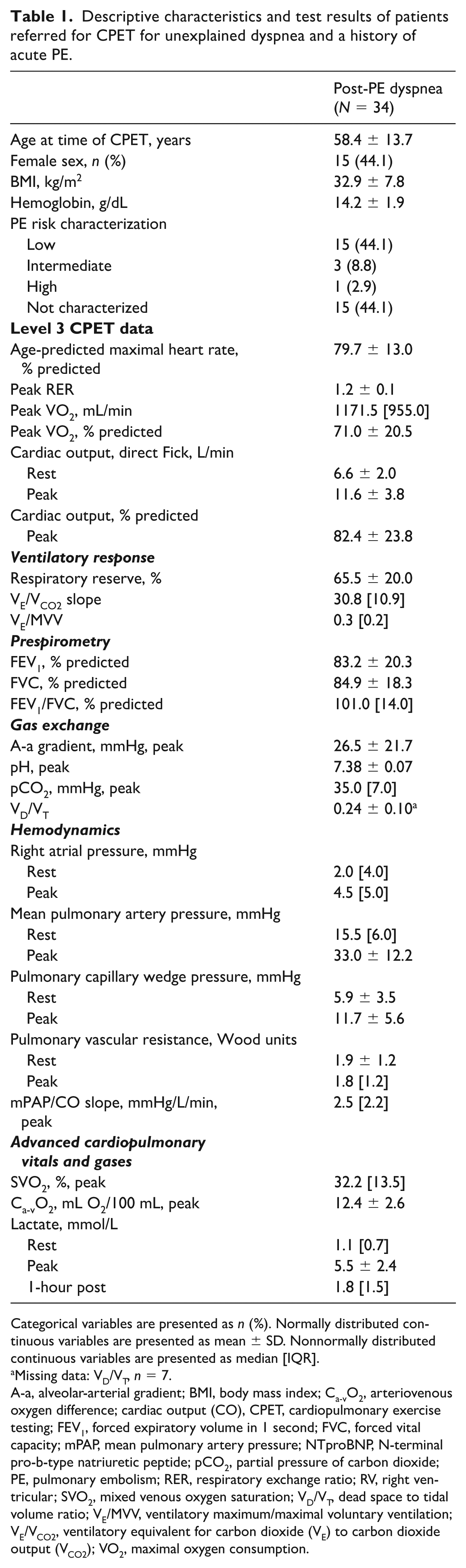
Categorical variables are presented as n (%). Normally distributed continuous variables are presented as mean ± SD. Nonnormally distributed continuous variables are presented as median [IQR].
Missing data: VD/VT, n = 7.
A-a, alveolar-arterial gradient; BMI, body mass index; Ca-vO2, arteriovenous oxygen difference; cardiac output (CO), CPET, cardiopulmonary exercise testing; FEV1, forced expiratory volume in 1 second; FVC, forced vital capacity; mPAP, mean pulmonary artery pressure; NTproBNP, N-terminal pro-b-type natriuretic peptide; pCO2, partial pressure of carbon dioxide; PE, pulmonary embolism; RER, respiratory exchange ratio; RV, right ventricular; SVO2, mixed venous oxygen saturation; VD/VT, dead space to tidal volume ratio; VE/MVV, ventilatory maximum/maximal voluntary ventilation; VE/VCO2, ventilatory equivalent for carbon dioxide (VE) to carbon dioxide output (VCO2); VO2, maximal oxygen consumption.
In this cohort, iCPET data were abnormal, demonstrating a central cardiac limit to exercise based on a mean peak exercise VO2 below 80% predicted (71.0 ± 20.5%). This cardiovascular limit was explained by PLF with a reduced RAP at peak exercise (4.5 [5.0] mmHg). 9 The mPAP to cardiac output slope excluded exercise pulmonary hypertension.3,17 There was no peripheral oxygen extraction defect based on a peak Ca-vO2 corrected by hemoglobin ratio > 0.80 and a normal 1-hour postexercise lactate. Among the subgroup of patients with reduced peak VO2 (n = 21), there was a discrepancy between peak VO2 (57.8% predicted) and CO (71.3% predicted). This was associated with an increased venous oxygen saturation (SvO2) at peak exercise (34.2 ± 8.6% ; normal < 28%) supportive of peripheral left-to-right shunting in the absence of an O2 saturation step-up on resting RHC. 15
Patients had normal spirometry without evidence of lung disease. There was no evidence of a pulmonary mechanical limit to exercise based on the VEpeak/MVV (maximal voluntary ventilation) < 0.70 and no evidence of ventilatory inefficiency (VE/VCO2 slope: 30.8 [10.9]). However, dead space ventilation at peak exercise was increased (0.24 ± 0.10), defined as VD/VT > 0.20. 19
Owing to our prior work establishing an association between SFN and PLF, we further evaluated the results of post-PE patients referred for skin biopsies at the providers’ discretion (32.4%, n = 11) (Table 2). 15 Of these, nine (81.8%) patients had evidence of SFN based on an epidermal small fiber nerve density below the age- and sex-specific lower limit of normal in at least one of two sites (proximal and/or distal leg).
Descriptive characteristics and CPET results for patients referred for skin biopy.
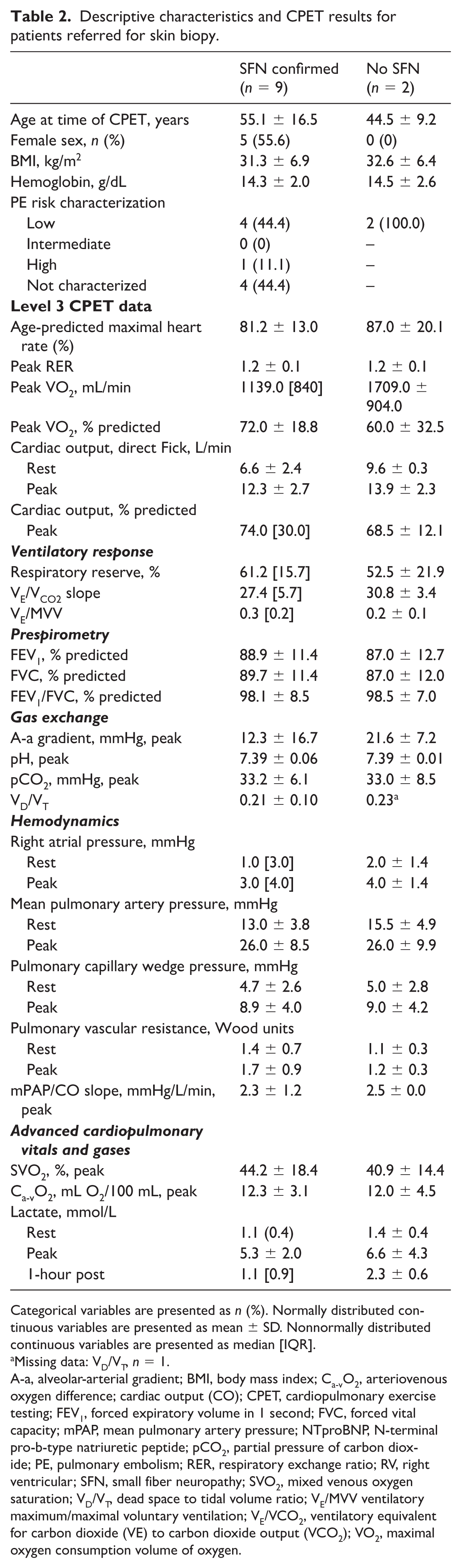
Categorical variables are presented as n (%). Normally distributed continuous variables are presented as mean ± SD. Nonnormally distributed continuous variables are presented as median [IQR].
Missing data: VD/VT, n = 1.
A-a, alveolar-arterial gradient; BMI, body mass index; Ca-vO2, arteriovenous oxygen difference; cardiac output (CO); CPET, cardiopulmonary exercise testing; FEV1, forced expiratory volume in 1 second; FVC, forced vital capacity; mPAP, mean pulmonary artery pressure; NTproBNP, N-terminal pro-b-type natriuretic peptide; pCO2, partial pressure of carbon dioxide; PE, pulmonary embolism; RER, respiratory exchange ratio; RV, right ventricular; SFN, small fiber neuropathy; SVO2, mixed venous oxygen saturation; VD/VT, dead space to tidal volume ratio; VE/MVV ventilatory maximum/maximal voluntary ventilation; VE/VCO2, ventilatory equivalent for carbon dioxide (VE) to carbon dioxide output (VCO2); VO2, maximal oxygen consumption volume of oxygen.
Among subjects with SFN, median peak VO2 was similarly reduced at 72.0 ± 18.8% predicted, with a commensurate decrement in peak CO (74.0 [30.0]% predicted) (Table 2). Within this group, RAP was more severely reduced at peak exercise as compared to the overall cohort and to those subjects with a skin biopsy negative for SFN. Unlike the mild evidence of dead space ventilation seen in the overall cohort, VD/VT was borderline normal at 0.21 ± 0.10, and the hemodynamic impairment appeared more germane to the exercise limitation. There was no strict evidence of a peripheral oxygen extraction defect; however, there was evidence of impaired oxygen extraction based on a reduced arteriovenous oxygen content difference (12.3 ± 3.1 mL O2/100 mL) and an elevated SVO2 (44.2 ± 18.4%).
Discussion
This is the first case series to utilize iCPET data to interrogate the cause of post-PE dyspnea. We describe PLF, characterized by a central cardiac limit to exercise and low RAP at peak exercise, in post-PE patients with otherwise unexplained symptoms. 9 These patients additionally have evidence of peripheral shunting and increased dead space ventilation. We additionally describe an association between PLF and SFN. Unlike prior studies, we did not identify evidence of ventilatory inefficiency, which may reflect our stringent exclusion criteria. Dyspnea in these patients may therefore reflect impaired venous return, an inability to recruit high V/Q areas in the lung apices during upright cycling, and resultant dead space ventilation due to multifactorial pathologies. Similar to the inflammatory insult implicated in impaired thrombus resolution and CTEPH, these data suggest that vascular pooling may occur due to SFN, as seen in dysautonomia.20–22 In addition, there may be structural impairment to venous return as a result of residual deep vein thrombus (DVT). 23 In fact, our findings mirror established post-PE dyspnea case reports that identify PLF on iCPET and ultimately inferior vena cava (IVC) filter thrombus.11,24 Our findings are unlikely to be explained by hypovolemia owing to our CPET protocol which does not require NPO status.
Dysautonomia, identified in up to 20% of cases of PLF, is characterized by peripheral neuropathy, abnormal venous pooling, and resultant volume dysregulation.16,25 The tilt table test and skin biopsy results consistent with SFN support the diagnosis. 22 Damage of distal nerves responsible for regulation of microvascular tone results in impaired venoconstriction with resultant ‘low-flow’ PLF due to impaired venous return. The ‘high-flow’ PLF phenotype is characterized by low peak VO2 but high pulmonary blood flow with impaired systemic oxygen extraction due to cutaneous microvasculature pathology and bypass of oxygenated blood across capillary beds.15,26 Pharmacologic work in PLF subjects supports the hypothesized neurovascular dysregulation, with improvements in right heart filling pressures, CO, and VO2 in subjects treated with pyridostigmine. 27 The high prevalence of SFN in our post-PE cohort suggests a potential mechanistic cause of PLF identified on iCPET; a similar phenomenon of large-fiber chronic inflammatory demyelinating polyneuropathy after PE is also described. 28
There are limitations to our study. Our hypothesized association of PLF and SFN may reflect selection bias, as skin biopsies were not available in all patients. Additionally, negative skin biopsy results (2 of 11 patients) may reflect sampling error due to the patchy nature of neuropathic involvement. The time from acute PE to iCPET was not standardized and PE risk characterization data were limited, which may limit generalizability. In addition, a follow-up V/Q scan and/or interval computed tomography data to evaluate for persistent clot were not available in all patients. However, iCPET data were not supportive of CTEPH or CTED. 29 Functional limitations prior to PE are associated with post-PE dyspnea; however, this was also not objectively assessed, which may limit interpretability.30,31 Finally, lack of access to care centers with iCPET expertise may limit the applicability of our findings to all clinicians evaluating post-PE dyspnea. In this setting, referral to a center with iCPET expertise is advised.6,7
Conclusion
In patients with unexplained following acute PE, we describe a novel pathophysiologic finding of a central cardiac limit to exercise characterized by PLF or a low RAP at peak exercise. We further identify a subset of patients in whom PLF is associated with skin biopsy confirmed SFN. We hypothesize a mechanistic link between cardiopulmonary physiology and peripheral nerve injury in the setting of a systemic inflammatory insult. Further investigation into the proposed link between PE, PLF, and SFN, including potential contributions from residual DVT or vena cava clot, is warranted to establish causality.
Footnotes
Declaration of Conflicting Interests
The authors declared the following potential conflicts of interest with respect to the research, authorship, and/or publication of this article. Aaron Waxman collaborates with Acceleron/Merck, United Therapeutics, OrphAI, and ARIA-CV as an investigator; and he is the DSMB chair for INSMED. The other authors have no conflicting interests.
Funding
The authors disclosed the following financial support for the research, authorship, and/or publication of this article. Arvind Pandey receives funding from the National Institutes of Health (K08177156) and American Heart Association (CDA1267729). David Systrom receives funding from the Open Medicine Foundation.