
Abstract
The aim of this review is to provide clinicians with an update on the evolution and current status of the carotid duplex criteria, as well as clinical applications for the diagnosis and follow-up of carotid disease. The ‘duplex concept’ combined real-time B-mode imaging and pulsed Doppler flow detection in a single instrument, which enabled direct visualization of blood vessels and analysis of flow patterns. The first application of the prototype duplex scanner built in the 1970s was the evaluation of extracranial carotid artery disease, and the validation studies performed at the University of Washington established the first carotid duplex criteria, including the 125 cm/s peak systolic velocity (PSV) threshold for ⩾ 50% internal carotid stenosis. Over time, those criteria have been widely adopted and modified, resulting in significant variability among vascular laboratories. In the 1990s, the randomized clinical trials of medical versus surgical management of carotid disease prompted further refinement of the carotid criteria to include a PSV threshold of 230 cm/s for the clinically important internal carotid stenosis category of ⩾ 70%. The Society of Radiologists in Ultrasound (SRU) proposed a set of criteria in 2003 that included both of these PSV thresholds. However, concern regarding a lack of standardization prompted the Intersocietal Accreditation Commission (IAC) to conduct an independent validation study of the SRU criteria. The main recommendation from the IAC study, published in 2021, was to increase the PSV threshold for ⩾ 50% internal carotid stenosis to 180 cm/s. Although the carotid duplex criteria have been validated primarily for atherosclerotic lesions, they have also been applied to nonatherosclerotic carotid conditions such as dissection, aneurysms, and fibromuscular dysplasia. Duplex scanning plays a major role in follow-up after carotid interventions, although modified velocity criteria must be used to avoid overestimating the severity of restenosis. There is growing evidence that certain histologic plaque features are associated with ischemic cerebrovascular events, independent of the degree of stenosis, and some of these, such as intraplaque hemorrhage, may be identified on B-mode imaging. Although velocity parameters have been the primary components of the carotid duplex criteria, it is likely that assessment of plaque morphology will increase the overall clinical value of carotid duplex scanning in the future.
Keywords
Introduction
In the late 1960s and early 1970s, the methods for noninvasive vascular testing were referred to as ‘indirect’ because they relied on detecting the physiologic changes produced by vascular lesions, rather than direct characterization of the lesions themselves. These included the periorbital Doppler examination and oculoplethysmography for identifying carotid artery stenosis.1–3 The result of an indirect test was either negative (normal) or positive (abnormal), with a positive test indicating the presence of hemodynamically significant carotid disease. Therefore, these tests could not classify stenosis in discrete categories, and they did not distinguish between severe stenosis and occlusion. Furthermore, the indirect tests could not provide any information about the exact location of carotid lesions or plaque morphology.
In the mid-1970s, Dr D Eugene Strandness, Jr and collaborators at the University of Washington built a B-mode ultrasound scanner for real-time imaging of blood vessels. 4 However, early experience with this system indicated that some thrombus and plaque had acoustic properties similar to those of flowing blood and could not be reliably identified by B-mode imaging alone. It became clear that the ability to detect and characterize blood flow would add significant diagnostic information, and a pulsed Doppler system seemed to be ideally suited for this purpose.5,6 The University of Washington B-mode scanning system was then modified to create a prototype ultrasound scanner with the B-mode and pulsed Doppler functions integrated in a single scanhead. 7 This combination of B-mode imaging and pulsed Doppler flow detection was referred to as ‘duplex scanning.’
The aim of this review is to provide clinicians with an update on the evolution and current status of the carotid duplex criteria and discuss the clinical applications of duplex ultrasound for the diagnosis and follow-up of carotid artery disease.
The first carotid duplex criteria
The flow waveforms provided by the prototype duplex scanner were produced by a Fast Fourier Transform (FFT) spectrum analyzer and were referred to as spectral waveforms. However, this instrument did not provide a velocity scale and only displayed the Doppler-shifted frequency in kilohertz (kHz) on the vertical axis, with time on the horizontal axis and the amplitude of the Doppler signal indicated by a rudimentary grayscale (Figure 1A). The actual flow velocities had to be calculated using the Doppler equation with the Doppler-shifted frequency obtained from the spectral waveforms and the cosine of the Doppler angle used during the examination, with the Doppler angle defined as the angle between the line of the ultrasound beam and the vessel wall. 8 An angle of 60° was usually easy to achieve, and the cosine of 60° (0.5) was easy to remember; consequently, that angle became a standard part of the carotid scanning protocol.
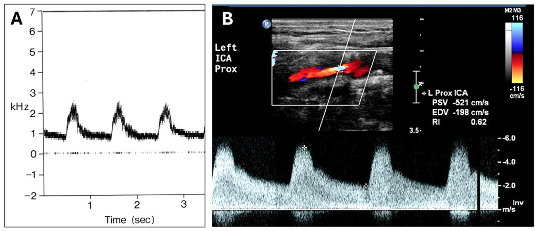
Examples of spectral waveforms from the prototype instrument and a modern duplex scanner.
Early experience with spectral waveform analysis suggested that there were characteristic changes associated with arterial wall abnormalities and stenotic lesions. 7 A normal center-stream flow pattern showed a narrow band with a clear area or ‘window’ under the systolic peak, consistent with laminar flow. Wall irregularities and stenoses of less than 50% diameter reduction produced a nonlaminar flow pattern with a widening of the waveform that was referred to as ‘spectral broadening.’ Stenoses of 50% diameter reduction or more were characterized by focal increases in peak systolic velocity (PSV) with spectral broadening distally (Figure 1B).
Beginning in the late 1970s and extending through the 1980s, Dr Strandness and colleagues carried out a series of studies to establish the first carotid duplex velocity criteria. In these studies, various spectral waveform parameters were correlated with the results of biplane arteriography, with percent diameter reduction of the internal carotid artery calculated using the estimated normal carotid bulb diameter as the reference site. 9 Among 135 patients (270 carotid bifurcations) who had both duplex scanning and arteriography, a peak systolic Doppler-shifted frequency of ⩾ 4.0 kHz was found to be predictive of 50–99% internal carotid artery diameter reduction, corresponding to a PSV threshold of 123.5 cm/s, which was rounded up to 125 cm/s. 10 This was the first specific velocity threshold to be established.
The term ‘carotid bulb’ refers to the slightly widened area at the bifurcation of the common carotid artery that typically includes the proximal internal carotid artery and the origin of the external carotid artery. This focal dilation results in a normal flow pattern referred to as ‘flow separation’ along the wall of the internal carotid artery across from the apical flow divider, consisting of low velocities and reversed or oscillating flow during each cardiac cycle (Figure 2).11,12 The absence of flow separation in an otherwise widely patent proximal internal carotid helps to identify those arteries with minimal plaque or wall thickening, features that define the category of 1–15% diameter reduction. 13 Internal carotid arteries with flow patterns showing no flow separation and extensive spectral broadening with a PSV less than 125 cm/s are classified as 16–49% diameter reduction.
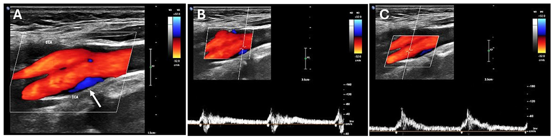
Color Doppler images and Doppler spectral waveforms illustrating an area of flow separation in a normal carotid bulb.
These initial validation studies created five categories for classifying the severity of carotid disease: normal, 1–15% diameter reduction, 16–49% diameter reduction, 50–99% diameter reduction, and occlusion. With very severe carotid stenoses, the peak systolic Doppler-shifted frequencies exceeded the Nyquist limit and could not be measured; however, it was observed that the end-diastolic frequencies also increased with higher degrees of stenosis, and these could be measured. A review of 98 internal carotid arteries with 50–99% arteriographic stenosis determined that an end-diastolic frequency of ⩾ 4.5 kHz distinguished between 50–79% stenosis and 80–99% stenosis with an accuracy of 87%, corresponding to an end-diastolic velocity (EDV) of approximately 140 cm/s. 14 This velocity threshold added a sixth category for the most severe (80–99%) internal carotid stenoses. These validation studies included patients with atherosclerotic lesions that typically involved the proximal to mid segments of the internal carotid artery, so the resulting velocity thresholds are based on the highest PSV and EDV obtained from those segments; therefore, they should only be applied to atherosclerotic stenoses in the proximal to mid internal carotid artery.
The duplex ultrasound criteria for the classification of internal carotid artery disease developed at the University of Washington up to 1984 are summarized in Table 1. 15 Color Doppler technology became available on duplex scanners in the mid-1980s. Early experience with color Doppler showed that it could identify areas of flow separation and flow disturbances associated with arterial lesions, but there are no specific carotid criteria based on color Doppler imaging alone. 16
The 1984 University of Washington criteria 15 for classification of internal carotid artery disease by duplex scanning.
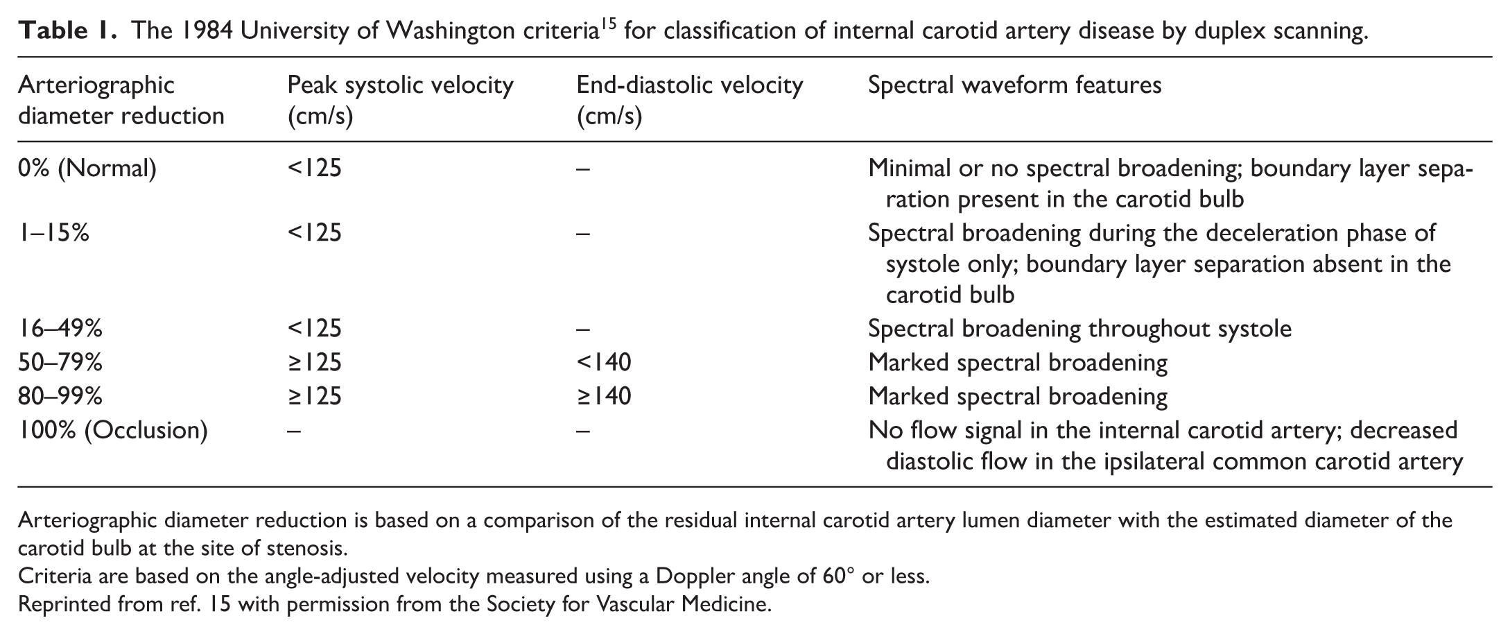
Arteriographic diameter reduction is based on a comparison of the residual internal carotid artery lumen diameter with the estimated diameter of the carotid bulb at the site of stenosis.
Criteria are based on the angle-adjusted velocity measured using a Doppler angle of 60° or less.
Reprinted from ref. 15 with permission from the Society for Vascular Medicine.
Evolution of the carotid criteria
The use of carotid duplex scanning increased through the mid to late 1980s, and the University of Washington criteria were adopted and modified by numerous vascular laboratories. Other criteria were developed independently, including the Bluth criteria published in 1988 (Table 2). 17 In a review of variation in velocity thresholds for carotid stenosis in 338 vascular testing centers accredited by the Intersocietal Accreditation Commission (IAC), 60 distinct PSV thresholds were identified for degrees of stenosis ranging from ⩾ 50% to ⩾ 90%. 18 These included 15 PSV thresholds for ⩾ 50% internal carotid artery stenosis ranging from 110 to 245 cm/s, with a median of 125 cm/s. This report also noted significant differences in the clinical management of patients with carotid disease based on these variations in velocity thresholds. For example, considering the various velocity thresholds for severe (⩾ 70%) internal carotid stenosis, in a sample of patients who underwent surgery for severe asymptomatic carotid stenosis, 9.8% of patients may not have been classified as having severe stenosis if their carotid duplex scan had been performed in another testing center with a different velocity threshold.
The 1988 Bluth criteria 17 for classification of internal carotid artery disease by duplex scanning.
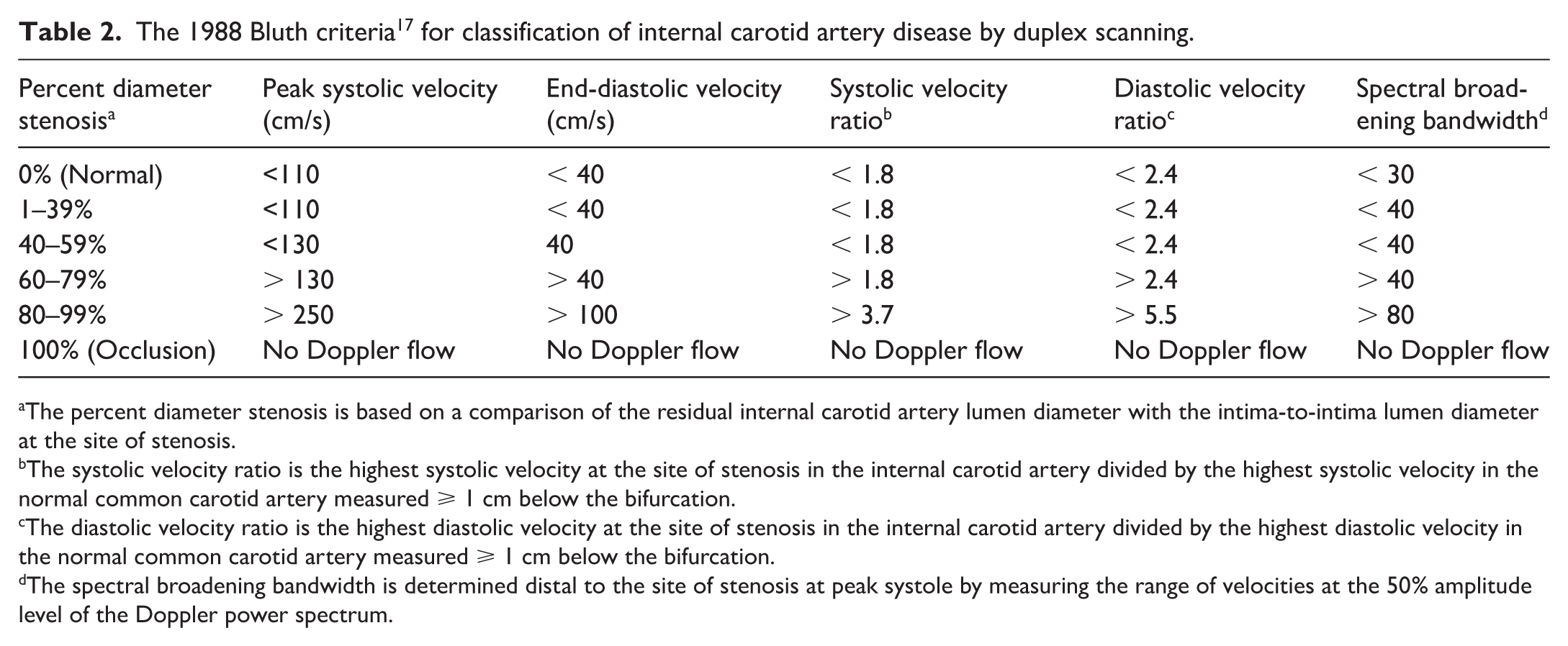
The percent diameter stenosis is based on a comparison of the residual internal carotid artery lumen diameter with the intima-to-intima lumen diameter at the site of stenosis.
The systolic velocity ratio is the highest systolic velocity at the site of stenosis in the internal carotid artery divided by the highest systolic velocity in the normal common carotid artery measured ⩾ 1 cm below the bifurcation.
The diastolic velocity ratio is the highest diastolic velocity at the site of stenosis in the internal carotid artery divided by the highest diastolic velocity in the normal common carotid artery measured ⩾ 1 cm below the bifurcation.
The spectral broadening bandwidth is determined distal to the site of stenosis at peak systole by measuring the range of velocities at the 50% amplitude level of the Doppler power spectrum.
The North American Symptomatic Carotid Endarterectomy Trial (NASCET) stenosis
In the 1990s, the results of the first randomized clinical trials comparing carotid endarterectomy with the prevailing best medical treatment were reported. These were the North American Symptomatic Carotid Endarterectomy Trial (NASCET) and the European Carotid Surgery Trial (ECST) for symptomatic patients and the Asymptomatic Carotid Atherosclerosis Study (ACAS) for asymptomatic patients.19–21 Each of these trials showed that carotid endarterectomy provided significant benefit in selected patients, with the severity of carotid stenosis expressed as a percentage reduction in vessel diameter as the major determining factor. However, there was variation among the trials in the clinically important diameter reduction thresholds and the methods used to measure the degree of arteriographic carotid stenosis.
Both the NASCET and ECST reported benefit from carotid endarterectomy in symptomatic patients with severe (70–99%) internal carotid stenosis. Both trials used the minimum diameter at the site of stenosis as the numerator for the stenosis calculation, but the denominator or reference diameter for determining the stenosis percentage was different. The NASCET used the normal distal internal carotid artery diameter, and the ECST used the estimated original diameter of the internal carotid at the site of stenosis. Because the ECST calculation typically relies on a larger reference diameter, it results in a higher percent stenosis value than the NASCET calculation. A study comparing these measurement methods concluded that the ECST method classified twice as many stenoses as severe compared with the NASCET, with a 70% NASCET stenosis being equivalent to an 82% ECST stenosis. 22 The ACAS reported benefit from carotid endarterectomy in asymptomatic patients with at least a 60% carotid diameter reduction, with a higher stroke risk reduction in men compared with women. The method used for arteriographic stenosis measurement in the ACAS was the same as that in the NASCET, and the 60% diameter reduction threshold was selected because that was regarded as the severity of stenosis required to produce a ‘positive’ or abnormal oculoplethysmography examination (the indirect tests for carotid artery disease were still being used in some laboratories at the time the ACAS protocol was designed).
In response to the NASCET, ECST, and ACAS reports, there was increased interest among clinicians in screening patients with suspected carotid artery disease to see if they might benefit from carotid endarterectomy. The approach to measurement of arteriographic carotid stenosis used in the original University of Washington validation studies was similar to the method used in the ECST, but there were no specific velocity criteria that corresponded to the clinically important arteriographic stenosis thresholds reported in the randomized clinical trials. The need for such criteria prompted additional validation studies of duplex scanning compared with arteriography, with the degree of stenosis measured by what is now referred to as the ‘NASCET method.’
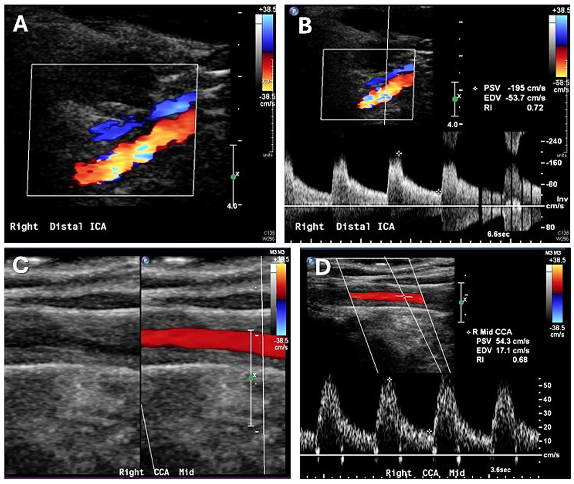
In a study of 100 patients with 184 patent internal carotid arteries who had carotid arteriography and duplex scans, an ICA/CCA ratio (highest internal carotid artery PSV at the site of stenosis divided by the normal mid-to-distal common carotid artery PSV) of ⩾ 4.0 had a sensitivity of 91.4%, specificity of 86.5%, positive predictive value of 75.7%, negative predictive value of 95.6%, and accuracy of 88.0% for identifying a 70–99% NASCET stenosis. 23 In the same study, an internal carotid artery PSV of ⩾ 230 cm/s had a sensitivity of 93.1%, specificity of 81.0%, positive predictive value of 69.2%, negative predictive value of 96.2%, and accuracy of 84.8%. A similar study was done to determine a velocity threshold for the 60–99% ACAS stenosis, which included 352 patent internal carotid arteries. 24 For that range of stenosis, the best velocity parameter was a combination of the highest PSV and the highest EDV in the internal carotid artery, with a PSV of at least 260 cm/s and an EDV of at least 70 cm/s resulting in a sensitivity of 84%, specificity of 94%, positive predictive value of 92%, negative predictive value of 88%, and accuracy of 90%. The velocity criteria for the 70–99% NASCET stenosis were widely adopted and eventually became part of the standardized criteria proposed by the Society of Radiologists in Ultrasound (SRU) and the IAC.
The Society of Radiologists in Ultrasound (SRU) consensus
In 2002, the SRU invited a group of experts in vascular ultrasound from a variety of medical specialties to participate in a conference ‘to develop recommendations for the performance of Doppler ultrasonography and interpretation of the results in the diagnosis of internal carotid artery stenosis,’ noting that there was considerable variation in the way carotid duplex scanning was being done, even in accredited vascular laboratories. 25 The SRU recommendations were reported in 2003 and are summarized in Table 3. It is important to point out that these velocity thresholds and other parameters were based on literature review, clinical experience, and ‘expert opinion,’ and did not represent a new rigorously validated set of carotid criteria. These included the original 125 cm/s threshold for ⩾ 50% internal carotid stenosis and the 230 cm/s threshold for 70–99% stenosis, thereby creating a 50–69% stenosis category.
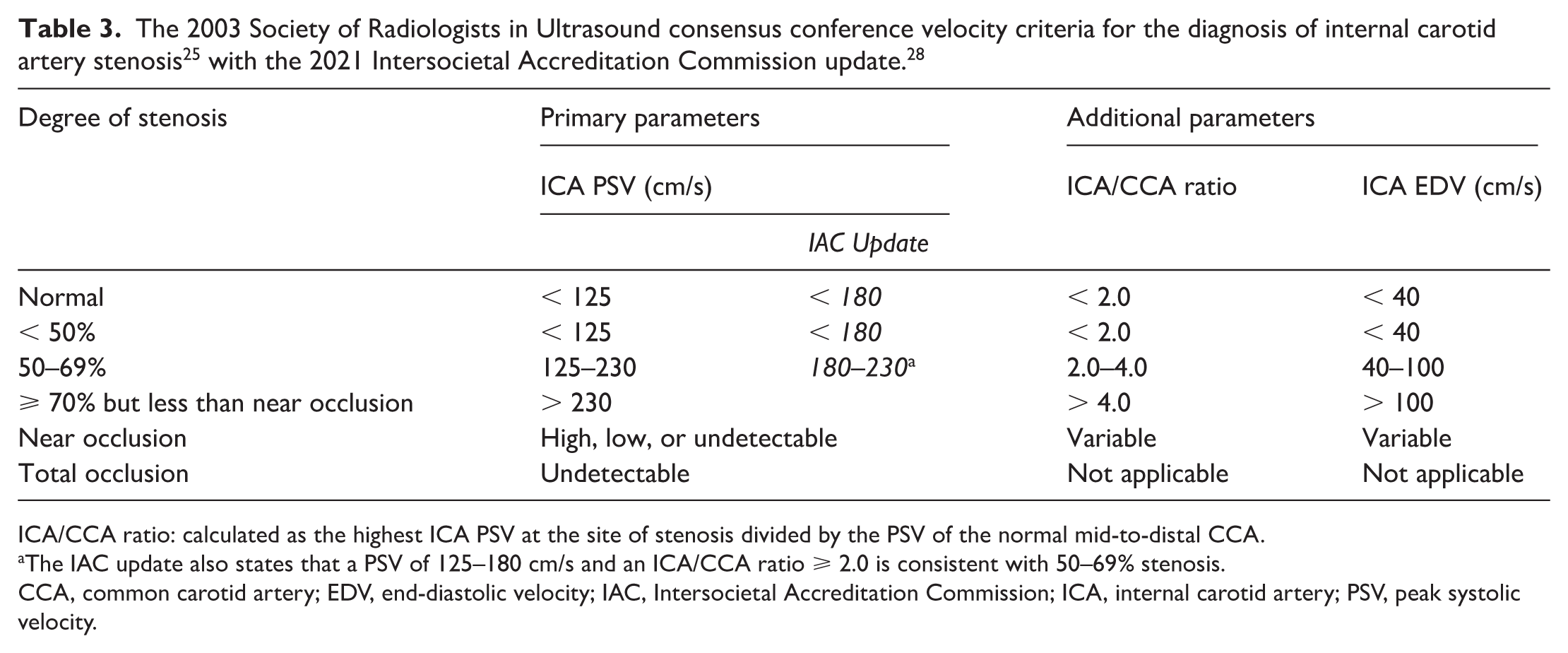
ICA/CCA ratio: calculated as the highest ICA PSV at the site of stenosis divided by the PSV of the normal mid-to-distal CCA.
The IAC update also states that a PSV of 125–180 cm/s and an ICA/CCA ratio ⩾ 2.0 is consistent with 50–69% stenosis.
CCA, common carotid artery; EDV, end-diastolic velocity; IAC, Intersocietal Accreditation Commission; ICA, internal carotid artery; PSV, peak systolic velocity.
A study reported in 2011 was performed to validate the SRU consensus criteria, with emphasis on the velocity thresholds for 50% and 70% internal carotid stenosis, and included 376 carotid arteries evaluated by duplex scanning and arteriography. 26 For 50–69% stenosis, the 125 cm/s threshold had a sensitivity of 93%, specificity of 68%, and accuracy of 85%. The 230 cm/s threshold had a sensitivity of 99%, specificity of 86%, and accuracy of 95% for 70–99% stenosis. The authors determined that the results for the 50–69% stenosis category could be significantly improved by increasing the PSV threshold to 140 cm/s, which provided a sensitivity of 94%, specificity of 92%, and overall accuracy of 92%. Increasing, the 125 cm/s PSV threshold for ⩾ 50% internal carotid artery stenosis would be a major recommendation of the IAC report on optimization of the carotid duplex criteria a decade later.
Intersocietal Accreditation Commission (IAC) optimization
The Intersocietal Commission for the Accreditation of Vascular Laboratories (ICAVL) surveyed a random sample of laboratories accredited in extracranial carotid testing to determine what criteria were being used to diagnose internal carotid artery stenosis, and the results were reported in 2011. 27 Among the 152 vascular laboratories surveyed, there were 17 distinct sets of carotid duplex criteria being used in 117 laboratories, with the remaining laboratories using locally developed criteria (six laboratories) or various combinations (hybrids) of published criteria (29 laboratories). The threshold for the most severe internal carotid stenosis category ranged from 70% to 91%, with 80% stenosis being the most common. Only 27% of laboratories were using the SRU consensus criteria. This extreme variability in carotid duplex criteria, and a subsequent survey indicating that more than two-thirds of the medical and technical staff of accredited vascular laboratories would support a requirement for the consistent use of a single set of validated carotid criteria, prompted the IAC to conduct an independent study to establish a standardized set of diagnostic criteria for carotid duplex scanning. (Note: due to organizational restructuring, the ICAVL became IAC Vascular Testing in 2008.)
The results of the IAC study were published in 2021 in a paper titled ‘Optimization of duplex velocity criteria for diagnosis of internal carotid artery stenosis.’ 28 The design of the IAC study was similar to the original validation studies performed at the University of Washington with comparison of carotid duplex ultrasound parameters to independently interpreted catheter arteriograms. However, the IAC study was multicentered, the arteriograms were read according to the method used in the NASCET, and the internal carotid artery stenosis thresholds and disease categories were taken from the SRU consensus criteria.
A total of 299 internal carotid arteries from 167 patients were included in the final analysis, with only moderate agreement for the categorization of internal carotid stenosis compared with arteriography (κ = 0.42). For predicting ⩾ 50% versus < 50% stenosis, the PSV threshold of 125 cm/s had a sensitivity of 97.8%, specificity of 64.2%, positive predictive value of 54.7%, negative predictive value of 98.5%, and accuracy of 74.5%. The combination of a PSV threshold of 125 cm/s and ICA/CCA ratio of ⩾ 2.0 produced a sensitivity of 94.3%, specificity of 84.3%, positive predictive value of 72.6%, negative predictive value of 97.1%, and accuracy of 87.4%. For a single PSV threshold, 180 cm/s had a sensitivity of 93.3%, specificity of 81.6%, positive predictive value of 69.2%, negative predictive value of 96.5%, and accuracy of 85.2%. For predicting ⩾ 70% versus < 70% stenosis, the PSV threshold of 230 cm/s had a sensitivity of 93.9%, specificity of 78.2%, positive predictive value of 35.6%, negative predictive value of 99.0%, and accuracy of 80.0%. Increasing the PSV threshold for ⩾ 70% stenosis to 250 or 260 cm/s improved specificity and accuracy, but all the threshold parameters were found to have a low positive predictive value, most likely due to the small number of cases with severe stenosis on arteriography.
Based on these results, the IAC study concluded that the diagnosis of ⩾ 50% internal carotid artery stenosis could be improved by increasing the PSV threshold from 125 cm/s to 180 cm/s as a single parameter, or by requiring a PSV of ⩾ 125 cm/s combined with an ICA/CCA ratio of ⩾ 2.0. The updated recommendations from IAC Vascular Testing include most of the original SRU consensus criteria, with the PSV threshold for ⩾ 50% stenosis changed to 180 cm/s (Table 3). The 230 cm/s PSV threshold for ⩾ 70% internal carotid stenosis remains, with the 50–69% stenosis category defined by a PSV in the range of 180–230 cm/s. However, the recommendations also state that the combination of a PSV of 125–180 cm/s and an ICA/CCA ratio of ⩾ 2.0 is consistent with 50–69% stenosis.
Throughout the evolution of the carotid duplex criteria, a principal assumption has been that there is a consistent relationship between internal carotid artery Doppler velocity parameters and the severity of stenosis as assessed by arteriography. The correlation between PSV and arteriographic percent stenosis was evaluated by creating a ‘scattergram’ of paired PSV and percent stenosis values from published articles for both native and stented internal carotid arteries. 29 Figure 3 shows the results for 2996 PSV versus percent stenosis pairs derived from 19 articles published between 1995 and 2010, and confirms that PSV increases with stenosis severity, although with wide variability. When the cumulative distribution of PSV was segmented by arteriographic percent internal carotid stenosis, the most predictive PSV thresholds were 165 cm/s for ⩾ 50% stenosis and 280 cm/s for ⩾ 70% stenosis. It is noteworthy that these PSV thresholds are quite close to those identified in the IAC study and included in the updated recommendations from IAC Vascular Testing.
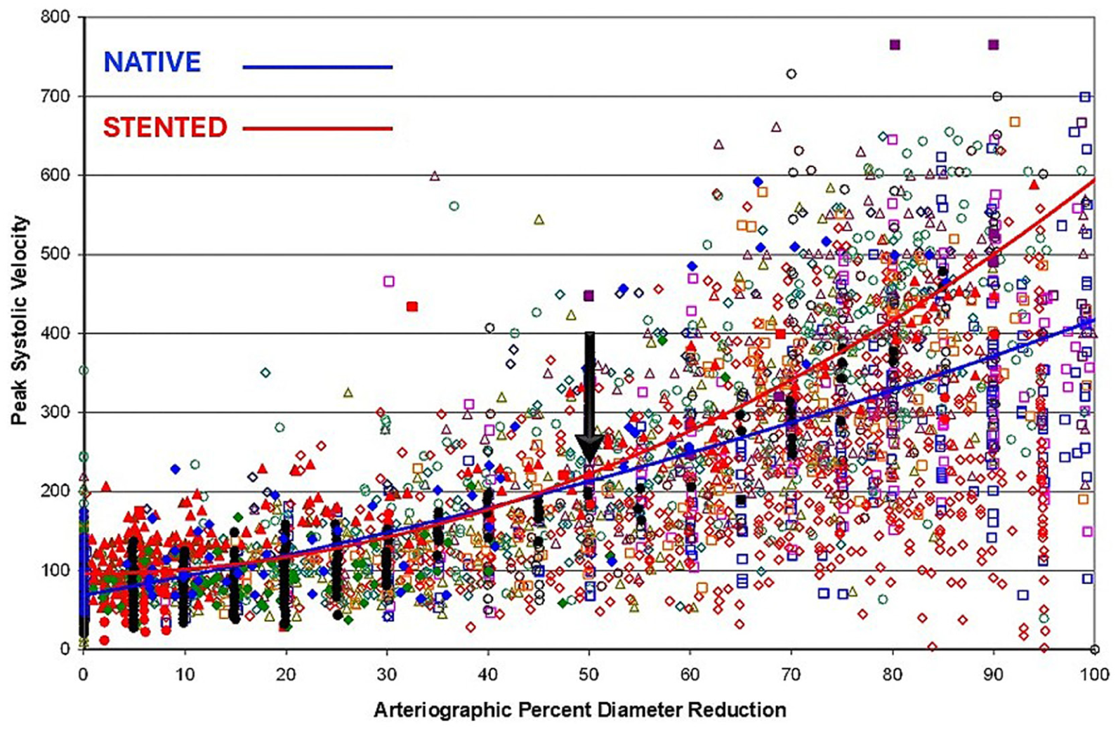
A ‘scattergram’ of 2996 paired internal carotid artery peak systolic velocity and arteriographic percent diameter reduction values showing that peak systolic velocity increases with stenosis severity, but with wide variability for both native and stented internal carotid arteries. The quadratic curves fitting the native and stented carotid artery data start to separate at a 50% diameter reduction (arrow).
Criteria for other vessels
The development and validation of the carotid duplex criteria have emphasized the diagnosis of internal carotid artery disease, but there are other arteries that are important to identify and evaluate during a carotid duplex scan, specifically the common carotid, external carotid, and vertebral arteries. Velocity thresholds and other parameters for classifying disease in these arteries have not been validated as extensively as those for the internal carotid artery, and the categories used are typically broader, such as normal, < 50%, ⩾ 50%, and occluded.
Common carotid artery
The diagnosis of common carotid artery stenosis relies on the typical findings of a focal high-velocity jet with increases in PSV and EDV; however, there are a small number of reports on specific threshold velocity criteria. A study published in 2010 included 62 patients who had both carotid duplex scanning and computed tomography (CT) angiography of the cervical vessels, providing 115 common carotid arteries for analysis. 30 The authors found better agreement between PSV and percent stenosis as determined by a reduction in the lumen area compared with percent stenosis determined by diameter reduction. A PSV of ⩾ 182 cm/s was the best threshold for ⩾ 50% area stenosis, with a sensitivity of 64% and specificity of 88% for all lesions, and a sensitivity of 76% and specificity of 89% when the stenosis was in the mid or distal segments of the common carotid artery. However, a 50% area reduction corresponds to only approximately a 30% reduction in diameter; therefore, the clinical relevance of this threshold is uncertain.
Another relatively small study published in 2014 identified 25 patients with isolated common carotid artery stenoses of ⩾ 60% diameter reduction and compared them with a control group of 74 patients with no common carotid stenosis. 31 All patients had carotid duplex scans and either carotid arteriography (21 patients) or CT angiography (four patients). The analysis showed that a PSV ⩾ 250 cm/s had a sensitivity of 98.7% and specificity of 95.7% for identifying ⩾ 60% common carotid stenosis, and an EDV ⩾ 60 cm/s had a sensitivity of 100% and specificity of 87%. The presence of both PSV ⩾ 250 cm/s and EDV ⩾ 60 cm/s had a positive predictive value of 100%.
External carotid artery
There are also only a few published reports on the validation of specific threshold velocity criteria for external carotid artery stenosis. A study published in 2018 included 314 patients with 536 external carotid arteries evaluated by duplex scanning and CT angiography. 32 There were 82 external carotid arteries with 50–99% stenosis on CT angiography using the NASCET method for determining percent diameter reduction, and these had a significantly higher mean PSV (294 cm/s) than those with < 50% stenosis (122 cm/s). A PSV ⩾ 200 cm/s provided a sensitivity of 84%, specificity of 93%, and accuracy of 91% for ⩾ 50% external carotid artery stenosis.
A similar study reported in 2020 compared carotid duplex scans and arteriograms in 140 patients, with the severity of external carotid stenosis measured using the NASCET method. 33 Among the 256 carotid bifurcations available for analysis, arteriographic ⩾ 50% external carotid stenosis was present in 60, and these were separated into two groups: those with < 50% arteriographic ipsilateral internal carotid stenosis and those with ⩾ 50% ipsilateral internal carotid stenosis. For the group without severe ipsilateral internal carotid stenosis, a PSV > 148 cm/s provided a sensitivity of 80%, specificity of 76.2%, and accuracy of 77.1% for ⩾ 50% external carotid stenosis. In patients with ⩾ 50% ipsilateral internal carotid stenosis, a PSV > 179 cm/s had a sensitivity of 50%, specificity of 79.6%, and accuracy of 71.3%.
Vertebral artery
Recognizing that the majority of atherosclerotic vertebral artery lesions are located at or near the origin from the subclavian artery, the validation of velocity criteria for identifying vertebral artery stenosis has concentrated on the proximal (V1) segment. For ⩾ 50% proximal vertebral artery stenosis, optimal PSV thresholds of 114 cm/s (sensitivity 71%, specificity 90%), 135 cm/s (sensitivity 74.1%, specificity 79.3%), and 140 cm/s (sensitivity 96.1%, specificity 96.4%) have been reported.34–36 Several studies found that a PSV ratio calculated as the highest PSV in the V1 segment divided by the PSV at a more distal site in the V2 segment was a strong predictor of ⩾ 50% vertebral artery stenosis, with a threshold of 2.1 providing a sensitivity of 92.1% and specificity of 89.1%, and a threshold of 2.2 yielding a sensitivity of 96% and specificity of 89%.36,37
In addition to velocity parameters for detecting vertebral artery stenosis, flow patterns and direction of flow are also important features that can be assessed during carotid duplex scanning. 13 A normal vertebral artery has a low-resistive flow pattern with pandiastolic antegrade flow that results from the low hemodynamic resistance of the cerebral circulation. A high-resistive flow pattern in a vertebral artery is abnormal and could indicate more distal vertebrobasilar disease. The diagnostic significance of a high-resistive vertebral artery flow pattern was evaluated in 79 duplex scans that showed unilateral or bilateral high-resistive antegrade vertebral artery waveforms and had correlative neuroimaging. 38 The mean PSV for high-resistive compared with low-resistive vertebral artery waveforms was 51.7 cm/s compared with 63.6 cm/s (p = 0.04), and the mean EDV was 4.6 cm/s compared with 17.3 cm/s (p < 0.001), respectively. Among all 90 high-resistive vertebral arteries, 18.9% were normal, 38.9% had distal vertebrobasilar stenosis or occlusion, 35.6% appeared congenitally small, and 6.7% had other abnormalities. Overall, approximately 46% of vertebral arteries with high-resistive flow patterns had some form of vertebrobasilar disease, with congenital nondominance accounting for most of the remaining vertebral arteries.
Partial or complete reversal of flow (retrograde flow) in a vertebral artery is another abnormality that can be identified on carotid duplex scanning. This finding is most often associated with a stenosis or occlusion in the ipsilateral subclavian artery proximal to the vertebral artery origin (or brachiocephalic artery on the right) and is commonly referred to as subclavian steal syndrome. During a 6-year period, patients having carotid duplex scans had bilateral arm pressure measurements, and 514 were identified with a > 20 mmHg brachial systolic pressure differential, with the lower pressure on the left side in 82%. 39 Vertebral artery flow reversal was complete (retrograde throughout the cardiac cycle) in 61%, partial or alternating (retrograde in systole and antegrade in diastole) in 23%, and absent (antegrade throughout the cardiac cycle) in 16%. Only 38 of the 514 patients (7.4%) had symptoms attributable to subclavian steal syndrome, and both the presence of symptoms and complete flow reversal were more common in patients with higher brachial systolic pressure differentials. Symptoms were present in 38.5% of the patients with a pressure differential of > 50 mmHg, compared with just 1.4% of patients with a pressure differential of 20–30 mmHg.
Nonatherosclerotic carotid disease
The duplex ultrasound criteria for classifying internal carotid disease, as well as disease in the other carotid and vertebral arteries, were developed and validated for atherosclerotic lesions in those vessels. Atherosclerosis is the most prevalent pathology encountered in carotid duplex scanning, but there are other conditions that can involve the extracranial carotid arteries. Although the velocity criteria for atherosclerotic lesions are often applied to nonatherosclerotic carotid disease, those criteria have not been rigorously validated for that purpose. Nonatherosclerotic conditions can produce stenotic lesions that cause flow disturbances, but they often result in structural abnormalities of the arterial wall, so the B-mode imaging component of duplex scanning plays a more prominent diagnostic role in this setting.
For example, dissections of the common carotid artery can occur as extensions of dissection in the aortic arch and appear on B-mode imaging as bright linear echoes in the vessel lumen, representing the intimal dissection flap. 40 Color Doppler and Doppler spectral waveforms may indicate two flow channels with different flow patterns (Figure 4). Dissection can also involve the internal carotid artery, typically starting 2–3 cm distal to the origin, and lead to a tapering occlusion or severe stenosis. Aneurysms and pseudoaneurysms of the carotid arteries appear as focal fusiform or saccular dilations surrounding areas of arterial flow and thrombus. A pseudoaneurysm may include a connecting neck with a typical ‘to-fro’ flow pattern.
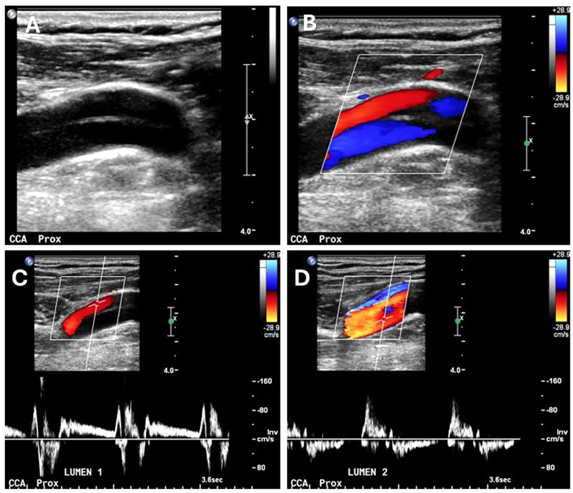
B-mode
Fibromuscular dysplasia (FMD) has been associated with a number of extracranial carotid lesions, but the most common multifocal type produces a typical ‘string-of-beads’ appearance with irregular wall thickening resulting in alternating segments of luminal narrowing and dilatation in the mid-to-distal internal carotid artery (Figures 5A and 5B). 41 A carotid web or diaphragm is characterized by a thin fibrous membrane originating from the wall of the proximal internal carotid artery (carotid bulb). Although these focal lesions have historically been classified as a variant of FMD, they may represent a different pathological process. Other carotid lesions that have been attributed to FMD include dissection, aneurysms, diffuse narrowing, and tortuosity.
Duplex ultrasound images of nonatherosclerotic carotid lesions.
Inflammatory conditions of the arterial wall can involve the extracranial carotid arteries, particularly the common carotid artery. Takayasu arteritis and giant cell arteritis are characterized by diffuse, concentric wall thickening resulting in varying degrees of luminal narrowing that is hypoechoic in the acute phase but becomes more echogenic as the disease progresses (Figures 5C and 5D). 40 Carotidynia is a rare lesion characterized by hypoechoic wall thickening with mild luminal narrowing. 42 Radiotherapy for head and neck cancer can also produce circumferential wall thickening and fibrosis, typically in the common carotid artery. 43
Follow-up after carotid artery interventions
Restenosis and neurologic symptoms are relatively uncommon following carotid endarterectomy or carotid artery stenting. In a systematic review and meta-analysis of randomized controlled trials, the prevalence of > 70% restenosis was 5.8% over a mean of 47 months after endarterectomy, and 10.0% over a mean of 62 months after stenting. 44 Duplex scanning was used to identify restenosis after the carotid interventions; however, there were multiple threshold criteria for > 70% stenosis. Over a mean surveillance period of 37 months, 9.2% of endarterectomy patients with > 70% restenosis or occlusion had a late ipsilateral stroke, compared with 1.2% of those without restenosis. Over a mean period of 50 months, 0.8% of patients with > 70% stenosis or occlusion after stenting had a late ipsilateral stroke, compared with 2.0% of those without restenosis. The majority of late ipsilateral strokes (85% after endarterectomy and 97% after stenting) occurred in patients without significant restenosis or occlusion.
Some specific risk factors for restenosis after carotid interventions have been identified. In a meta-analysis including over 17,000 patients, diabetes, dyslipidemia, female sex, chronic kidney disease, hypertension, and pretreatment stenosis > 70% were associated with a significantly increased risk of restenosis. 45 The subgroup analysis showed that female sex and smoking increased the risk of restenosis after carotid endarterectomy but not stenting. Hypertension was associated with restenosis after stenting, but not after endarterectomy.
Based on the low but clinically important incidence of restenosis and late stroke, as well as the frequent presence of untreated contralateral carotid disease, regular follow-up with duplex scanning has been recommended after carotid endarterectomy and stenting.46–48 However, the original rigorous surveillance protocols have been replaced by recommendations for more selective and less frequent follow-up imaging. An early (1–3 months) postintervention duplex scan is recommended to establish a baseline for comparison with subsequent examinations and to differentiate between residual and recurrent stenosis. Annual duplex surveillance is then recommended until no restenosis is observed on two consecutive annual scans. 47 More frequent or longer-term surveillance should be considered for patients with significant risk factors for restenosis, including the more aggressive forms of in-stent restenosis after carotid stenting. 49 Regular follow-up imaging of contralateral carotid disease has also been recommended for patients with > 50% stenosis in an untreated carotid artery; however, the appropriate frequency is unclear due to variations in the management of asymptomatic carotid disease. 48
It is recognized that the standard duplex criteria for classification of stenosis in native carotid arteries may overestimate stenosis severity after carotid interventions, and modified velocity criteria should be used after endarterectomy and stenting.46,48 In a study of 195 patients undergoing carotid endarterectomy with patch closure and a mean follow-up of 25 months, the optimal PSV thresholds were 213 cm/s for ⩾ 50% restenosis (overall accuracy 99%) and 274 cm/s for ⩾ 70% restenosis (overall accuracy 98%). 49 Higher PSV thresholds have been proposed for restenosis after carotid stenting, including 220 cm/s for ⩾ 50% restenosis and 340 cm/s for ⩾ 80% restenosis. 50 A PSV of 300 cm/s was used as the threshold for significant (⩾ 70%) restenosis in the Carotid Revascularization Endarterectomy versus Stenting Trial (CREST), as well as other clinical trials. 51
In addition to velocity thresholds for restenosis, B-mode image findings play an important role in duplex scanning after carotid interventions. 46 Early restenosis, defined as occurring within 24 months of the intervention, is attributed to intimal hyperplasia and can be focal or diffuse. This appears on B-mode imaging as smooth hypoechoic wall thickening (Figure 6). The patterns of neointimal hyperplastic lesions after carotid stenting have been described and classified. The diffuse proliferative (type IV) lesion, which is more than 10 mm long and extends beyond the ends of the stent, is associated with a particularly aggressive clinical course requiring reintervention. 52 Other findings that may be apparent on B-mode imaging after carotid stenting are poor stent apposition to the arterial wall (incomplete stent expansion) and residual plaque. If a transcarotid artery revascularization (TCAR) procedure has been done, the access site in the common carotid artery should be evaluated. B-mode image findings after carotid endarterectomy may include intimal flaps (especially at the endarterectomy endpoints), narrowing associated with the arterial closure, and residual plaque. The external carotid artery should be evaluated for patency, as plaque is typically removed from the orifice of this vessel during a carotid endarterectomy, or it is perfused through the interstices of the stent after carotid stent placement.
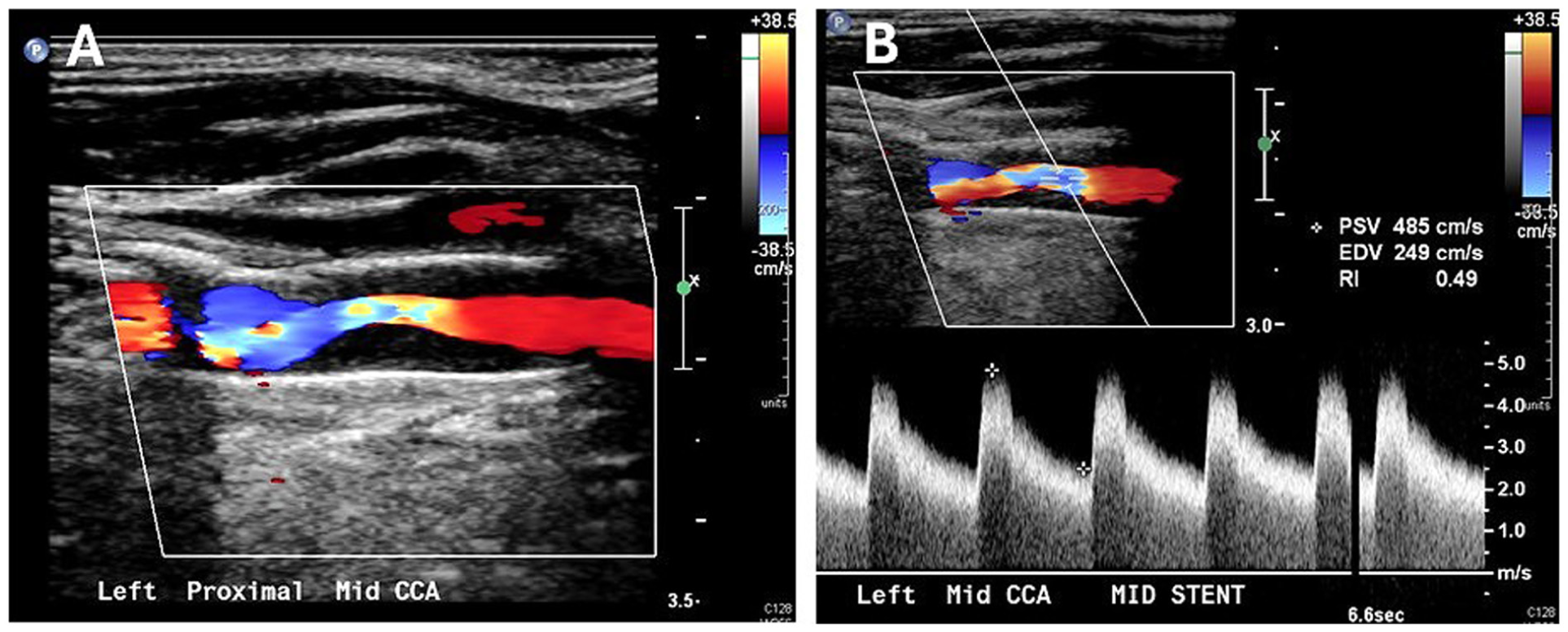
Color Doppler image
Beyond velocity: Plaque morphology
The development, validation, and clinical applications of carotid duplex scanning have primarily focused on assessing the extent of luminal narrowing produced by atherosclerotic plaques. This emphasis on quantifying carotid stenosis has been driven by the clear relationship between flow velocity and degree of stenosis, as well as the ability to measure various velocity parameters with Doppler ultrasound. It is also generally recognized that the risk for stroke increases with increasing stenosis severity. 53 However, patients with mild-to-moderate carotid stenosis can also present with carotid territory ischemic events. In the NASCET, 61% of the 2226 patients with recent neurologic symptoms were found to have < 50% carotid stenosis. 54 The ECST reported that 43% of 3018 symptomatic patients had < 30% stenosis. 55 These findings indicate that the severity of atherosclerotic carotid stenosis is not the only determinant of stroke risk.
Histological studies have shown that there are particular features of atherosclerotic plaques that are associated with an increased risk for stroke and other cerebrovascular symptoms, independent of the degree of stenosis.56,57 These features can be used to describe a high-risk or ‘vulnerable plaque’ and include a lipid-rich necrotic core, thin or ruptured fibrous cap, intraplaque hemorrhage, plaque neovascularization, and vessel wall inflammation. The use of morphological plaque assessment in clinical practice has been hindered by the lack of an accurate, low cost, and readily available imaging method to identify vulnerable plaques. Atherosclerotic lesion size and some compositional features can be quantified by CT, and positron emission tomography (PET) can identify vessel wall inflammation, but magnetic resonance imaging (MRI) is currently the best method for characterizing atherosclerotic plaque composition. 56 Based on histological confirmation, and using specialized protocols, MRI can reliably determine the status of the fibrous cap, plaque composition, neovascularization, and wall inflammation. The plaque feature that is most strongly correlated with stroke is intraplaque hemorrhage, and MRI is particularly well-suited for detecting these hemorrhagic plaques. 57
Experience with B-mode ultrasound has shown that carotid plaques with echolucent, heterogeneous, or ulcerated areas are associated with cerebrovascular symptoms.56,58 However, the accuracy of B-mode imaging for characterizing atherosclerotic plaques is limited by acoustic shadowing from calcification, anisotropic effects (where lesion properties vary with the angle of insonation), and operator or reader variability. B-mode imaging also has a low sensitivity and specificity for identifying intraplaque hemorrhage. The presence of hypoechoic or echolucent areas near the plaque surface (referred to as juxtaluminal black areas) is suggestive of intraplaque hemorrhage; however, the lipid-rich necrotic core has a similar hypoechoic appearance on ultrasound, making it difficult to reliably differentiate between these plaque features. Contrast-enhanced ultrasound, with scanning performed following intravenous (IV) injection of microbubble contrast agents, is a valuable method for demonstrating plaque neovascularization. The use of ultrasound contrast also improves the ability of B-mode imaging to detect plaque ulceration by clearly showing the boundary between flowing blood and the plaque surface. 57
Summary
The original duplex scanner was based on the ‘duplex concept’ of combining B-mode imaging and Doppler flow detection in a single diagnostic instrument to provide both anatomic and physiologic information on blood vessels. Although the extracranial carotid system was the first clinical application of duplex scanning, subsequent experience and advances in technology have expanded the capabilities of duplex ultrasound to the evaluation of arteries and veins in the extremities and abdomen. Criteria for classification of carotid artery disease have been based on the relationship between various velocity parameters and the severity of stenosis. The first carotid criteria developed at the University of Washington, including the 125 cm/s PSV threshold for ⩾ 50% internal carotid stenosis, are now more than 40 years old and were developed with instrumentation that is no longer used. Recent updates to the carotid criteria, such as the IAC recommendation for a PSV threshold of 180 cm/s for ⩾ 50% internal carotid stenosis, have been advocated to improve diagnostic accuracy and consistency in reporting.
Although not as well-studied as atherosclerotic carotid disease, duplex scanning has been used to identify and characterize nonatherosclerotic conditions, including dissection, aneurysms, FMD, and arteritis. Duplex scanning also plays a major role in follow-up after carotid endarterectomy and stenting, although modified velocity criteria must be used to avoid overestimating the severity of restenosis after carotid interventions. Finally, duplex criteria for carotid disease have been based primarily on velocity parameters and the severity of stenosis; however, there is clear evidence that morphological plaque features, particularly intraplaque hemorrhage, are also associated with ischemic cerebrovascular events. It is likely that future developments in carotid duplex scanning will include more emphasis on plaque morphology along with the well-established velocity criteria. A list of key points to remember about carotid duplex scanning is given in Table 4.
Key points to remember about carotid duplex scanning.
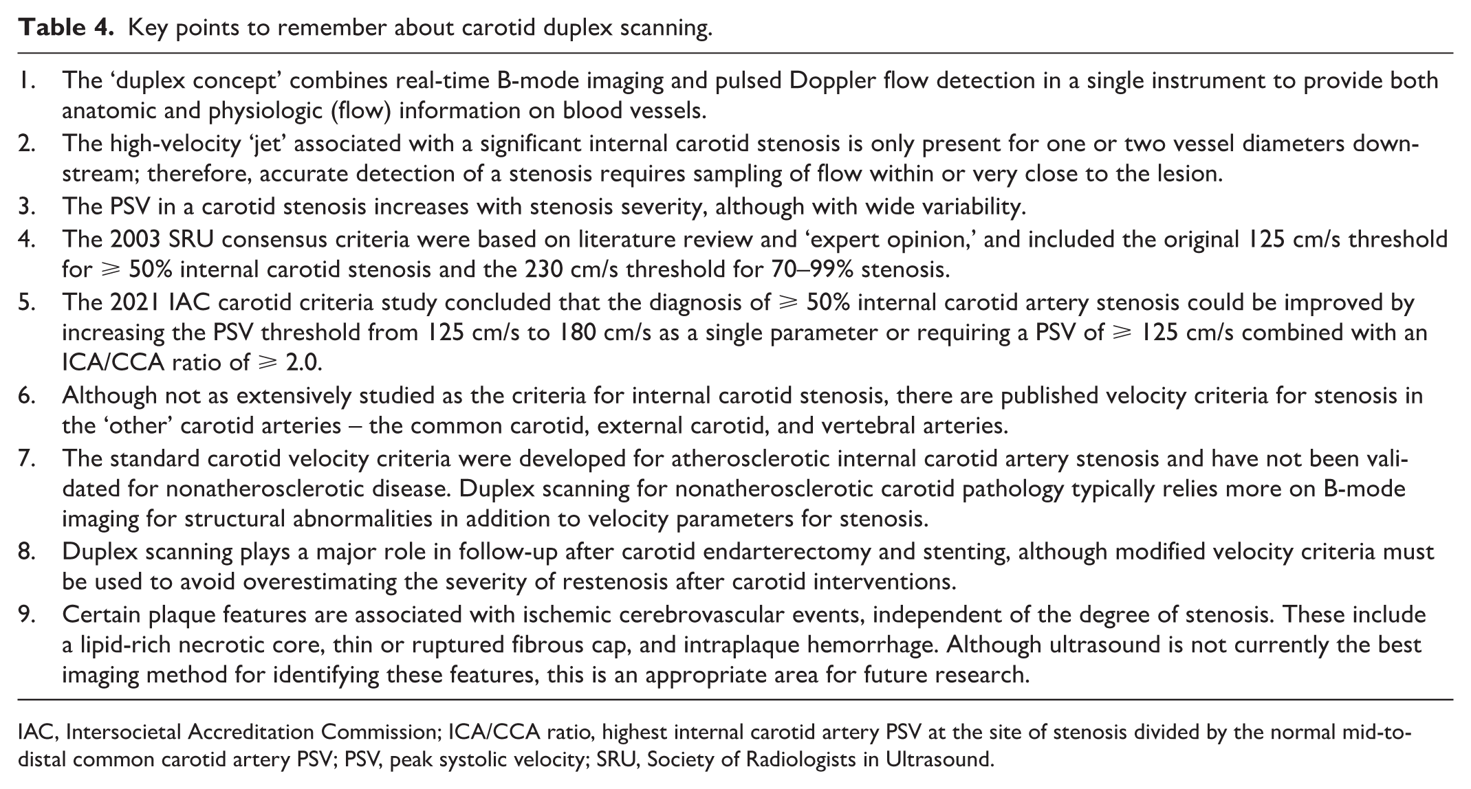
IAC, Intersocietal Accreditation Commission; ICA/CCA ratio, highest internal carotid artery PSV at the site of stenosis divided by the normal mid-to-distal common carotid artery PSV; PSV, peak systolic velocity; SRU, Society of Radiologists in Ultrasound.
Footnotes
Declaration of conflicting interests
The author declared no potential conflicts of interest with respect to the research, authorship, and/or publication of this article.
Funding
The author received no financial support for the research, authorship, and/or publication of this article.