
Abstract
Background
Sacubitril is an important and effective agent for cardiovascular remodeling. This study aims to evaluate its proangiogenic effects in an experimental model.
Methods
The chorioallantoic membrane (CAM) model was created for the investigation of the proangiogenic potential of sacubitril with different therapeutic doses (10−7 M, 10−6 M, 10−5 M) and compared with drug-free pellet group (sham) and normal morphology (control) of chick embryo development in drug-free chick embryos. The developing embryo's vascularity and the pellets’ effect were evaluated under a stereoscopic microscope. The density of vascular shoots and newly formed vascular nodules were not recorded.
Results
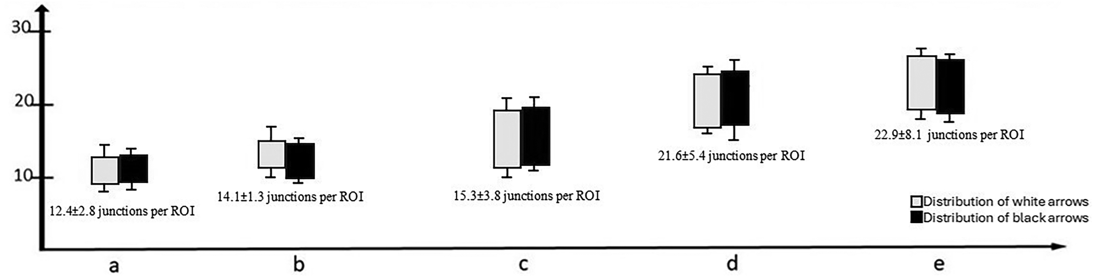
There was no significant difference between the control and drug-free pellet groups (12.4 ± 2.8 vs. 14.1 ± 1.3 junctions per ROI, p = 0.48). The incremental angiogenic properties were detected in drug groups as follows: 15.3 ± 3.8 per ROI in Group I (10−7 M concentration); 21.6 ± 5.4 per ROI in Group II (10−6 M concentration); 22.9 ± 8.1 per ROI in Group III (10−5 M concentration) (p < 0.001).
Conclusion
Our findings support that sacubitril provokes angiogenesis in a dose-dependent manner. Investigating these properties can be useful for understanding further effects of this agent in other cardiovascular diseases. Therapeutic angiogenesis is important for ameliorating the results.
Key Messages
The effects of Sacubitril plus valsartan therapy on myocardial angiogenesis were investigated through biomarkers. However, there are no clear studies showing its direct effect on the angiogenic process.
According to our knowledge our study is the first data showing the dose-dependent angiogenic effects of sacubitril.
The results were interpreted in favor of sacubitril increases angiogenesis with dose dependent manner.
Introduction
Angiogenesis is critical for preserving myocardial functions in myocardial diseases. Studies have shown that therapeutic angiogenesis improves myocardial functions in conditions such as hypertrophic cardiomyopathy and heart failure (HF). Thus, the effect of drugs used in cardiovascular diseases on angiogenesis has become an important issue for researchers.1,2
Neprilysin (NEP), an integrated membrane metalloproteinase, is an important biotarget for substrates with multiple physiological regulations. 3 This zinc-dependent metalloproteinase is found in many cells and plays an active role in renal proteolytic processes, myocardial regulation, immune responses, and cellular events.3,4
Sacubitril plus valsartan therapy is also a combination with NEP inhibition restriction in addition to angiotensin II receptor blockade (ARB).5,6 Sacubitril plus valsartan treatment also exerts antihypertensive and myocardial remodeling effects through NEP inhibition and angiotensin receptor blockade using the catabolization of natriuretic peptides. These effects increase fluid excretion from the body, decreasing systemic vascular resistance and body fluid load. Thus, the symptoms of HF regress and blood pressure decreases.5–7 Studies have emphasized that this also increases fat breakdown and insulin sensitivity by increasing lipolysis in obese individuals, increasing energy consumption with natriuretic catabolism. 7 The effects of this combination on myocardial angiogenesis have been investigated through biomarkers. However, no clear studies are showing its direct effect on the angiogenic process.6–8 Previous studies claim that ischemic conditions due to cardiac hypertrophy can trigger the proangiogenic effects of the angiotensin receptor NEP inhibitor. Suematsu et al. demonstrated significantly higher expression levels of myocardial angiogenic factors, such as CD34, VEGFA, ATP2a2, and MCP1, in apolipoprotein E-knockout mice fed a high-fat diet. 8 In another study, Poglajen et al. found that NEP inhibitors promoted the circulation of CD34 + cells, which plays a key role in angiogenesis in HF patients. 9 Neprilysin inhibition provides higher expression levels of proangiogenic markers such as CD34, and endothelial growth factors. Angiogenesis is induced as a response to increased oxygen demand. Another approach is that neprilysin inhibition independently induces angiogenesis by improving calcium reuptake. Previous studies have also shown that it contributes to the angiogenic response induced by natriuretic peptides.8,10,11 When all these processes are considered, it can be said that neprilysin inhibition has a regulating effect on myocardial angiogenesis together with cardiac remodeling. It has been predicted that neprilysin inhibitors may have effects on angiogenesis through oxidative stress and inflammation regulation. It provides beneficial results in preventing endothelial dysfunction with its positive effect on nitric oxide synthesis, and studies have indicated that it may increase endothelial NO bioavailability by regulating matrix metalloproteinases and pro-inflammatory cytokines.12–14 The chick embryo chorioallantoic model (CAM) is a well-described in vivo vasculature model that presents inflammation, hemorrhage, thrombosis, endothelial damage, and angiogenesis during or vessel wall destruction in vivo or angiogenesis/vasculogenesis.15,16 However, although the positive effects of sacubitril plus valsartan treatment on endothelium and vascularity have been reported, there is no previous study on this model and the subject has not been studied comprehensively.
The current study investigated the dose-dependent angiogenic effects of sacubitril. Using different molar concentrations of sacubitril, the CAM model was created and compared with normal physiological processes.
Material and method
Angiogenesis research on the CAM model for this study was conducted using Ross 308 chicken embryos. As was already issued before, the CAM model did not require ethical approval. 17 Nevertheless, the study was designed taking into account the Animal Welfare Act and the Guide for the Care and Use of Laboratory Animals.
Ross 308 eggs were placed in a classical chicken incubator after wiping with alcohol. They were removed on the 4th day of incubation, and 10 ml of amniotic fluid was aspirated from the non-pointed flat region with a sterile needle. The viability of the eggs was observed from a 1.5–2 cm hole drilled with sterile forceps from the pointed convex tip. Sterile agar with or without the drug was then dripped onto the viable eggs. The open tips were then closed with sterile films. The eggs were placed back in the incubator and kept until day 8. On the 8th day, all eggs were removed, and the control group and the groups containing agar were compared in terms of vascularity. The study protocols are shown in Figure 1. The sample size was determined according to the protocols.17,18 Each egg was randomly numbered for randomization when first placed in the incubator. Working groups were created by choosing random numbers. Each test was repeated twice for confirmation. Drug doses between molar concentrations reach large proportions, and in previous pharmacological studies, different molar concentrations were determined according to daily pharmacological use and applied to CAM. In our study, each dose was determined by this principle.17,18
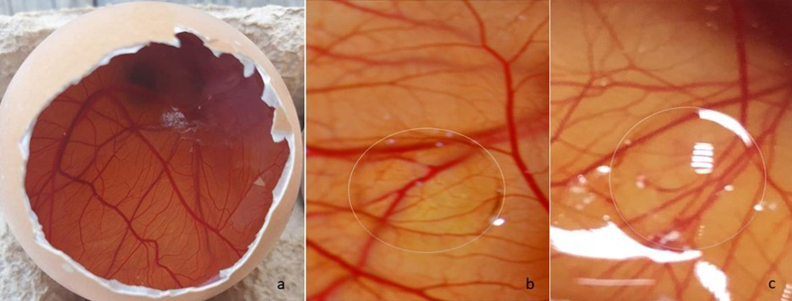
a. The developing healthy egg embryo and vascular bed, b. Placement of drug-free agar on the vascular bed of the chorioallantoic membrane (CAM), c. Placement of drug embed agar on the vascular bed of the CAM.
Grouping:
Control group (n: 8): Normal morphology of chick embryo development was determined. (All steps were applied without agar application).
Sham group (n: 8): A drop of agar without the drug was applied on the 4th day and the vascularity was evaluated on the 8th day.
Group I (n: 8): A drop of agar containing 10−7 M concentration of sacubitril was applied on the 4th day and the vascularity was evaluated on the 8th day.
Group II (n: 8): A drop of agar containing 10−6 M concentration of sacubitril was applied on the 4th day and the vascularity was evaluated on the 8th day.
Group III (n: 8): A drop of agar containing 10−5 M concentration of sacubitril was applied on the 4th day and the vascularity was evaluated on the 8th day.
Each step was repeated twice with different CAM groups to confirm the results.
Classifying of Angiogenesis:
The vascularity of the developing embryo and the effect of pellets were evaluated under a stereoscopic microscope. The density of the vascular shoots and newly formed vascular nodules was noted according to how they were previously described in the literature.17,18 This system depends on photographing the development of vascular beds during the incubation period and classifying capillary formation and vascular density that vary from the normal form.
Statistical analysis:
The commercially available statistical analysis program SPSS (ver. 15.0 software, SPSS Inc., Chicago, IL, USA) was used for statistical analysis. The number of newly formed vascular junctions and nodules was expressed as the mean ± standard deviation (SD) and subjected to one-way ANOVA followed by Tukey's range test.
Results
The effects of sacubitril on angiogenesis during embryonic growth in the CAM model were calculated by assessing the density and number of vascular budding and vascular sprouting junctions formed in two equally sized rectangular regions of interest (ROI). First, the chick embryo group with normal morphology and without any intervention (control group) was compared with the chick embryo group dripped with empty agar (sham group) (Figure 1 a,b). There was no significant difference between the two groups (12.4 ± 2.8 vs. 14.1 ± 1.3 junctions per ROI, p = 0.48) (Graph 1). The drug-free agar group (sham group) was compared with groups that administered sacubitril at different concentrations. The average junctions were found to be as follows: 15.3 ± 3.8 per ROI in Group I (10−7 M concentration); 21.6 ± 5.4 per ROI in Group II (10−6 M concentration); 22.9 ± 8.1 per ROI in Group III (10−5 M concentration) (p < 0.001) (Figure 1 c,d,e, and Graph 1). The CAM model examination, where the angiogenesis areas are compared, is presented in Figure 2. The graph below shows the distribution graph of the newly formed vascular nodules and group comparisons. These results show that no significant vascularity change was observed between the sham group and the group with low molar concentration sacubitril (10−7 M), while a considerable increase in angiogenic activity was detected at high concentrations (10−6 M and 10−5 M) (Graph 1, p < 0.001).
a. The distribution of newly formed vascular junctions and nodules in the control group, b. The distribution of newly formed vascular junctions and nodules in the sham group, c. The distribution of newly formed vascular junctions and nodules in groups I, d. The distribution of newly formed vascular junctions and nodules in group III, e, The distribution of newly formed vascular junctions and nodules in the control group.
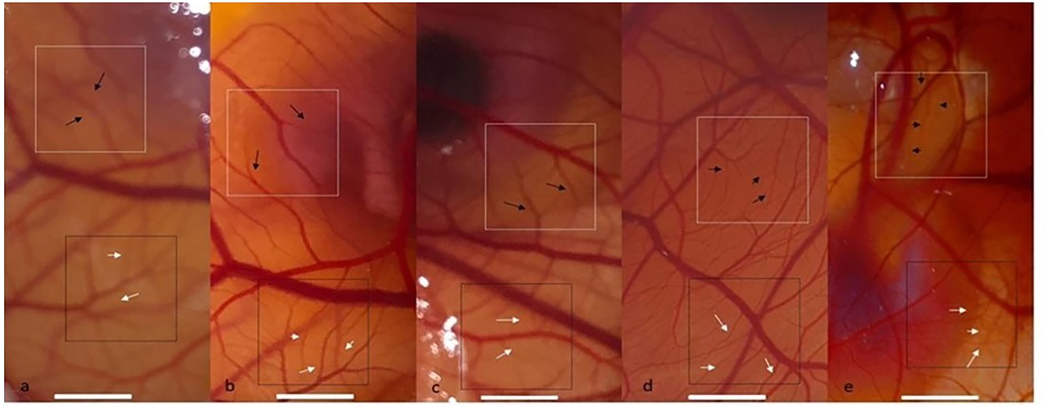
a. The control group on the 8th day, b. Sham group (with drug-free agar) on the 8th day, c. Group I (10−7 M concentration of sacubitril) on the 8th day, d. Group II (10−6 M concentration of sacubitril) on the 8th day, e. Group III (10−5 M concentration of sacubitril) on the 8th day; The proangiogenic properties were evaluated by evaluating the number of vascular junctions and nodules in two rectangular equally sized regions of interest (ROI) (Black and White rectangles).
Discussion
Our results indicate that sacubitril provokes angiogenesis in physiological conditions. Marked angiogenic effects were observed at the 10−6 M and 10−5 M concentrations. Our results are in line with existing literature, and these are the first data demonstrating the dose-dependent effects of sacubitril. Moreover, to our knowledge, this data is the first macroscopic evaluation of sacubitril on vascular density and newly formed vascular nodules.
The CAM model is an experimental research model that has been proposed to examine the positive and negative effects of medical preparations on angiogenesis.19,20 In this manner, it has been used in the evaluation of several cardiovascular drugs. 21 For example, the dose-dependent antiangiogenic effects of these drugs were demonstrated in a study investigating the effects of anticoagulant drugs on physiological angiogenesis. 22 In addition, the angiogenesis potentiating effect of cilastazol, which is also used in vascular occlusive diseases, has been demonstrated using this method. 17 The CAM method, while evaluating antiangiogenesis, is based on the reduction of capillary density on the membrane and the comparison of areas devoid of vascular bed. Contrastingly induced angiogenesis is evaluated by looking at newly formed vascular budding and the density of the vascular bed.19,21,22 In our study, we studied the effects of sacubitril on angiogenesis with the CAM model and further observed that sacubitril promotes new vessel formation and the development of vascular bed density with increasing molar concentrations.
The purpose of NEP inhibition in the treatment of HF is to prevent a decrease in the levels of these substances, prevent neurohumoral overactivation resulting in water and sodium retention, and vasoconstriction and inappropriate remodeling due to the decrease of these substances. 23 NEP inhibitors have become the focus of attention in diseases such as Alzheimer's due to their analgesic and anti-inflammatory effects as well as their decomposition of the amyloid-β peptide. 3 The decrease in NEP activity after hypoxia may also explain its relationship with events such as hypoxia-induced vasculogenesis. 24 Studies on NEP inhibitors after corneal injury have concluded in favor of increasing regeneration. In this case, it may be a reason for its positive effects on angiogenesis. 25 Previous studies have experimentally demonstrated that NEP inhibits fibroblast growth factor-2-induced angiogenesis in the murine cornea. In this context, it was predicted that neutral endopeptidases have an effect on growth hormone-dependent angiogenesis. 26 Similarly, in the same study, they found that NEP did not affect VEGF. Studies have thus been conducted to show that the inhibition of neutral endopeptidases can positively impact angiogenesis. This hormone-dependent effect might be related to a dose-dependent sacubitril effect, and this could be the reason for 10−6 M and 10−5 M concentrations being more effective on angiogenesis. The effects of many peptidases on the migration, proliferation, life cycle, metastatic tendency, and angiogenesis of cells have been studied. In this regard, its effects on metastasis have also been questioned. 27 When the effects of NEP on the heart were investigated, it was further emphasized that its myocardial remodeling might be due to supporting perfusion by increasing angiogenesis. These studies, based on the results of experimental planning, did not reveal any dose-dependent effects. 22 Using an experimental model, Suematsu et al. concluded that apolipoprotein E-knockout mice fed a high-fat diet preserved left ventricular systolic function through the induction of myocardial angiogenic factors. 8 They found sacubitril plus valsartan to increase the expression of CD34, VEGFA, MCP1, ATP2a2, and VCAM-1 on the myocardium, but it did not significantly affect β-catenin or VE-cadherin levels. Based on these results, they suggested that this molecule has a positive effect on myocardial angiogenesis. 8 While these potential effects are clear, the role of NEP inhibitors on multicellular and multisystemic effects remains uncertain and requires further explanation. Therefore, macro effects can be clearly understood by describing microcellular effects.
Studies have suggested that sacubitril has a more pronounced effect at high doses compared to low doses and has more positive effects on mortality and morbidity.28,29 Previous data have determined that sacubitril has a more positive effect on natriuretic peptides, which are also effective in angiogenesis, at high doses. 24 We believe that one of the reasons for the more pronounced effect on angiogenesis at higher concentrations (10−5 M concentration) detected in our study is related to this process.
Dose-dependent effects were investigated on the CAM model in our study. The results were interpreted in favor of sacubitril increasing angiogenesis with an increase in dose. An improved understanding of this dose-dependent effect needs to be investigated through more comprehensive cellular studies.
Limitations of Study:
The main limitation of this study concerns the experimental model of the study. It aimed to investigate vascular nodule screening during embryo development. Moreover, cellular mechanisms cannot be comprehensively clarified. The other limitation is related to the long-term outcomes of sacubitril on angiogenesis and cardiovascular remodeling. These results should therefore be confirmed with additional studies, and the mechanism of action should be clarified using cellular investigations.
Footnotes
Contribution
Mustafa OZGUL: Study design, main researcher, text writing
Eyup AYDOGAN: Researcher, Data collection and analysis
Declaration of conflicting interests
The authors declared no potential conflicts of interest with respect to the research, authorship, and/or publication of this article.
Funding
The authors received no financial support for the research, authorship, and/or publication of this article.
Ethical Approval
This study is in-vitro in terms of design and does not require ethics committee approval.