
Abstract
Background
This study aims to explore the causal relationship between five selected factors—lysosome, migrasomes, macrophage, fibroblast, and endothelium cell—and early-onset Alzheimer's disease (EOAD) through related genes, providing clues for EOAD research.
Methods
Using genes related to the five selected factors as exposure variables and EOAD as the disease outcome, significant genes were screened through Mendelian randomization (MR) analysis using the inverse-variance weighted (IVW) method, based on the OpenGWAS database. The selected genes were intersected with genes related to the exposure factors to assess the causal relationships between the five factors and EOAD.
Results
MR analysis identified 13 genes in total. Six of these genes were protective factors for EOAD, with LYST being the most significant (OR = 0.4259, 95% CI: 0.2218–0.8178, p = 0.0103). Seven genes were risk factors for EOAD, with NCF4 being the most significant (OR = 2.7207, 95% CI: 1.0229–7.2362, p = 0.0449). A total of 1925 genes related to lysosome, migrasomes, macrophage, fibroblast, and endothelium cell were analyzed. After intersection, 10 lysosome-related genes (NCF4, VIPAS39, LYST, SORT1, ARSB, EPDR1, SYNGR1, ANXA11, PYGB, CLN5) and 3 endothelium cell -related genes (ADM, NFIB, NKTR) were found to have significant causal relationships with EOAD.
Conclusions
There are genes related to lysosome and endothelium cell that have significant relationships with EOAD, while no causal relationships were found between migrasomes, macrophage, fibroblast, and EOAD. This study provides an important basis for further EOAD research.
Introduction
Alzheimer's disease (AD) is a complex neurodegenerative disorder. Its prevalence is gradually rising with the global increase in the aging population. Early-onset Alzheimer's disease (EOAD) whose patients are commonly below 65 years, has an early onset and is more aggressive.1,2 Given the current lack of highly effective and safe treatment options, the primary objective of this study is to identify genes associated with the pathogenesis and therapeutic targets of EOAD. By exploring the risk and protective factors for EOAD, we aim to provide a foundational reference for the early diagnosis and treatment of this condition.
Lysosome, as crucial cellular organelles, are involved in the degradation of toxic substances in neural tissues of the brain. For instance, the lysosome in neural cells participate in the endocytosis and degradation of amyloid-β (Aβ) plaques in EOAD. 3 Furthermore, lysosome interact with other organelles, such as mitochondria, thereby influencing neuronal functions. Dysfunction of lysosome leads to various brain diseases, including neurodegenerative disorders. 4 Investigating the characteristics and degradative functions of lysosome may pave the way for the development of EOAD therapeutics.5–7 Therefore, exploring the genes related to lysosome and EOAD is particularly important, as it can provide clues for potential drug targets in subsequent EOAD treatment development. Endothelium cell is essential components of the tissue matrix. Together with various brain cells, endothelium cells form the neurovascular network and maintain the stability of the internal environment of the nervous system. Endothelium cell is involved in maintaining the stability of the blood-brain barrier and the inflammatory response process, 8 as well as in angiogenesis during the progression of AD and brain cell aging. 9 Consequently, studying the relationship between endothelium cell and EOAD is a worthwhile endeavor. Macrophage in the central nervous system can transform into microglia under certain conditions and mediate the phagocytosis of toxic substances. 10 Although macrophage and microglia act as a double-edged sword, they positively influence neuronal regeneration. 11 Studies in mice have shown that Aβ plaques can induce the formation of macrophage-derived migrasomes, which can damage endothelial cells and disrupt the blood-brain barrier. 12 Under certain conditions, macrophage in AD brain tissue can transform into neuroglial cells and then target Aβ plaques for phagocytosis. 13 These findings suggest that macrophage may act as “scavengers” of Aβ plaques, prompting efforts to elucidate the relationship between macrophage and EOAD and to develop immunotherapeutic drugs for AD. Migrasomes, which are small bodies produced during cell movement, can mediate communication between brain cells and influence the functions of organelles.14,15 Thus, they may have unknown impacts on the progression of EOAD. Fibroblast, as vital cells in the neurovascular system unit, can secrete or modify extracellular matrix proteins, providing support to surrounding vessels and cells. However, brain inflammation induces fibroblast to form fibrotic scars,16,17 potentially promoting neuronal damage and the occurrence of AD. Research indicates that in AD brain tissue, fibroblast exhibit reduced mitochondrial calcium levels, sustained activation of the mitochondrial permeability transition pore, and increased oxidative stress. This leads to mitochondrial damage, functional failure, and a decrease in number, exacerbating the inflammatory response in the brain and promoting the progression of EOAD. 18
Mendelian randomization (MR) is a data analysis method for the evaluation of disease inference, which uses genetic variations as instrumental variables to estimate the causal relationship between exposure factors and outcomes. MR analyses for EOAD have clear causal direction and is reliable without the influence of confounding factors.
Methods
Data sources
This study utilized the following datasets from the OpenGWAS database (https://gwas.mrcieu.ac.uk/): outcome data from the finn-b-AD_EO_EXMORE dataset, which provides information on 112,058 European samples, including 587 disease cases and 111,471 controls. The exposure-related genes were sourced from the following data: fibroblast and endothelium cell genes were obtained from the GeneCards database, while genes for migrasomes (PMID: 36405728), lysosome (PMID: 37275878), and macrophage (PMID: 35967352) were sourced from literature. Their corresponding GWAS data were taken from the eqtl-a dataset. For example, the exposure factors NCF4 and LYST correspond to GWAS IDs: eqtl-a-ENSG00000100365 and eqtl-a-ENSG00000143669, respectively. All studies within the OpenGWAS database included informed consent from participants.
MR analysis method
Three assumptions and three principles for MR analysis
The three assumptions are: (1) Relevance assumption – the selected instrumental variables (SNPs) should have a strong association with the exposure factors; (2) Independence assumption – the selected SNPs should not be associated with confounding factors; (3) Exclusion restriction assumption – the selected SNPs should not be directly associated with the outcome, but should influence it only through the exposure factors.
The three principles for MR method selection are: (1) If there is no heterogeneity or pleiotropy, the IVW result is prioritized; (2) If heterogeneity exists but no pleiotropy, the weighted median method is prioritized (or the random effects model of IVW may be used); (3) If pleiotropy is present, the weighted regression method (MR-Egger) is prioritized.
Main steps in MR analysis
The key steps include: (1) Reading the exposure GWAS data and selecting suitable instrumental variables (clumping if necessary); (2) Extracting the selected SNPs from the outcome GWAS data; (3) Harmonizing the formats of exposure and outcome GWAS data; (4) Conducting MR analysis using the selected method; (5) Visualizing the results (scatter plots, forest plots, funnel plots). MR methods include IVW, MR-Egger, weighted median, weighted mode, and simple mode, with IVW as the primary method. IVW is characterized by disregarding the intercept term during regression and using the inverse of the outcome variance (the square of the standard error, se²) as weights for fitting. It requires the SNPs to fully comply with the three core principles of MR studies to obtain accurate causal estimates.
Statistical methods
The R package TwoSampleMR function extract_instruments was used to extract exposure data and filter instrumental variables. The criteria for filtering include: p < 5 × 10⁻⁶, clump = TRUE, r² = 0.001, and kb = 10,000. A p-value of < 5 × 10⁻⁶ indicates that only SNPs with a p-value below this threshold are retained for further analysis. If too few SNPs meet this criterion, the threshold can be relaxed (e.g., p < 1 × 10⁻⁵), but additional sensitivity and robustness checks are required. The clump parameter ensures that genetically correlated SNPs (linkage disequilibrium) are removed. If r² between two SNPs exceeds 0.001, one of them will be excluded. The clumping distance (kb) is set to 10,000, meaning SNPs within 10,000 kb of the most significant SNP that exceed the LD threshold will be removed. The strength of the association between instrumental variables and exposure factors was assessed using the F-test, and SNPs with F < 10 were removed to avoid weak instruments. SNPs directly associated with EOAD were also excluded to ensure instrument independence. The harmonise_data function in R was used to align effect alleles and effect sizes for consistency across all SNPs. If there is inconsistency, primarily multiply the beta value by −1 (-beta), and secondarily change the eaf to (1-eaf). MR analysis used the inverse-variance weighted (IVW) algorithm, and Steiger directionality tests were applied to verify whether the effect direction of the exposure factor on the outcome through the SNP is consistent with expectations. If consistent, it supports the causal effect of the exposure factor on the outcome. Significant factors associated with EOAD were screened at a threshold of p < 0.05, and sensitivity analyses were conducted using the leave-one-out method.
Results
Overview of gene data
A total of 1925 genes related to lysosome, migrasomes, macrophage, fibroblast, and endothelium cell were collected from the respective datasets. The number of genes is displayed through an upset plot, showing that lysosome-related genes are the most numerous, with a total of 875. There are 7 migrasome-related genes, 240 macrophage-related genes, 571 fibroblast-related genes, and 232 endothelial cell-related genes, with some overlap between these gene sets (Figure 1).
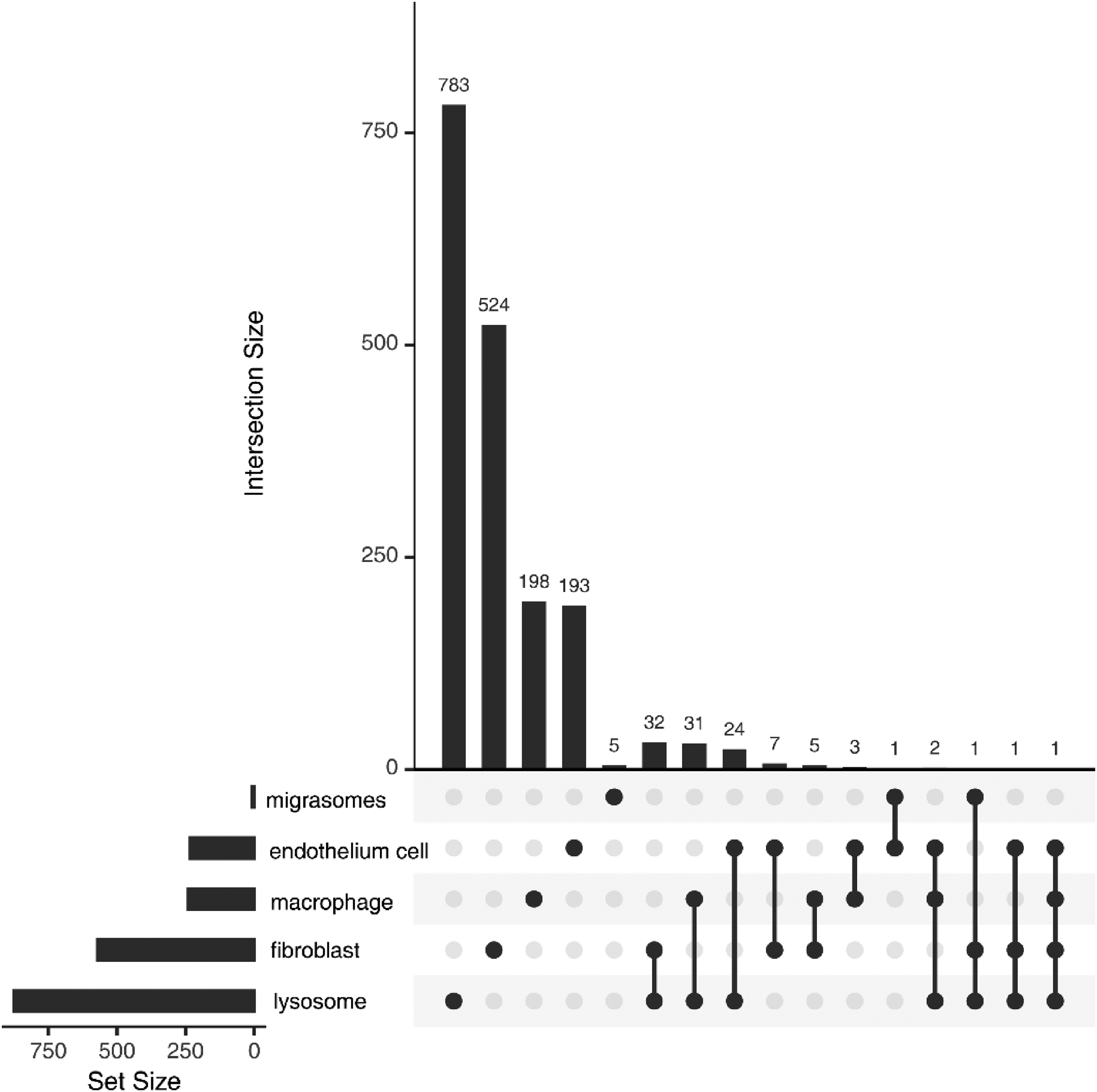
Upset plot of related genes of different types.
Mendelian randomization (MR) analysis
Using the above statistical methods, 13 genes (EPDR1, SYNGR1, NCF4, PYGB, CLN5, ARSB, ANXA11, SORT1, LYST, VIPAS39, NFIB, ADM, NKTR) were identified through the Steiger direction test, IVW p-value < 0.05, and horizontal pleiotropy test. The hazard ratio (HR) from the forest plot helps to determine the risk rate of EOAD caused by each gene. An HR of 1 indicates equal risk, HR > 1 indicates a higher risk, and HR < 1 indicates a lower risk. Of the identified genes, 6 genes (LYST, ADM, ARSB, NKTR, NFIB, PYGB) were protective factors against EOAD, with LYST being the most significant (HR = 0.43, 95% CI: 0.22–0.82). The other 7 genes (EPDR1, SYNGR1, ANXA11, CLN5, SORT1, VIPAS39, NCF4) were risk factors, with NCF4 being the most significant (HR = 2.72, 95% CI: 1.02–7.24) (Figure 2). By intersecting the significant genes with those related to the five selected factors, it was found that 10 lysosome-related genes (NCF4, VIPAS39, LYST, SORT1, ARSB, EPDR1, SYNGR1, ANXA11, PYGB, CLN5) and 3 endothelium cell-related genes (ADM, NFIB, NKTR) had a significant causal relationship with EOAD (Figure 3). The results indicate that lysosome and endothelium cell are involved in the progression of EOAD through specific genes, with NCF4 and LYST being particularly notable.
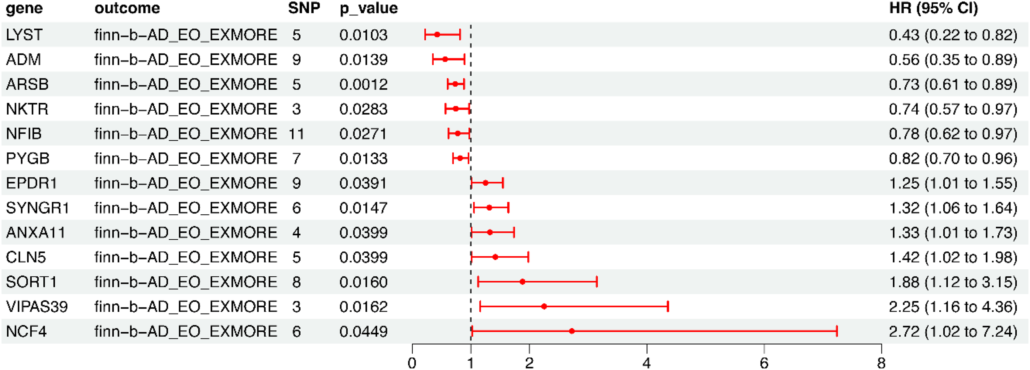
Mendelian randomization forest plot.
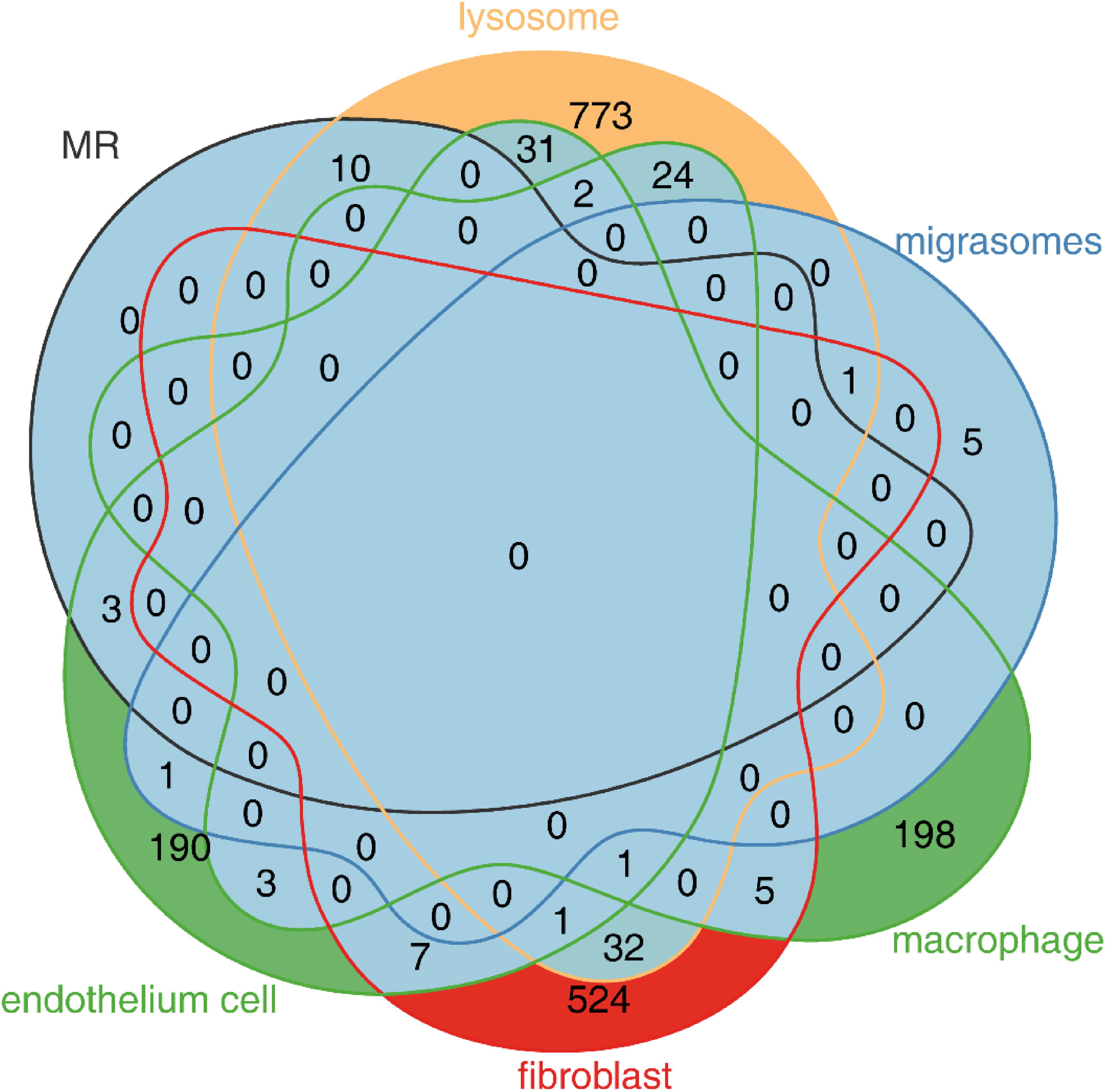
Intersection of genes with significant causal relationships.
Mendelian randomization analysis of NCF4 and LYST genes
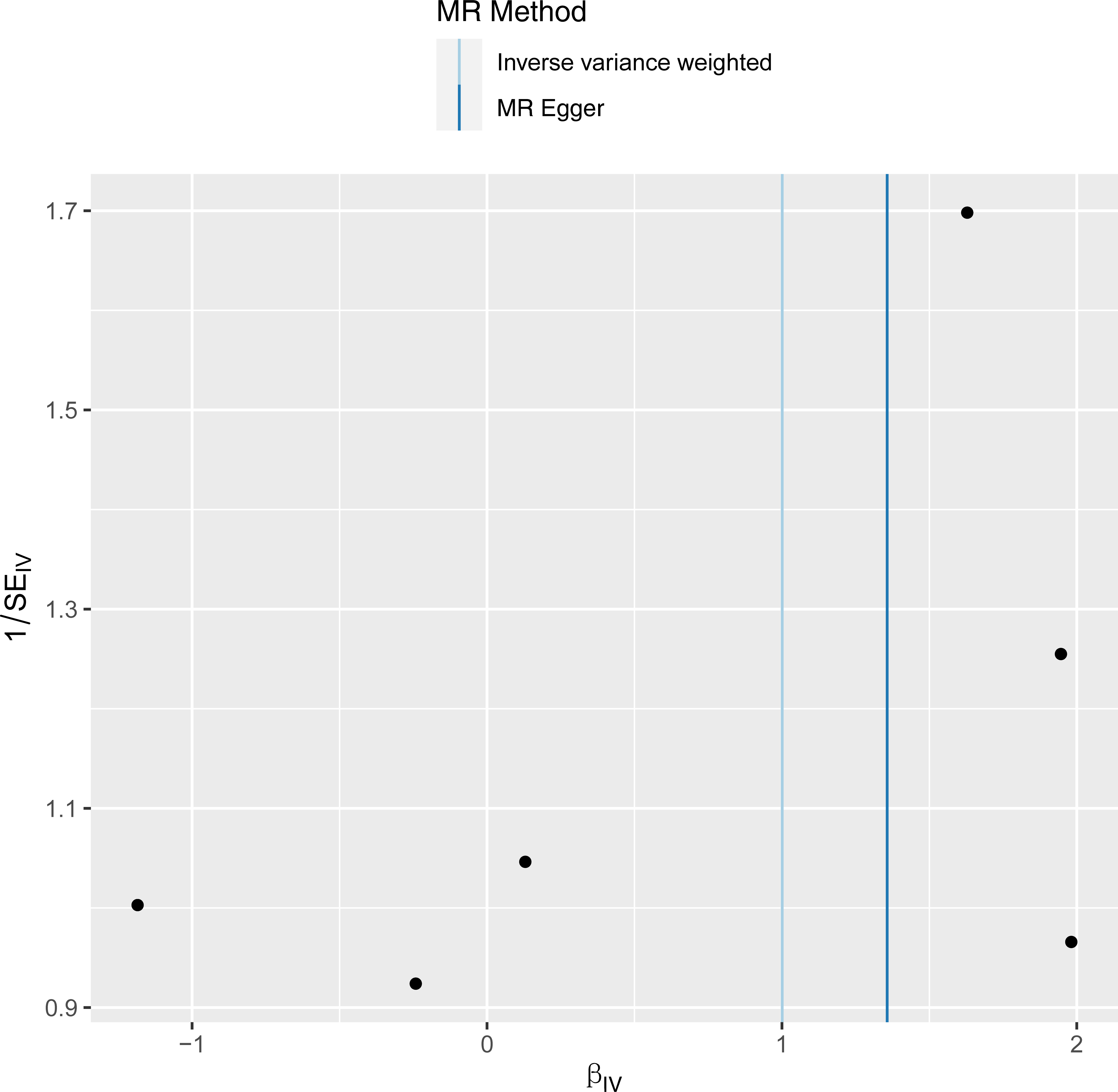
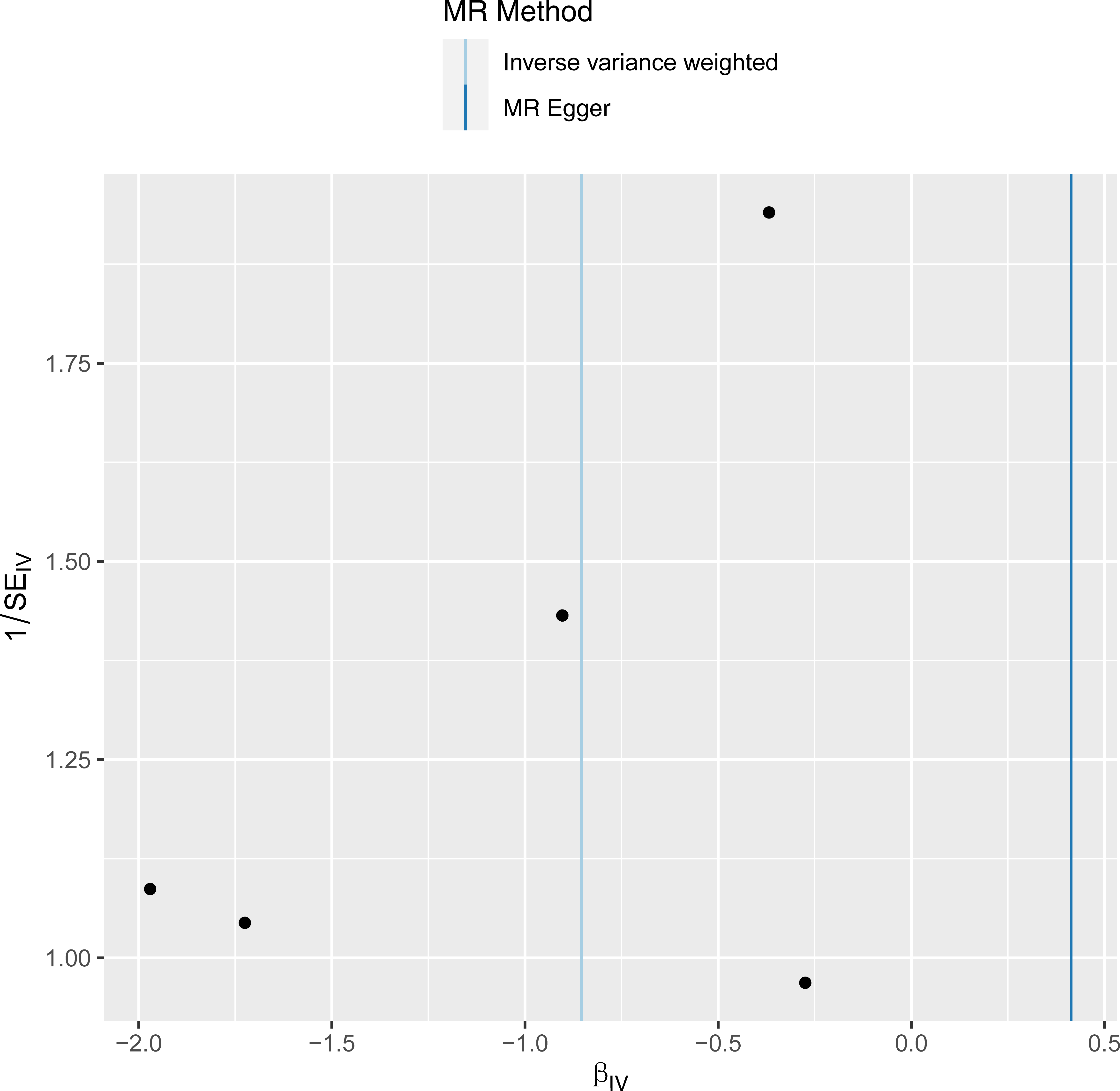
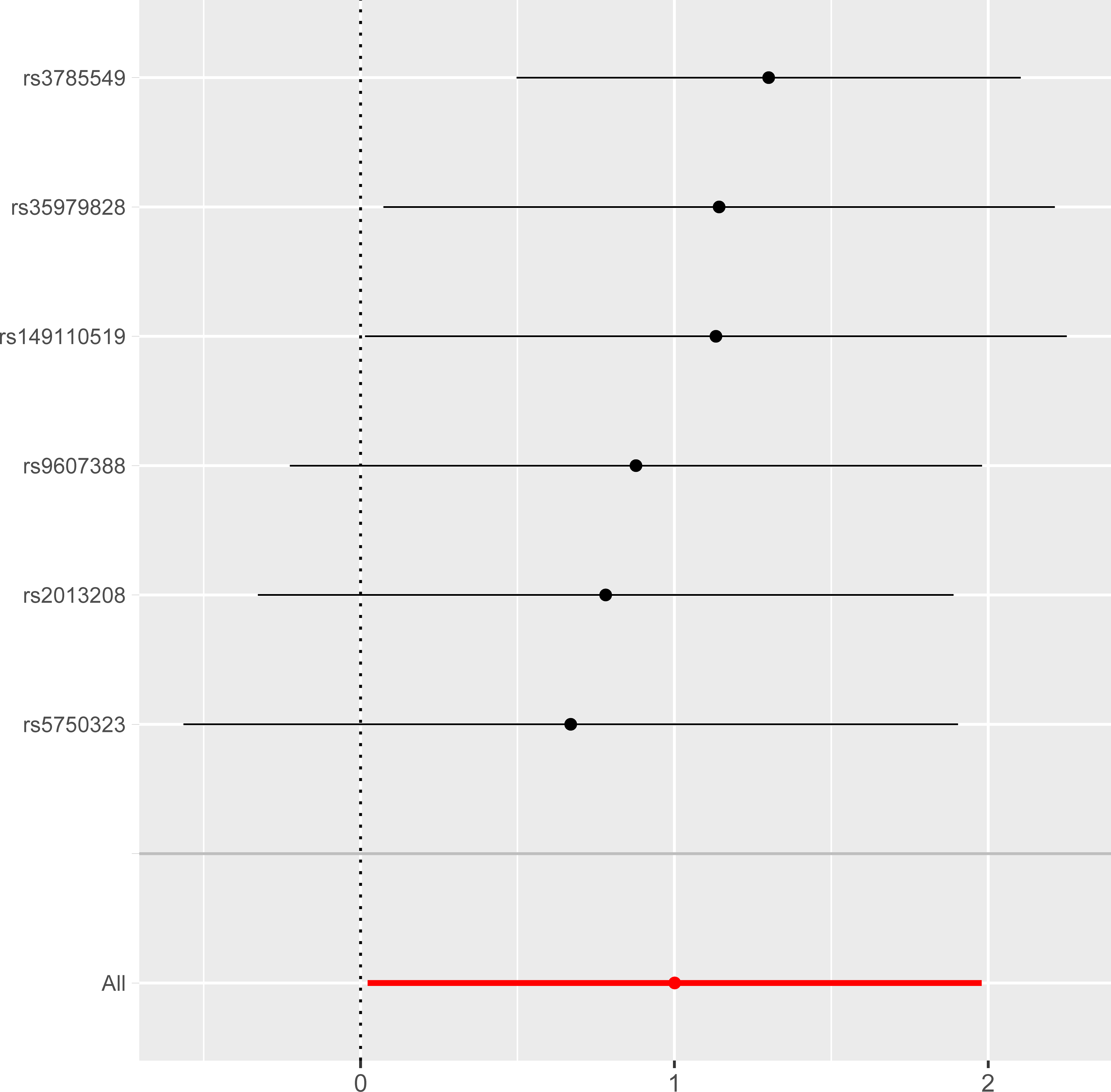
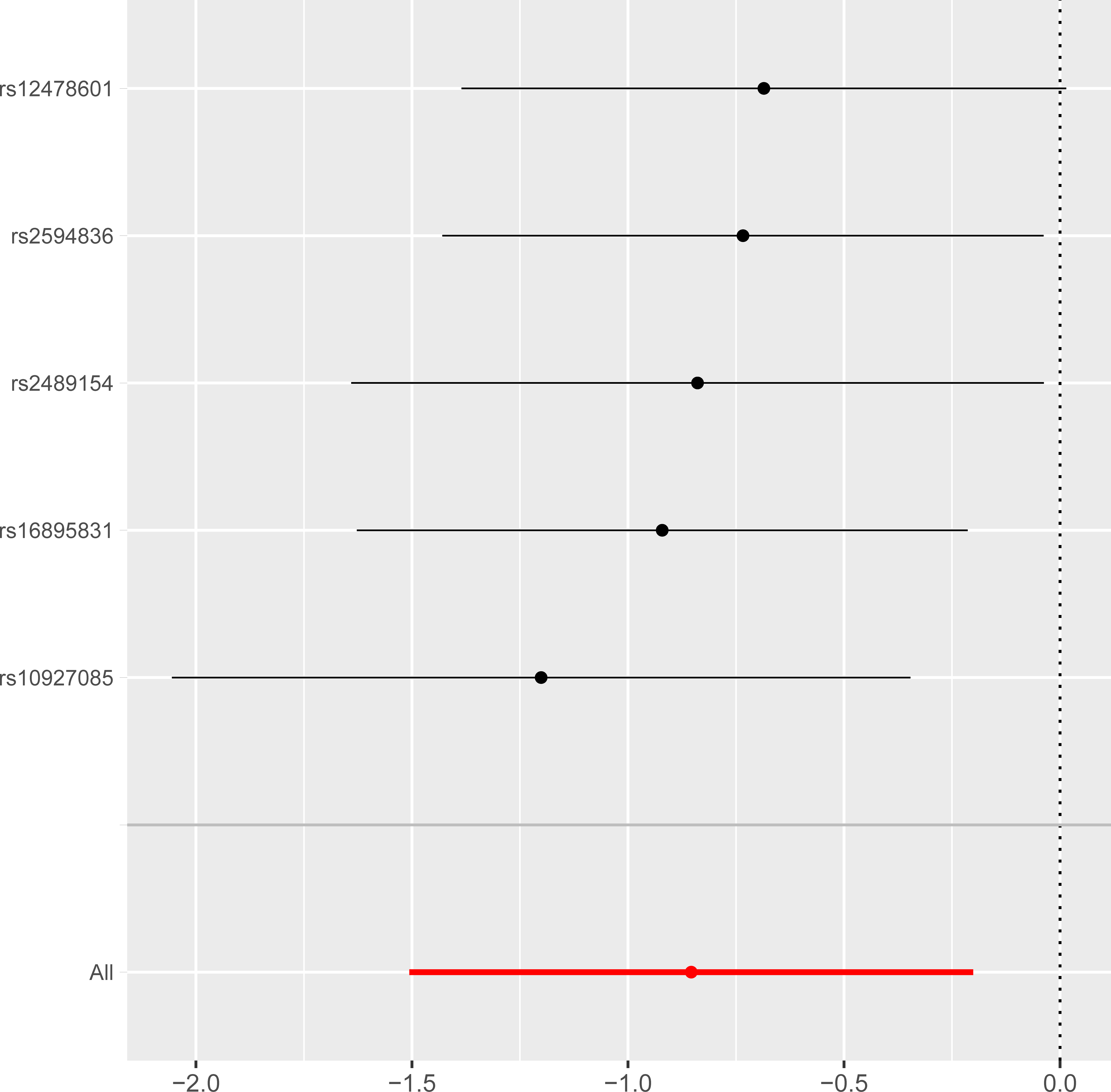
MR analysis results: (1) The IVW method revealed NCF4 as the most significant risk factor for EOAD (OR = 2.7207, 95% CI: 1.0229–7.2362, p = 0.0449) (Table 1), and LYST as the most significant protective factor (OR = 0.4259, 95% CI: 0.2218–0.8178, p = 0.0103) (Table 2). The OR > 1 for NCF4 indicates a positive association with EOAD risk, while the OR < 1 for LYST suggests a negative association. The accuracy of the estimates is evident from the b and SE values in the tables. (2) A scatter plot illustrates the relationship between the exposure and outcome. Each point represents an SNP, and the line shows the 95% confidence interval. The x-axis is the SNP effect on the exposure, and the y-axis is the SNP effect on the outcome. For NCF4, all slope lines are positive, indicating it as a risk factor, with no confounding factors suggested by the slope passing through zero (Figure 4). For LYST, except for the MR-Egger method, all slope lines are negative, confirming LYST as a protective factor (Figure 5). (3) The forest plot shows the diagnostic efficiency of each SNP in estimating the outcome. If the line is entirely to the left of 0, the SNP is a protective factor; if to the right, it is a risk factor. For NCF4, most SNPs indicate it is a risk factor for EOAD (Figure 6), while for LYST, most SNPs suggest it is a protective factor (Figure 7). (4) The funnel plot checks for bias. The points are mostly symmetrical around the IVW line, suggesting no significant publication or other biases for NCF4 and LYST (Figures 8 and 9). (5) Sensitivity analysis was performed using leave-one-out analysis, which involves excluding each SNP one by one. NCF4 and LYST analysis results showed no significant deviation, with no SNP exclusion affecting the overall result. This indicates that the findings are stable and reliable, free from heterogeneity and bias (Figures 10 and 11). The MR results for NCF4 and LYST, as well as the other 11 significant genes, are robust and free from bias.
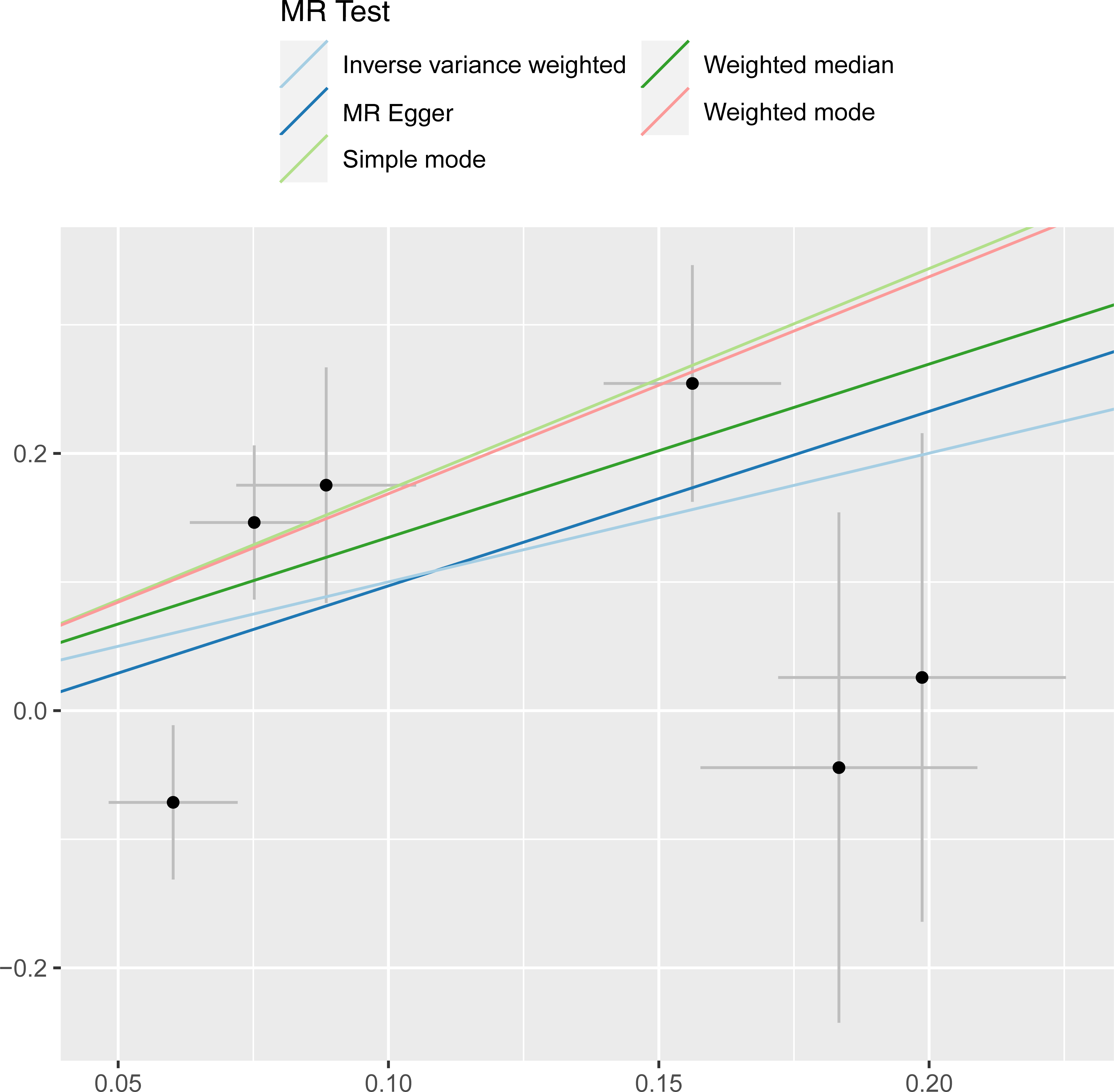
NCF4 scatter plot.
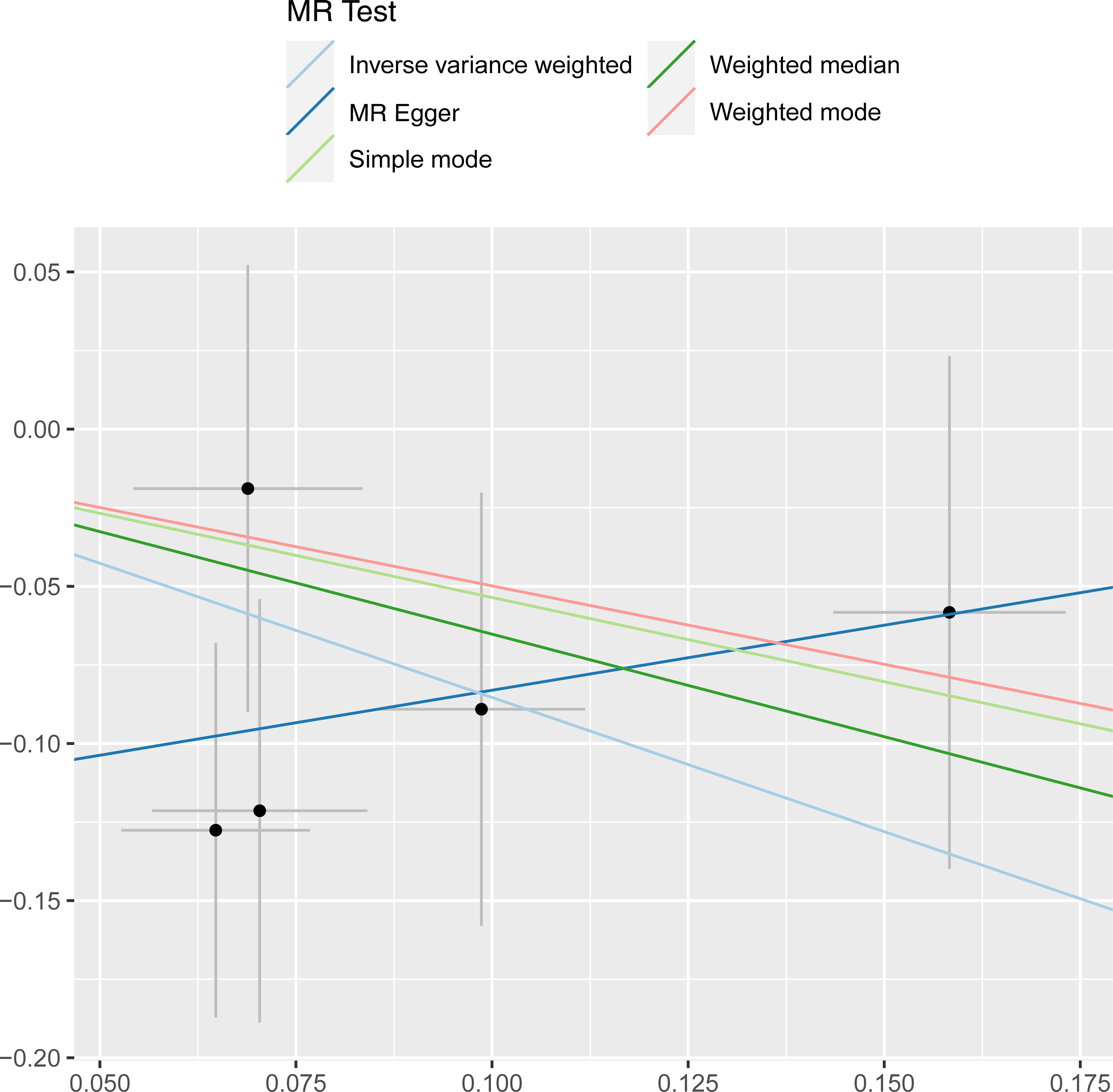
LYST scatter plot.
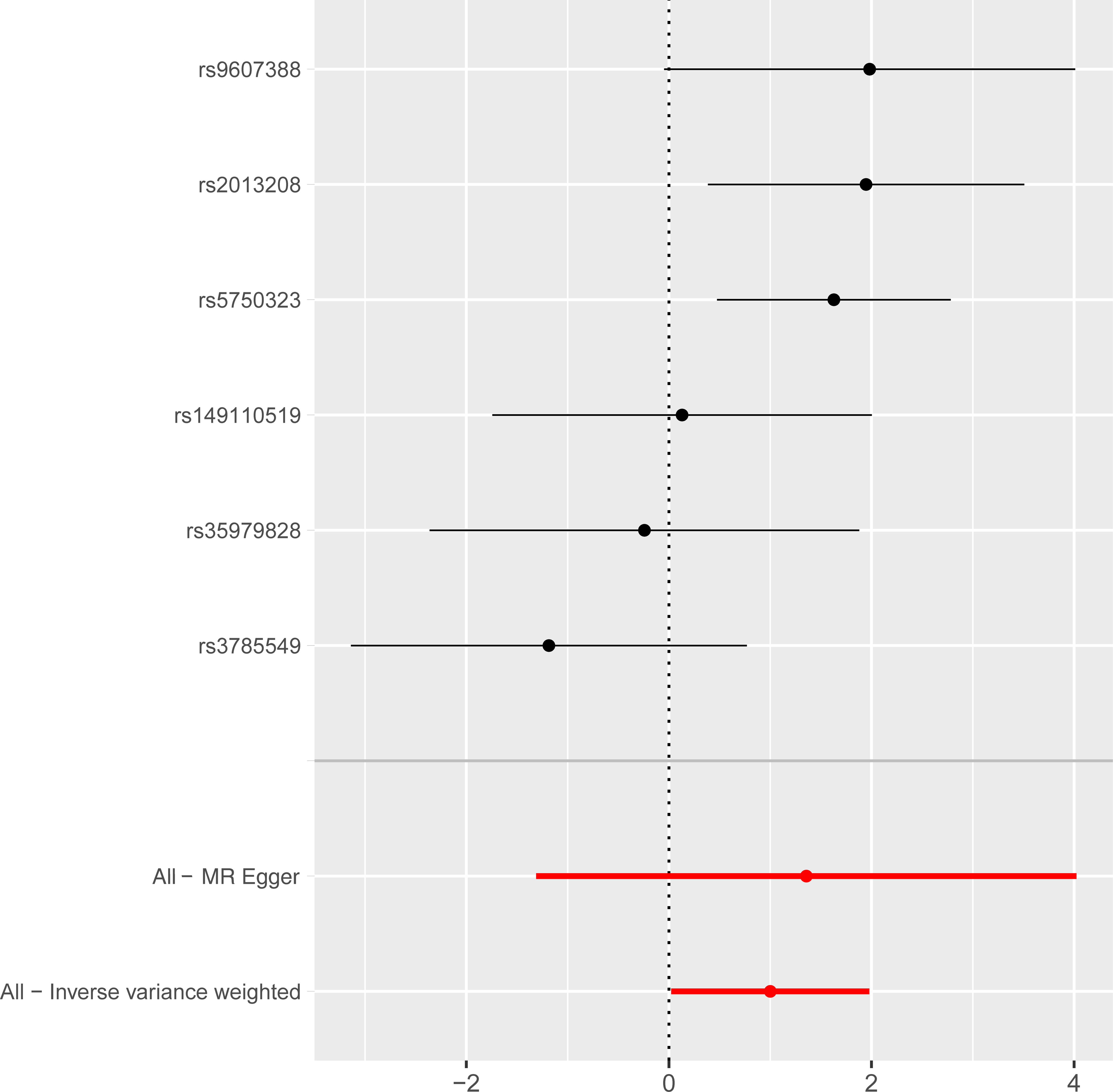
NCF4 forest plot.
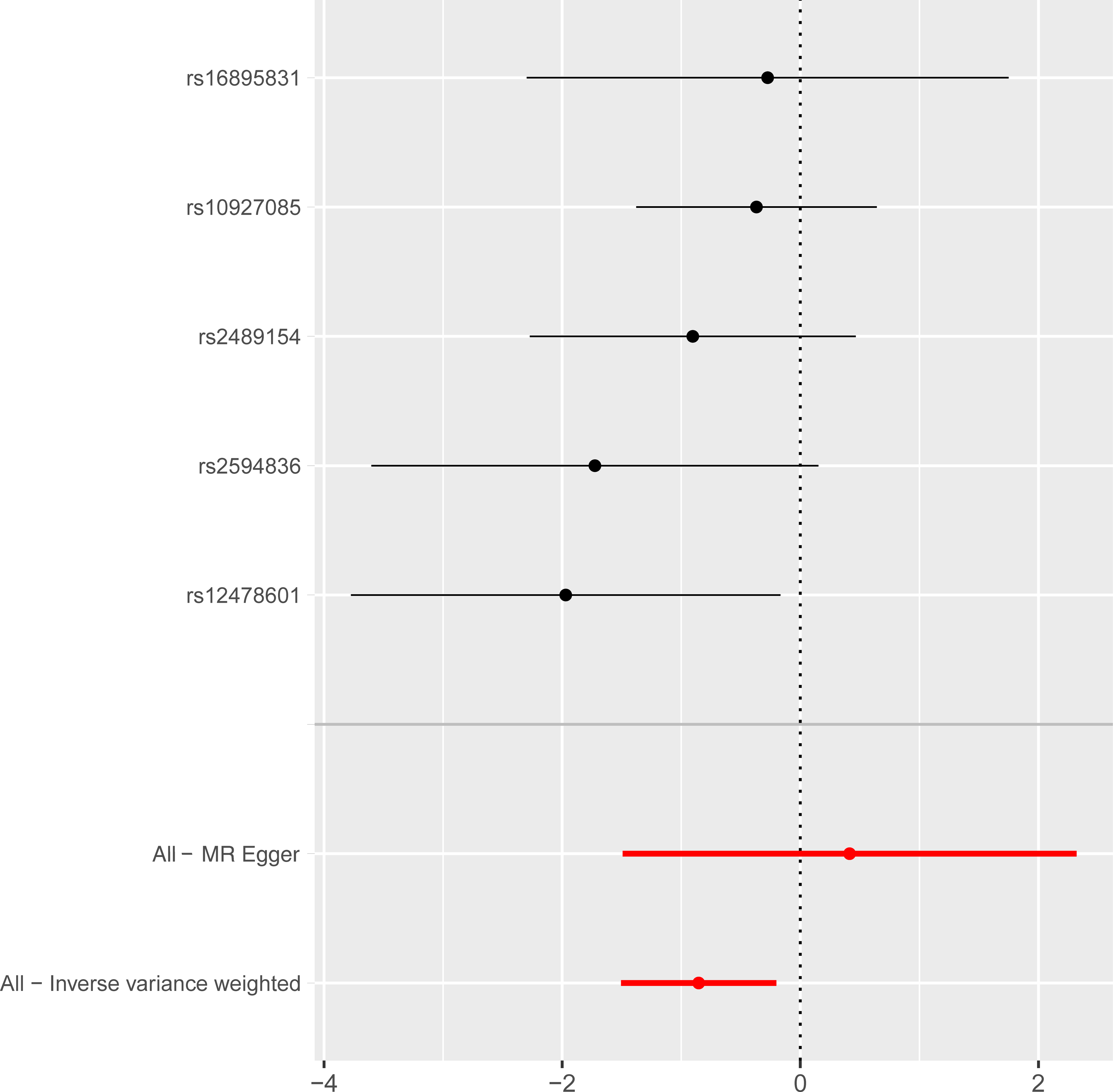
LYST forest plot.
NCF4 funnel plot.
LYST funnel plot.
NCF4 leave-one-out forest plot.
LYST leave-one-out forest plot.
Mendelian randomization analysis results of (NCF4) gene.
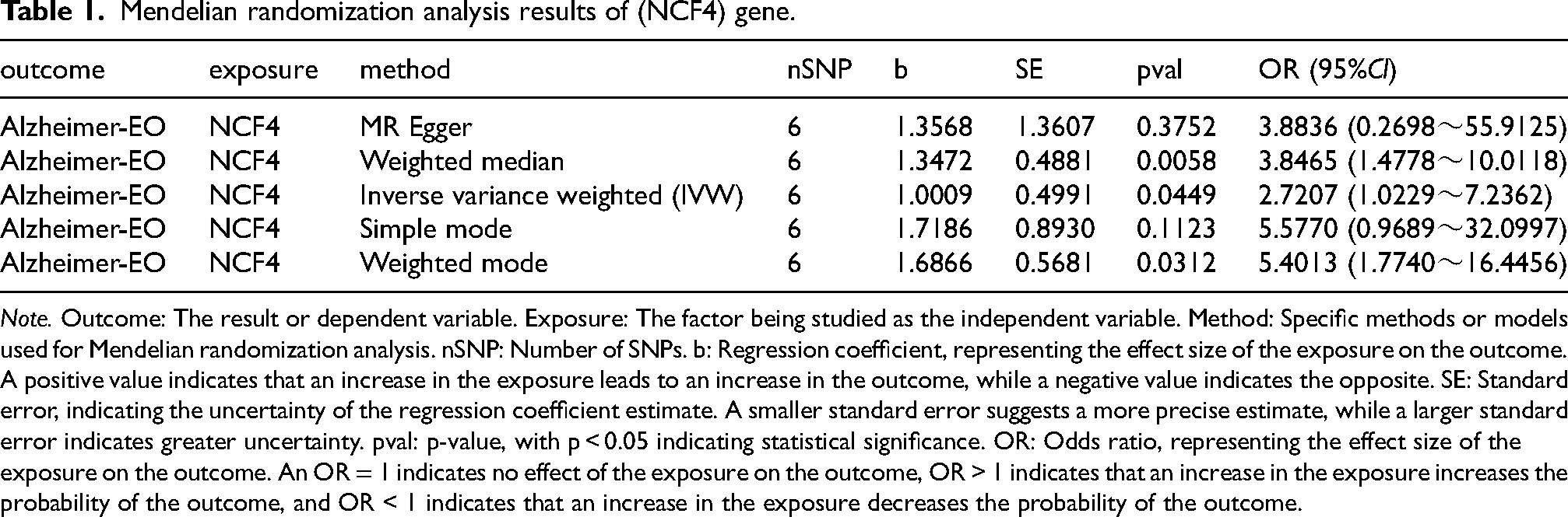
Note. Outcome: The result or dependent variable. Exposure: The factor being studied as the independent variable. Method: Specific methods or models used for Mendelian randomization analysis. nSNP: Number of SNPs. b: Regression coefficient, representing the effect size of the exposure on the outcome. A positive value indicates that an increase in the exposure leads to an increase in the outcome, while a negative value indicates the opposite. SE: Standard error, indicating the uncertainty of the regression coefficient estimate. A smaller standard error suggests a more precise estimate, while a larger standard error indicates greater uncertainty. pval: p-value, with p < 0.05 indicating statistical significance. OR: Odds ratio, representing the effect size of the exposure on the outcome. An OR = 1 indicates no effect of the exposure on the outcome, OR > 1 indicates that an increase in the exposure increases the probability of the outcome, and OR < 1 indicates that an increase in the exposure decreases the probability of the outcome.
Mendelian randomization analysis results of (LYST) gene.
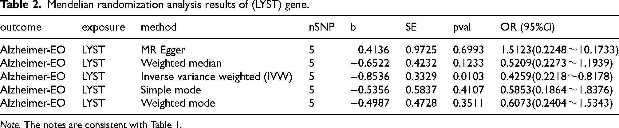
Note. The notes are consistent with Table 1.
Discussion
EOAD is characterized by pathologies such as aggregation of amyloid plaques and neurofibrillary tangles. Compared with late-onset Alzheimer's disease (LOAD), it progresses more rapidly and is associated with shorter survival times. Beyond the typical memory impairments, EOAD patients exhibit non-memory symptoms, including language, visuospatial, or executive dysfunctions.19–21 However, interaction is found among brain neurons, amyloid plaques, and other resident cells, which influences the onset and progression of the disease. Therefore, exploration of the relationship between EOAD and related genes can offer helpful guidance for its diagnosis and treatment.
As the human body ages, various harmful substances accumulate in organs, including brain tissue, while the activity and digestive function of lysosome also decline.22,23 Repairing damaged lysosome or even enhancing their degradative functions poses significant challenges.24–27 Given that lysosome is distributed across various tissues and organs, associating them with brain cells is a crucial step in studying the relationship between lysosome and neurodegenerative diseases. Endothelium cell, which is vital components of the brain vascular unit, play significant roles in hematopoiesis and vascular inflammation.28,29 They also assist in mediating immune cell targeting of Aβtoxic plaques, thereby playing a critical role in the brain's vascular microenvironment.
The lysosomal trafficking regulator (LYST) engages in the formation of lysosome and the vesicular transport of proteins within the lysosomal system. Variations in LYST may lead to neurological dysfunction. 30 Studies have shown that the expression of ADM gene increases under the stimuli of inflammatory factors such as TNF-α and IL-1β, and hypoxia, stabilizing the barrier function of vascular endothelial and epithelial cells. 31 In addition, ADM may facilitate the migration of astrocytes. However, abnormal overexpression of ADM and aberrant activation of the STAT-3 pathway can lead to tumor cell development.32,33 ARSB deficiency can lead to lysosomal storage disorders due to the inability to degrade lysosomal material, resulting in the accumulation of harmful proteins that accelerate the progression of EOAD. 34 ARSB can also encourage neurite growth in astrocytes. 35 EPDR1 is a lysosomal protein which involves in the lipid transport and degradation when combined with negatively charged liposomes. Its high expression in neural tissues can affect the excretion and transport of lipids in glial cells of the brain tissue. 36 ANXA11 acts as a mediator in transporting RNA granules to cellular substructures for protein translation. It mainly employs active lysosomal transport to mediate RNA transport in neurons. Knockout of ANXA11 leads to a reduction in important mRNAs in organelles far from the cell nucleus. This disturbs the normal homeostasis and synaptic activity of neurons, which contributes to the occurrence of EOAD. 37 Additionally, mutations in ANXA11 can also impact the transport of critical RNA granules, resulting in intracellular RNA metabolic disorders. The loss of CLN5 can impair the fusion process of lysosome, causing the ineffective degradation of endocytosed proteins and autophagic dysfunction in lysosome. Lysosome fail to degrade lipids and neurotoxic plaques in the brain, which can cause and aggravates EOAD.38,39 VPS35 (vacuolar protein sorting 35) is an essential component of brain neurons, and its decreased expression can lead to the onset of AD. Elevated expression of SORT1 fusion protein in lysosome of neurons can damage lysosomal function and collaborate with VPS35 deficiency-induced neuronal dysfunction. The Vps35-Sort1 pathway may influence the EOAD development. 40 The α-granules of platelet granules are considered to play an important role in angiogenesis and inflammation. Multivesicular bodies (MVB) within megakaryocytes interact with lysosome to form α-granules. Vacuolar protein sorting protein VPS33B in megakaryocyte α-granule-related vesicular transport interacts with VIPAS39, microtubules, etc., indirectly affecting the digestive functions of neuronal lysosome. This will accelerate EOAD plaque accumulation and promote disease progression.41,42 SYNGR1 encodes synaptic glycoprotein 1, which participates in vesicle transport and synaptic plasticity regulation. Its expression increases in AD mouse models. 43 Neutrophil cytosolic factor 4 (NCF4) is primarily expressed in cellular endosomes and phagosomes. Its increase can enhance the activity of NADPH oxidase, elevate ROS, exaggerate the inflammatory response, and encourage the progression of AD. 44 Increased expression of NKTR can inhibit tumor cell proliferation and migration, 45 and it may act through endothelium cells. NKTR upregulation could be applied to prevent further spread of Aβ plaques in brain neurons. In vitro models show that overexpression of NFIB can convert human pluripotent stem cells into functional astrocytes, 46 facilitating the clearance of Aβ plaques in EOAD. The expression of PYGB in brain tissue can inhibit the production of cellular ROS and provide a transient energy supply to the organism. The significant increase in PYGB in AD patients 47 is conducive to suppressing excess production of ROS in inflammatory cells, which damages glial cells and neurons, thus playing a positive role in the clearance of toxic plaques in EOAD.
From this perspective, it is evident that the ten lysosome-related genes are almost all involved in the processes of metabolism, autophagy, and degradation within neural tissue lysosome. Additionally, three endothelial cell-related genes participate in the generation of astrocytes and the inflammatory response of brain vasculature. These biological processes are clearly linked to the onset and progression of EOAD. Targeting overexpression or inhibition of these genes may enhance lysosomal phagocytic degradation of Aβ plaques, reduce lysosomal depletion damage, decrease local brain inflammation, and improve the survival rate and phagocytic capacity of functional astrocytes. These factors collectively contribute to the most favorable conditions for treating EOAD, ultimately aiming to alleviate disease progression and achieve a cure.
However, genes related to macrophage, fibroblast, and migrasomes are not associated with EOAD. Therefore, in the absence of influence from migratory body substances, immunotherapy can be employed to clear toxic plaques. Studies indicate that CAR-macrophage targeting Aβ plaques can absorb these plaques to a certain extent. 48 To minimize inflammatory interference and the challenges posed by the surrounding matrix of Aβ plaques, fibroblast inhibitors can be considered in conjunction with CAR-macrophage therapy for EOAD. In AD, the phagocytic function of macrophage lysosome diminishes, making it difficult to effectively clear Aβ plaques. 49 Maintaining the expression of the NEU1 gene can improve lysosomal function, thereby enhancing the macrophage's ability to phagocytose Aβ plaques. 50 Knocking out the BHLHE40/41 gene can improve macrophage lysosomal quality and enhance the lysosomal degradation capability. 51 Multiple studies have demonstrated that macrophages modified in vitro can traverse the blood-brain barrier and target lesions52,53 through cellular interactions and migration 54 within specific temporal and spatial constraints. Thus, designing macrophage capable of crossing the blood-brain barrier and targeting Aβ plaques can achieve the goal of phagocytosing these plaques. For macrophage whose lysosome cannot degrade Aβ plaques, a method of guiding target macrophage through the blood-brain barrier for external blood circulation can be used to directly clear macrophage that have phagocytosed Aβ plaques.55–57 This approach aims for effective and low-side-effect clearance of Aβ plaques. Leveraging the inherent characteristic of macrophage receptor-ligand binding, during external blood circulation, macrophage can bind to specific ligands to facilitate the clearance of macrophage that have phagocytosed Aβ plaques. Thus, macrophage can act as external transport and clearing tools for Aβ plaques. 58 Therefore, overexpression or knockout of the aforementioned ten lysosome-related genes should be considered to improve macrophage lysosomal activity, enhancing their endocytic and degradative functions, and thereby strengthen the macrophage's capacity to clear Aβ plaques.
In summary, discussion of the protective and risk genes associated with the five exposure factors in EOAD can provide important clues for the study of the disease's onset, progression, as well as its diagnosis and treatment.
Conclusion
This study demonstrates that lysosome and endothelium cell have a significant causal relationship with EOAD, while migrasomes, macrophage, and fibroblast do not. The analysis of the five selected factors provides theoretical support and reference value for EOAD research. However, due to the limitations of the data, such as geographical, age, race, and dietary differences, the results may be influenced. Future research should account for these factors to ensure a more comprehensive analysis.
Footnotes
Acknowledgments
The authors would like to acknowledge GWAS database, GeneCards database and the authors who published the publicly available literature data, as well as all the authors for their valuable support in the completion of this work.
Authors contributions
Chunxin Lu (Supervision; Writing – review & editing)
Jiechao Zhou (Conceptualization; Data curation; Formal analysis; Funding acquisition;
Investigation; Methodology; Project administration; Resources; Software; Validation;
Visualization; Writing – original draft)
Funding
The authors received no financial support for the research, authorship, and/or publication of this article.
Declaration of conflicting interests
The authors declared no potential conflicts of interest with respect to the research, authorship, and/or publication of this article.
Data availability
The data supporting this study's findings are available on request from the corresponding author. The data are not publicly available due to privacy or ethical restrictions.