
Abstract
After smoking bans were enforced in most public spaces, residences became the major places for exposure to second-hand smoke. Since cigarette smoke contains mainly submicron and ultrafine particles (UFPs), the harmful effects of the particles in cigarette smoke might be correlated more with number and surface area concentrations of UFPs instead of their mass concentration. To assess these harmful effects, we investigated the size distribution of particles (20 nm to 600 nm) released from sidestream smoke. An experiment was carried out in a two-zone house which was divided into smoking and non-smoking areas. Particle concentrations were measured in both zones and the exposure doses of people in both zones were calculated. The lung cancer risk of UFPs was then analysed by a modified surface area-based risk assessment scheme, which was found to be much closer to the incidence rate of lung cancer. In most of the studied scenarios, the excess lifetime cancer risks to non-smoker occupants exceeded an acceptable level. Although closing a door separating the two zones is a common isolation measure, adequate protection for the non-smokers cannot be achieved unless sufficient ventilation in the smoking zone is provided.
Keywords
Introduction
Indoor air pollution due to particulate matter (PM), especially submicron particles and ultrafine particles (UFPs), is considered to be one of the most critical environmental risks to human health.1–4 Various illnesses, including respiratory and cardiovascular diseases, are related to exposure to PM.4–6 The toxicity of particles has a correlation with their size.7,8 Small particle size allows UFPs to enter human cells, and their large surface area renders them more active biologically. The characteristics of submicron particles and UFPs and their associated health risks have been important research topics recently.9–11
Cigarette smoking and exposure to the environmental tobacco smoke (ETS) are widely accepted as major causes of lung cancer.1,12–14 ETS can be divided into second-hand smoke (SHS) and third-hand smoke (THS). The SHS is the combination of smoke escaping from the burning end of the tobacco product (sidestream smoke (SSS)) and smoke exhaled by the smoker, while THS is the re-suspension or re-emission of the residue from SHS that settles on indoor surfaces.15,16 Although the THS has attracted attention in recent years, its hazard is difficult to quantify due to the complexity of indoor environments. SHS can be divided into mainstream smoke (MSS) and SSS. The MSS is the smoke inhaled to the lung of the smoker and then exhaled into the environment, and the SSS is the smoke emitted from the burning end of a cigarette. Both MSS and SSS contain more than 4000 chemical compounds, with more than 60 known carcinogens including the important respiratory carcinogens PAHs and the tobacco-specific nitrosamine. 17 Previous research has shown that smaller particles can adsorb greater quantities of pollutants, 18 and since the surface areas of UFPs are huge, the above carcinogens could be more easily adsorbed on UFP surfaces. SSS accounts for 80% of the total cigarette smoke, and particles in it are usually smaller in size and contain more pollutants than MSS. 19 Most of the particles in SSS are submicron particles which usually account for more than 80% of the mass of all particles generated from the cigarette burning, while the number fraction of UFPs is usually over 90% of all particles generated. 20
Human health risk associated with smoking has been studied based on the composition and contents of volatile organic compounds (VOCs), 21 PAHs 22 and heavy metals 6 in SSS. However, few of these studies analysed the exposure of people to UFPs during the smoking process,22,23 or attempted to establish the harmful health effects of UFPs. Considering the insignificant mass concentration of UFPs, their toxicity was probably underestimated using the conventional health risk assessment method7,24 which assessed the risk by analysing the mass concentration of particles. Sze-To et al. 7 developed a risk assessment scheme based on the surface area concentration of cooking-generated UFPs to assess their cancer risk. Using this approach, the lung cancer risks from an industrial plant ash 22 and from cigarette-generated mainstream particles 24 were assessed. Stabile et al. 24 compared their assessments with the actual incidence rates for the Italian smokers.24,25 The lifetime lung cancer risk calculated using the surface area-based risk assessment scheme was verified to be much closer to the actual incidence rate than the risk assessed from the conventional assessment scheme based on particle mass concentration.
Following smoking bans enforced in most public spaces in different countries, the family residence with one or two smokers has been the main place for exposure to ETS. In general, closing a room door and opening a window are common strategies to protect non-smoking family members by isolating the cigarette smoke and enhancing ventilation. However, the effectiveness of these measures has not been investigated in terms of risk reduction. This study focuses on SSS, which accounts for more than 80% in mass of the SHS, 20 and the lung cancer risk due to UFPs in SSS was assessed. Different scenarios in a residence were investigated, and the UFP exposures in both smoking and non-smoking zones were measured in a field experiment. Different from previous research, a surface area-based scheme was modified and employed to assess lung cancer risk in different zones.
Methods
Previous research has found that most particles emitted from SSS are UFPs. 26 A two-zone apartment (smoking and non-smoking) was employed to investigate the dispersion of UFPs from SSS. Four scenarios were investigated, corresponding to the different isolation measures of opening and closing windows and doors. Due to the small size of UFPs, the SSS in the smoking zone may transport to the non-smoking zone in the residence through a door gap even when the door is closed. The concentrations of the UFPs in both zones were measured. Combined with a modified risk assessment scheme, the lifetime lung cancer risk of non-smokers exposed in both zones was estimated.
Field experiments
The two-zone apartment had a smoking zone (25.5 m³) and a non-smoking zone (38.4 m³) (Figure 1). Both zones were equipped with three-vane fans to mix the air. The face velocity of fans was about 1.70 m/s at a 50 cm distance. The apartment was located on the second floor of a building at the intersection of two roads with moderate vehicle flow. During experiments, the temperature and the relative humidity ranged from 13°C to 22°C and from 48% to 66%, respectively. A cigarette smouldered in the middle of the smoking area and was the only combustion source of UFPs in the residence, except for background pollution from outdoors. Four cases that commonly happen when smoking in a house were investigated to determine the effect of different isolation measures on UFP dispersion (Figure 1).
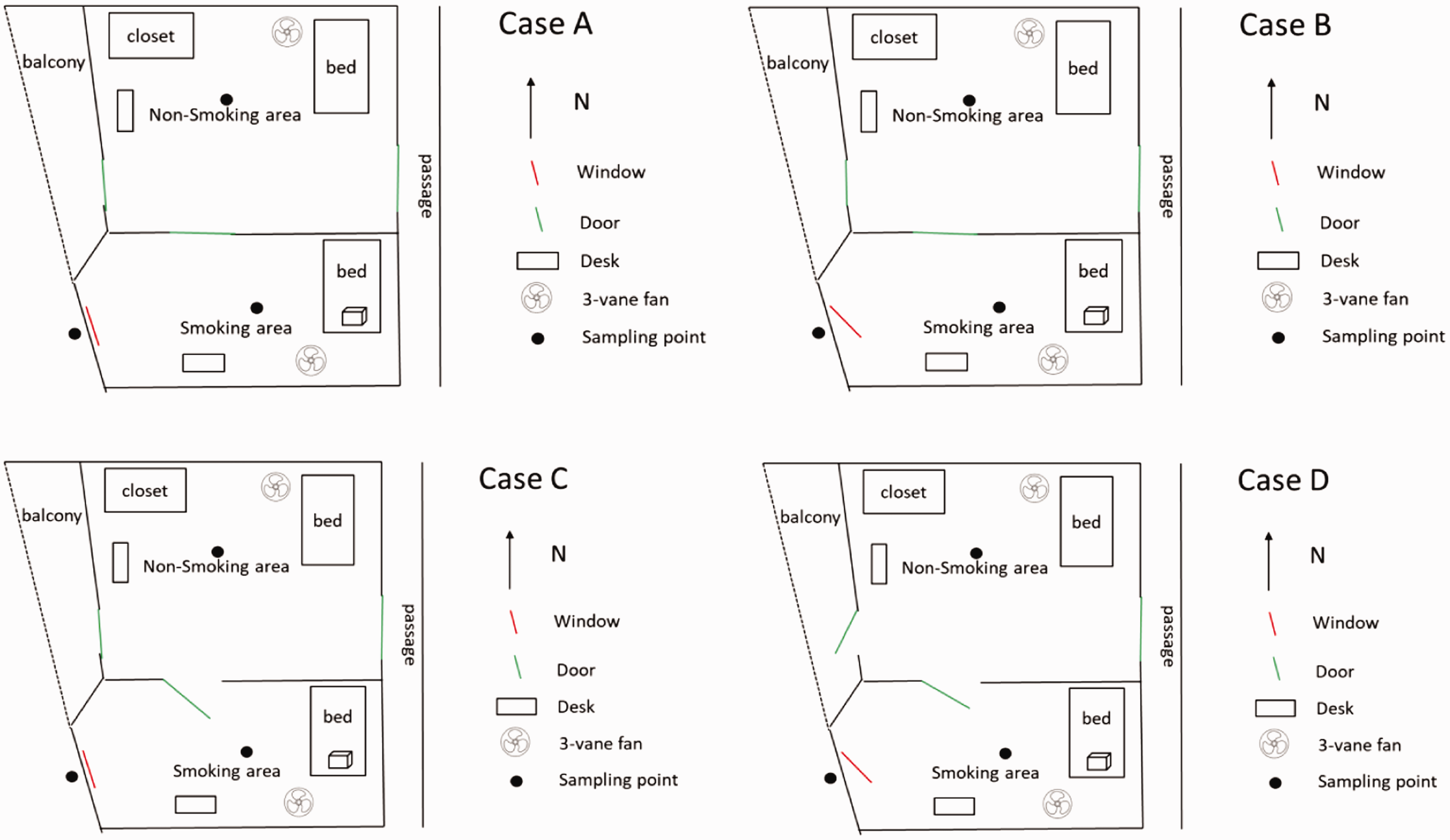
Experimental setup of the study.
Case A: all windows and doors of the house were closed.
Case B: the window in the smoking area was open and all doors were closed.
Case C: the door between the smoking and non-smoking areas was open, and all other doors and windows were closed.
Case D: all windows and doors were open.
In all cases, the main entrance door of the house was closed. By comparing Cases A and C, the effectiveness of smoke isolation by closing the internal door could be studied. Cases A and B investigated the effect of outdoor ventilation and leakage of UFPs through the door gap. Case D assessed the UFPs in the non-smoking area when the ventilation was maximum in the house, under prevailing conditions.
SSS is dominated numerically by particles with diameters less than 100 nm, but our measurement showed that particles from 100 to 600 nm may contribute 10% of the total number concentration. Particles from 100 to 600 nm can also reach the alveoli, and the chemical components adhered to particles may be dissolved and absorbed into the bloodstream. 6 This effect should not be ignored in health risk assessment. Wu et al. 12 determined that UFP emissions from cigarette smouldering were in the range of 14.6–661.2 nm. In the present study, UFPs from 20 nm to 600 nm were counted in both the smoking and non-smoking areas and their number concentrations were measured by an Aerosol Generator and Monitor (Model 1500, Copley Scientific Ltd, UK). The indoor sampling points were 1.2 m above the floor in the middle of each area, and the outdoor sampling point was the same height as that indoors and 20 cm away from the window. A 2.0 m long stainless steel tube with plastic connectors was used as the sampling pipe. By comparing concentrations of background UFPs with and without the sampling pipe, the average percentage loss of particles was determined to be 30% (±0.5%). In this paper, all concentrations were calibrated and adjusted for this percentage loss. Before each experiment, the fans were turned on to mix the indoor air. The background concentrations of UFPs in indoor and outdoor air were measured for 30 min, with samples taken every 2 min. After lighting up a cigarette, concentration of UFPs was measured continuously until it was reduced to the background level. For cases with an open door or windows, the background concentrations of UFPs indoors and outdoors were found to be essentially the same. All experiments were repeated at least three times.
The air changes per hour (ACH) between the outdoor and indoor areas were estimated by using the tracer gas decay method.
27
Before experiments, the door and window of the smoking area were closed, and the three-vane fan was turned on to mix the air. Then, a dose of sulphur hexafluoride (SF6) was released in the room for 5 min. The decay of the tracer gas (SF6) concentration was measured in the middle of the smoking area for at least 30 min by a gas detector (Model MS400-SF6-IR, Shenzhen Eranntex Electronics Co., Ltd, China). The ACHs were calculated by the equation:
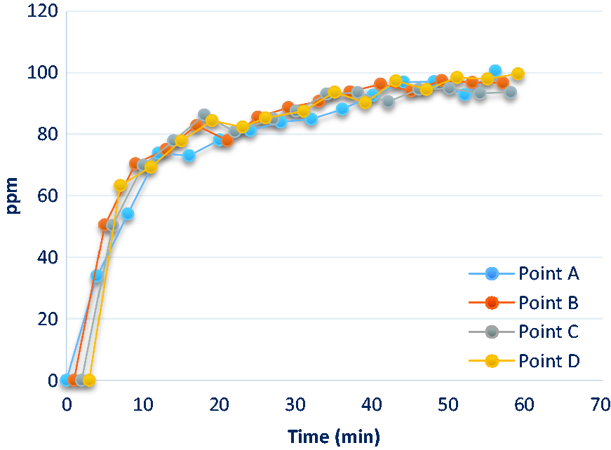
The concentration of SF6 at different sampling points in the smoking area for Case A.
The ACH in the smoking area with the window open (Case B) was also measured by this method, and found that the ACH was related to the outdoor wind speed, as shown in Table 1. Due to limited data, a correlation was not determined for these data. The average ACH of the smoking area was 6.24 per hour, with a standard deviation of 0.95 per hour. During the smoking experiments, the outdoor wind speed fluctuated within the range as shown in Table 1.
The outdoor wind speed during ACH measurement.
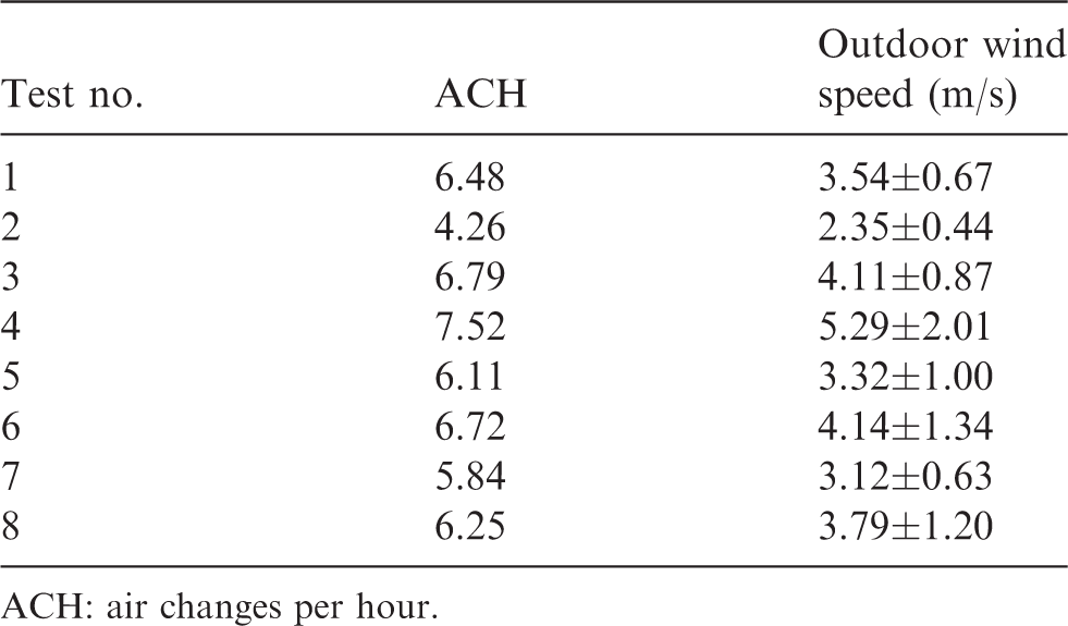
ACH: air changes per hour.
It was difficult to obtain the ACHs for Case C and Case D, and the standard deviation for repeated measurements was quite large. Possibly, the connection between the smoking area and the non-smoking area by the door opening complicated the SF6 decay process and the decay method was no longer suitable for estimating ACH. ACH results for Cases C and D are not reported.
Risk assessment method
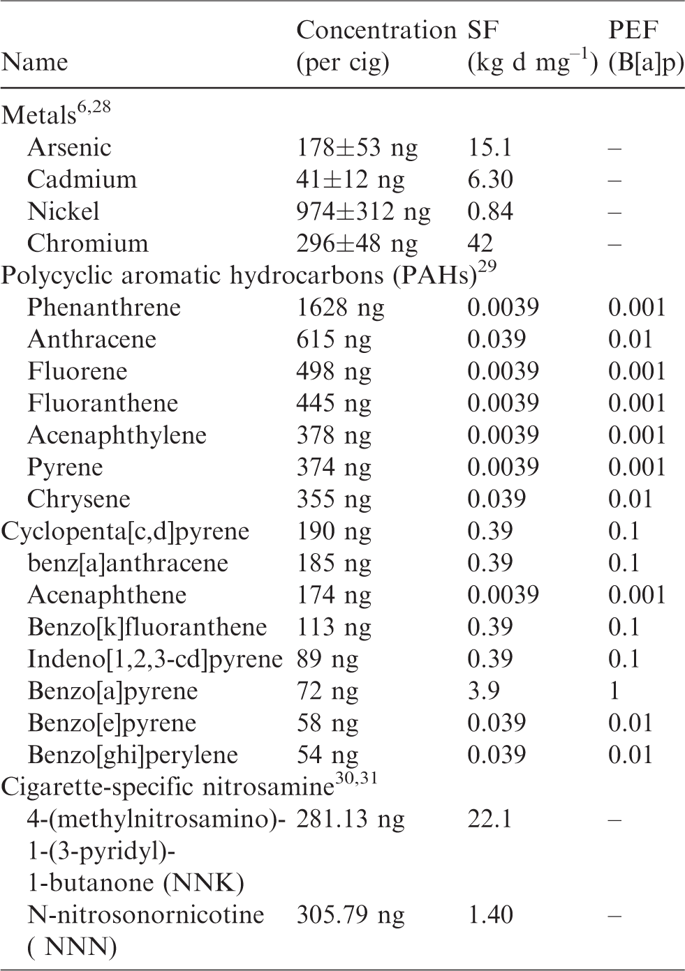
Based on the UFP concentrations in the smoking and non-smoking areas, the health risk for non-smoking people exposed in these two different zones was assessed. The risk assessment consisted of four steps. The first step was hazard identification. The adsorption of carcinogens on UFPs was considered, as listed in Table 2. The second step was exposure assessment (equation (1)). The intake of hazardous substances identified in the first step was calculated based on the UFP concentration distributions. The third step was to assess the dose–response effects. The effect on an organ (lung) caused by exposure to hazardous substances after a certain exposure time was assessed. Finally, by combining the dose–response relationship and the exposure data, the health risk could be estimated (equations (3) and (4)).
Chemical concentrations of SSS particles and their inhalation slope factor (SF) and potency equivalent factors (PEF) relative to benzo[a]pyrene).
The dose of UFPs deposited in the alveolar and tracheobronchial regions of the lung for each smouldering of one cigarette was evaluated in terms of particle number, particle surface area and particle size distribution. The concentration of UFPs changed over time, which should also be considered. A previous surface area-based risk assessment scheme32,33 had no consideration of the concentration change and used the average concentration of UFPs to calculate exposure. With the improvement of detection technology, real-time measurement of UFP concentration has become possible and temporal effects can be considered. In this study, an improved calculation of deposited dose received by an exposed person per day (

Summary of the parameters used to calculate the exposure dose and ELCR of UFPs in SSS.
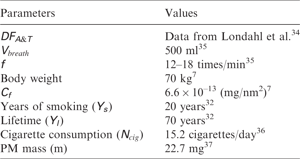
The excess lifetime cancer risk (ELCR) is the additional or extra risk of developing cancer due to exposure to a toxic substance over the lifetime of an individual. Cigarette smouldering smoke contains many toxic compounds such as heavy metals,28,38 PAHs29,39 and some cigarette-specific nitrosamine (N-nitrosonornicotine (NNN) and 4-(methylnitrosamino)-1-(3-pyridyl)-1-butanone (NNK)).30,31 Most of the above are known or suspected carcinogens. In this study, the typical carcinogens in the SSS were selected as the hazardous substances for risk assessment. The key parameter in calculating the ELCR is the cancer slope factor (SF) of each carcinogenic element, which was an upper bound, approximating a 95% confidence limit, on the increased cancer risk from a lifetime exposure to an agent by ingestion or inhalation. In order to estimate the SF of multi-component PAHs, the individual compound’s potency equivalency factor (ranging from 0 to1) relative to Benzo[a]pyrene (PEF (B[a]p)) was used. 29 Mass concentration, SF and PEF(B[a]p) of these substances are listed in Table 2.
After having identified the hazardous substances, the lung cancer risk due to the inhalation of UFPs in the SSS was calculated by applying the modified surface area-based risk assessment scheme. Based on the exposure dose data, the ELCR due to UFPs exposure per day (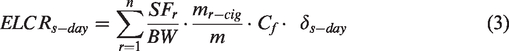

Results and discussion
Distribution characteristics of UFPs
The measured peak number concentrations and peak surface area concentrations of UFPs in the SSS are summarised in Table 4, averaged from three experiments. In all cases, the UFP concentrations by the cigarette smouldering were higher in the smoking area than in the non-smoking area. For the smoking zone, the highest levels were measured in Case A where all windows and doors of the smoking area were closed, while the lowest levels were measured in Case D where all windows and doors were open. Although the windows were closed in both Case A and Case C, the UFP level of Case C was lower than that of Case A, because the two zones were connected together to form a larger zone that diluted the UFPs. The UFP level of the smoking area in Case B was found to be lower than that of Case C, but still higher than that of Case D.
Peak concentrations of UFPs during the cigarette burning process.
For the non-smoking zone, Case B had the lowest UFP level with peak number and peak surface area concentrations one to two orders of magnitude lower than the other cases. In fact, Case B levels were close to the background concentrations which were (1.08 ± 0.12) ×104 particles/cm³ and (2.91 ± 0.22) ×102 μm2/cm³, respectively. Although the doors between the smoking and non-smoking zones were closed in Cases A and B, the UFP concentrations in the non-smoking zone of Case A were three times higher than those of Case B. The UFP level in Case A of the smoking zone was also about two to three times higher than that of Case B. Therefore, a good ventilation in the smoking area plays a more dominant role than an isolation measure by closing the door to protect the people in the non-smoking zone from UFPs. The UFP level of the non-smoking zone in Case C was the highest because the two zones were actually joined together and the UFP level was half of that of the smoking zone. Without a sufficient air exchange to outdoors, there was no significant reduction in the indoor UFP concentration.
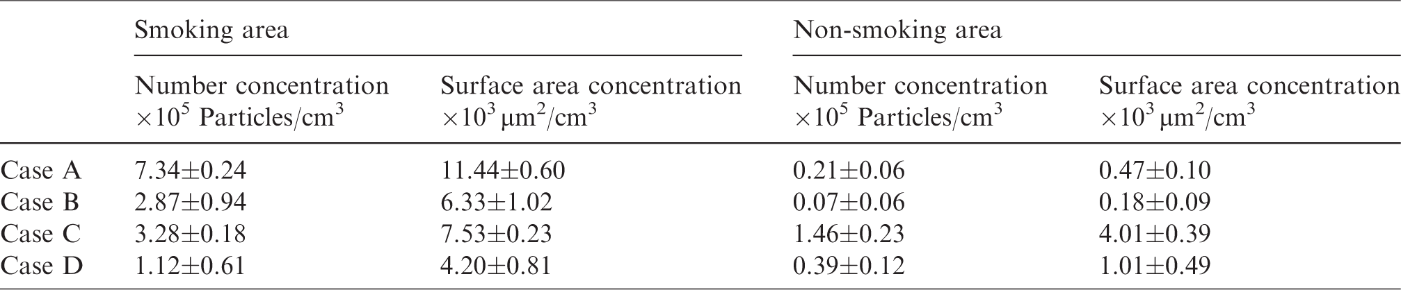
The trend of the surface area concentrations during the smouldering experiment is presented in Figure 3. In order to have a better comparison for the decay process, the curves for different Cases were shifted so that for each Case, time zero in the horizontal axis corresponded to the time when the concentration in the smoking zone reached the peak (close to the point of smouldering completion). When all the windows and doors were closed (Case A), the decay process to the background level in the smoking zone took over 2 h, while it took about 30 min when all the windows and doors were open (Case D). In Case A, the exponential decay coefficients of UFP were found to be 2.90 ± 0.03 per hour (according to number concentration) and 0.94 ± 0.20 per hour (according to surface concentration). 34 These values are larger than the ACH calculated by the tracer gas method (0.36 ± 0.02 per hour), which represents the ACH in the smoking area of the room. This implies that the decay of UFP concentration is faster than that of the tracer gas because the decay of UFP concentration is not only due to air change through the air gap of a door or a window or infiltration through the wall but also due to the deposition of particles on the wall and furniture of the room. Agglomeration of particles is also a possible explanation for the more rapid concentration decay of UFP.
Variations of UFP concentrations with time in (a) smoking area and (b) non-smoking area.
For the non-smoking zone, there were delays for the concentrations to reach the peaks. The delays were similar for Cases C and D where the doors separating the two zones were open. The delay for Case A was the longest showing that the door did not prevent but only delayed the transport of UFPs from the smoking zone to the non-smoking zone. The decay time in the non-smoking zone of Case A was very similar to that of Case C, both cases having limited outdoor air exchange.
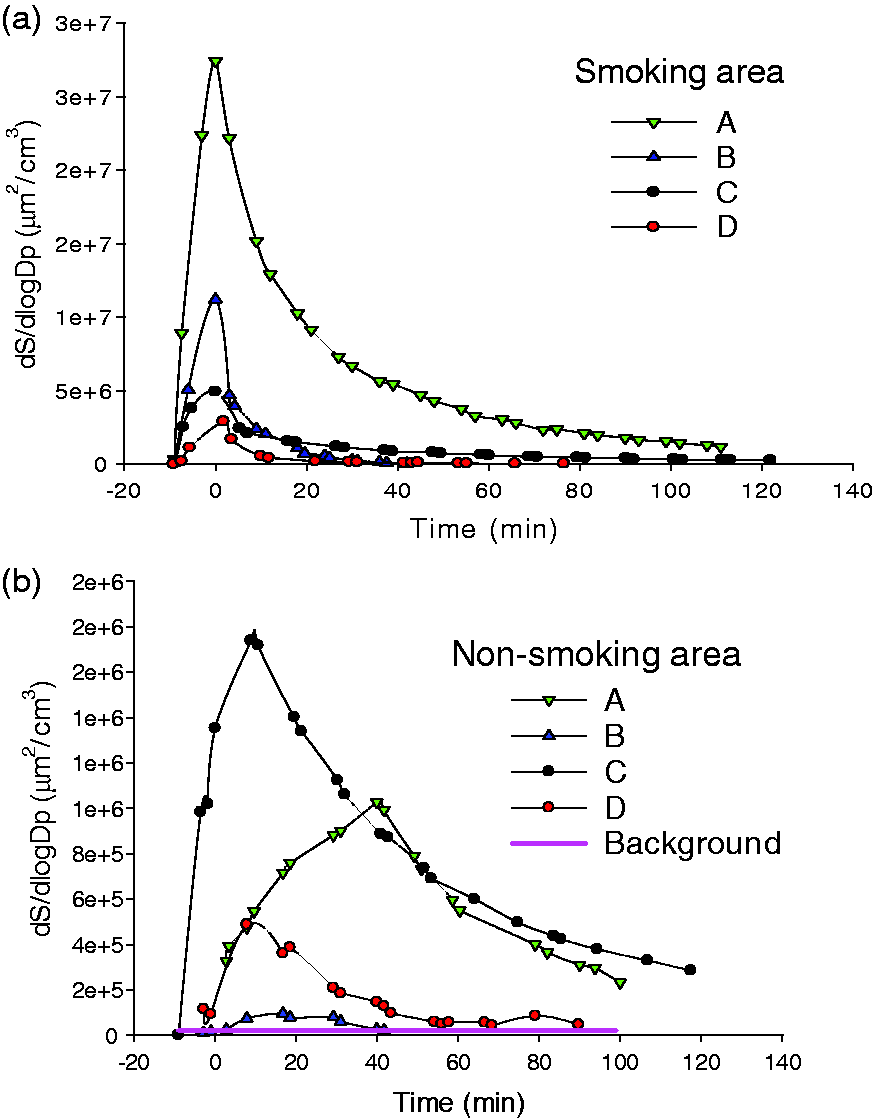
The size distributions of the UFPs at the peak concentrations were also measured (Figure 4). From Case A to Case D, the values of the surface area median diameters (SMDs) at the peak concentrations were 54.8 ± 3.4, 116.2 ± 7.8, 92.5 ± 5.7, 128.2 ± 14.7 for smoking area and 70.9 ± 4.2, 132.2 ± 9.0, 96.8 ± 5.6, 118.4 ± 10.6 for non-smoking area, respectively. The SMDs at the peaks ranged from 52 nm to 112 nm in closed condition (Case A and Case C) which were similar to other reported results,40,41 and in Case B and Case D, the SMDs were larger. When the UFP levels decayed to the background levels, the SMDs changed to 305 nm, while the SMDs of the background levels were about 300 nm. For Cases A and C, with negligible air exchange between the outdoor air and the indoor air during the whole smouldering and decay process, the SMDs increased slightly but still remained below 200 nm. In these cases, the effect of coagulation is considered to be the major reason for the increase of UFP diameters. 12 For Cases B and D, with possible entry of UFPs from outdoors, the SMDs increased to values similar to the background levels within an hour, which possibly indicates that the SSS has been replaced by the outdoor air. For the non-smoking zone, it was found that the peak of SMD of Case A was the smallest, the peak of SMD of Case C was a little larger, and that of Case D was the largest.
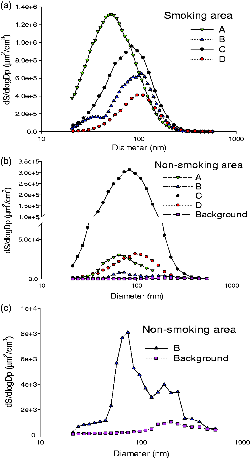
Particle size distributions in terms of surface area in (a) smoking area, (b) non-smoking area and (c) non-smoking area (enlarged) for Case B.
Exposure dose and ELCR
Based on the surface area concentration data and the risk assessment method, the exposure dose per day and the ELCR were calculated (Table 5). The ELCR calculated by the modified scheme was of the same order of magnitude as the risk of being diagnosed with lung cancer by Age 65 (1.38 × 10⁻² for male) and over a lifetime (1.06 × 10⁻² for male) 42 in the UK. One study reported the lung cancer incidence rates to be 4.92 × 10⁻³ for heavy smokers (>15 cigarettes/day) and 3.00 × 10⁻⁴ for non-smokers. 43 The ELCR means the risk of developing a disease during one’s lifetime, and generally it is larger than the incidence rate. 44
Exposure dose and ELCR for people exposed in SSS.
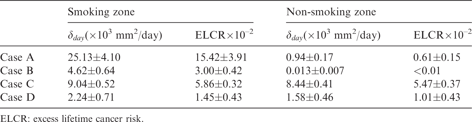
ELCR: excess lifetime cancer risk.
Since the ELCR caused by coarse particles was shown to be negligible compared to that of UFPs, 7 only the exposure dose and ELCR of UFPs have been evaluated. As reported in Table 5, the highest exposure dose per day for people exposed in the smoking area was 2.51 × 104 mm2/day, and the lowest exposure dose was 2.24 × 103 mm2/day, while the ELCR for people exposed in the smoking area were ranged from 1.45 × 10⁻² to 1.54 × 10⁻¹. Such values are slightly lower than results obtained by Stabile et al., 24 because their research studied the health hazard of cigarette smoke inhaled through the mouth of smokers, while the SSS is the focus of the current study. For the people exposed in the non-smoking area, the highest ELCR (5.47 × 10⁻²) occurred in Case C where the door between the smoking area and the non-smoking area was open. The UFPs dispersed to the non-smoking area quickly and the UFP concentrations of the two zones reached the same level in a few minutes after the cigarette was burnt out. Even though peak concentrations of UFPs in the smoking and non-smoking areas were different (Table 4), the exposure dose and the ELCR for the people exposed in these two zones were very similar. This illustrates that the exposure time plays a critical role in assessing the risk caused by cigarette smoking. The exposure dose and ELCR for people exposed in the non-smoking area in Case B were 13.01 ± 7.05 mm2/day and (8.44 ± 4.42) ×10−5, respectively, which were about two orders of magnitude lower than the other cases. In this case, the SSS in the smoking area dispersed quickly, and the concentrations of UFPs reduced to the background level within 20 min, which shortened the exposure time and minimised the exposure level. Except in Case B, all values of ELCR assessed in the other three situations exceeded the acceptable level (∼10−5).22,45,46 This shows that the isolation measure (closing door) must be performed with a sufficient ventilation in the smoking zone, so as to achieve adequate protection for the people exposed in the non-smoking zone.
The findings of this study are based on very limited experiments in one apartment and may not be generalised for various residential applications. This study demonstrates a risk assessment method using UFP measurements. A risk assessment for children should also be conducted in future work because children are the most susceptible population to harmful effects of ETS. The same procedure can be followed with different parameters. Moreover, the risk due to re-suspension of deposited hazardous particles on indoor walls, furniture, clothing, etc., or the so-called THS has not been considered in this study. This should be included in the evaluation in the future to estimate a more accurate risk of ETS.
Conclusion
This study modified a surface area-based risk assessment scheme by considering the temporal effect of UFP concentration change during cigarette smouldering and analysed the lung cancer risk due to cigarette-generated UFPs of SSS in a selected residence. The effectiveness of different measures to isolate the cigarette smoke was also investigated. The exposures in both smoking and non-smoking zones were measured in the field experiment, and the lung cancer risk was assessed by the modified surface area-based scheme. The findings show that the modified scheme can assess the ELCR for exposed persons more realistically, and the risk values, in most cases, exceeded the acceptable level. Adequate protection for people exposed in the non-smoking zone was achieved by closing the door only when sufficient ventilation was provided in the smoking zone.
Footnotes
Authors’ contribution
All authors contributed equally in the preparation of this article.
Declaration of conflicting interests
The author(s) declared no potential conflicts of interest with respect to the research, authorship, and/or publication of this article.
Funding
The author(s) disclosed receipt of the following financial support for the research, authorship, and/or publication of this article: This work was supported by the General Research Fund granted by the Research Grants Council of the Hong Kong Special Administrative Region, China (Project Nos. 16200315 and 16207817) and Natural Science Foundation of Guangdong Province, China (No. 2015A030310447).