
Abstract
Background
Supracondylar humeral fractures (SCHFs) are among the most common pediatric fractures. Closed reduction and percutaneous pinning are considered the established gold standard. Biomechanically, cross-pinning is resilient for any axial rotation, but the medial pin increases the risk of iatrogenic injury to the ulnar nerve.
Objective
A systematic review was conducted to provide an evidence-based analysis of the literature on the management of iatrogenic ulnar nerve injury caused by the medial pin during closed reduction and percutaneous fixation of displaced SCHF in children.
Methods
MEDLINE, SCOPUS, and ScienceDirect databases were searched to identify all articles that reported ulnar nerve injury caused by the medial pin during closed reduction and percutaneous fixation of displaced SCHF in children and suggested the management. Reference lists from the articles retrieved were further scrutinized to identify any additional studies of interest.
Results
One thousand six hundred and six articles on SCHF treated by closed reduction and cross-pinning were identified initially with 25 studies included in the analysis after screening. Four thousand six hundred and seventy-five children sustained SCHF with a median age of 7 years. Of 3036 children treated by closed reduction and cross pinning, 205 (6.75%) were diagnosed with iatrogenic ulnar nerve injury. The management involves observation only, removal of the medial pin, or exploration. The average recovery time in the group treated by removal of wire was statistically shorter than the other two groups.
Conclusion
The evidence suggests surgical exploration of the ulnar nerve can be delayed for up to 7 months, with most studies favouring observation only. In selected cases, immediate removal of the medial pin should be considered.
Introduction
A supracondylar humeral fracture (SCHF) occurs through the thin part of the distal humerus above the level of the growth plate and is divided into either extension or flexion types according to the mechanism and direction of displacement of the distal fragment. The most common type is extension which occurs as a result of falling on an outstretched hand and comprises up to 98%.1,2 These fractures can be further subclassified based on the Wilkins-modified Gartland classification. 3 The Gartland classification is based on the radiological appearance of fracture displacement. Type I is a non-displaced fracture, Type II is a displaced fracture with an intact posterior cortex, whereas Type III is a displaced fracture with no cortical contact.
SCHF are common in children and their treatment can be challenging if they are completely displaced. 4 However, closed reduction with percutaneous pinning is considered a gold standard treatment since it was described by Swenson in 1948, 5 but there is always a debate about the method of placement of a pin. 6 Most authors favour the placement of cross pins as they are considered biomechanically more stable but have an increased risk of ulnar nerve injury5,6 which could range from 5–30%.7–10 The factors considered in causing iatrogenic injury are gross swelling, excessive manipulation, and insertion of the pin at an acute angle. 11 The management of these injuries is also highly controversial and ranges from early surgical exposure of the ulnar nerve,12–16 removal of the medial pin,13,15,17,18 or just observation.4,19–22
The purpose of this systematic review is to evaluate the management of iatrogenic ulnar nerve injuries by the medial pin in the treatment of displaced SCHF in children.
Methods
A systematic review of the literature was conducted and reported in accordance with Preferred Reporting Items for Systematic Reviews and Meta-Analysis (PRISMA) guidelines. The study protocol was prospectively registered on the International Prospective Register of Systematic Reviews (PROSPERO with ID CRD42022264445).
Search strategy
A preliminary search was done to confirm that there were no published systematic reviews on the management of the iatrogenic ulnar nerve injury that occurred by cross-pinning during the treatment of displaced SCHF in children. Three authors (MAI, HSZ, and JVG) independently conducted electronic searches of
The Mesh terms and their combinations used in the search were as follows: “supracondylar fractures” AND “children” AND “closed reduction” AND “ulnar nerve injury”. Only articles originally written in English or translated into English were considered. The bibliography of each paper identified electronically was searched manually for further potential references. We identified prospective and retrospective observational studies of displaced, closed SCHF in children treated with closed reduction, and cross-percutaneous pinning, reporting iatrogenic ulnar nerve injury post-operatively.
All articles were initially screened by title, abstract, and keywords. The articles were selected for further reading if ulnar nerve injury was caused by the medial pin.
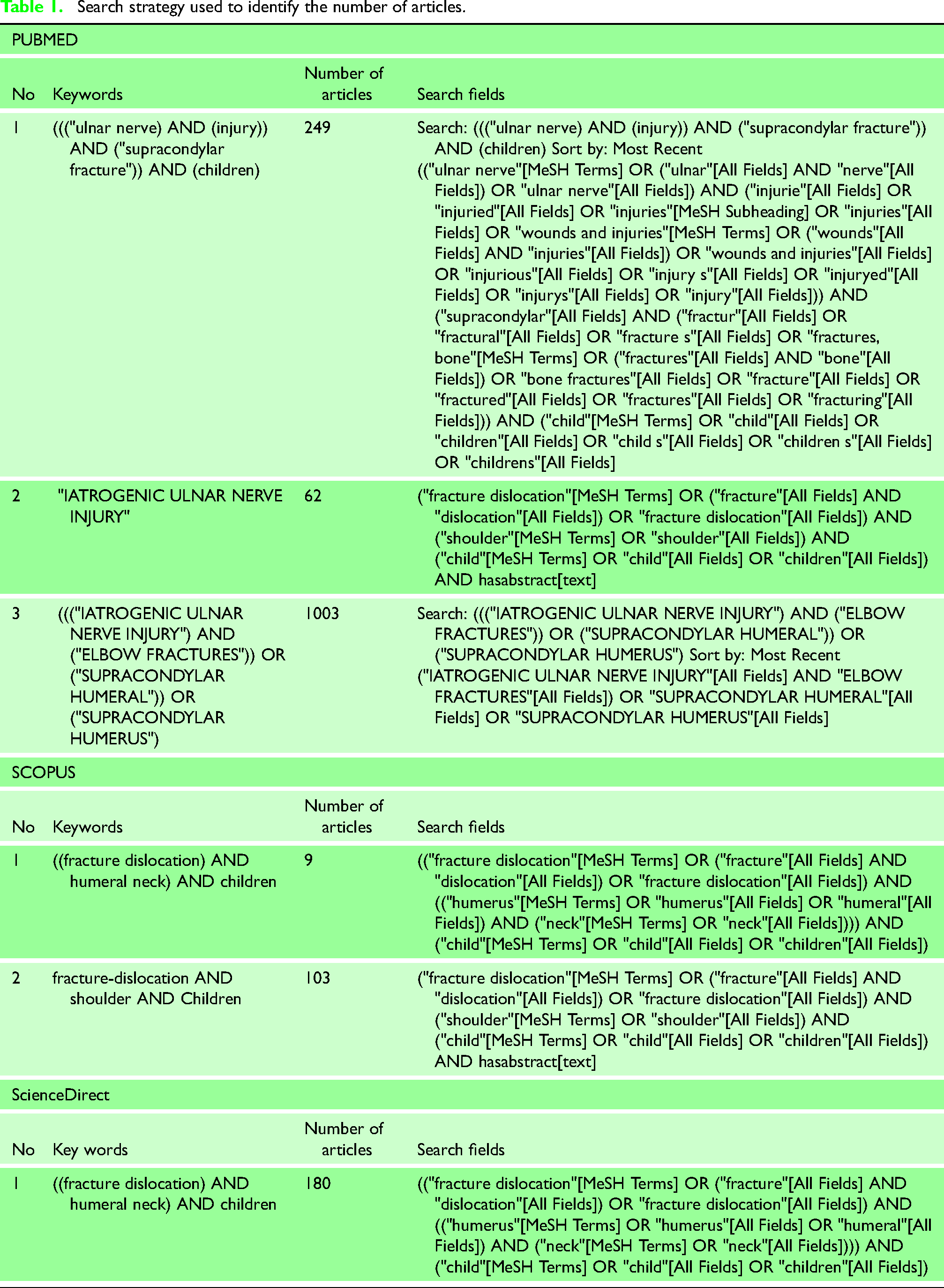
The combined searches identified 1606 potentially eligible studies as shown in Table 1.
Identification and eligibility of study
Articles were considered eligible if they met the following inclusion criteria
The target population consisted of children with totally displaced SCHF
Cross-pinning fixation using closed, open, or mini-open techniques.
Studies reporting iatrogenic ulnar nerve injuries and suggesting the management
clusion
The excusion criteria were:
Open fractures
Fractures treated by only lateral pinning
Ulnar nerve injury by the fracture and reported preoperatively
Studies in which the status of the ulnar nerve postoperatively was not discussed
Review articles
Data extraction
Three authors (MAI, HSZ, and JVG) scanned for trial selection through the titles and abstracts from the searches. From there, we obtained full-text articles when they appear to meet the eligibility criteria or when there was insufficient information to assess the eligibility. The authors assessed the eligibility of the trials independently and we documented the reasons for exclusion. The title and abstracts of the identified studies were screened after removing the duplicates from the search results. Any disagreements about the inclusion or exclusion of a study were solved by discussion or consultation within the group. Three authors then, independently, extracted data from all relevant reports using a standardized collection form with Microsoft Excel (Microsoft Corporation, Washington, 2020). The data were grouped into the following categories: patient demographics, method of treatment, clinical outcome, radiological outcome, and any complication.
Results
The initial literature search identified 1606 relevant articles on SCHF treated by closed reduction and cross-pinning. Titles and abstracts were screened, and 52 articles were retrieved for full-text evaluation and 25 studies met the inclusion criteria and were included in the systematic review.4,12–15,17,19,20,22–38 The PRISMA flow chart (Figure 1) shows the study selection.
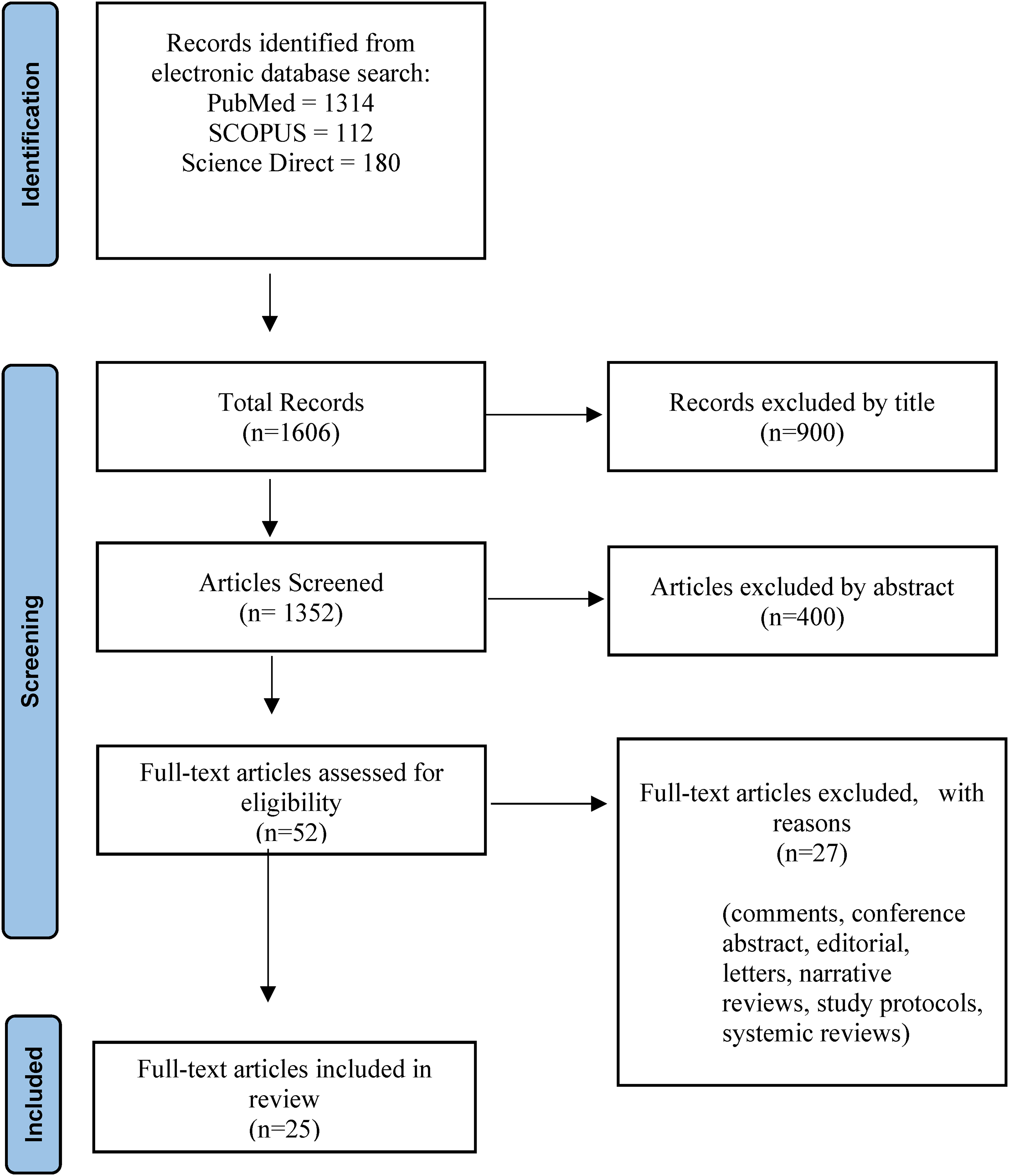
Reporting items for systematic reviews and meta-analyses (PRISMA) study flow diagram.
Synthesis of results
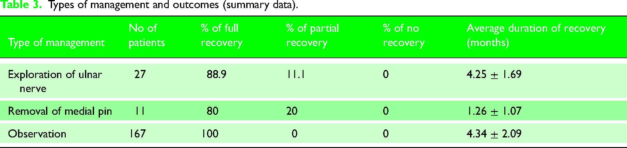
This study yielded 4675 children with ages ranging from 5 to 8.4 years (median 7 years); of 3036 children who underwent fixation of the SCHF by cross pins, 205 showed clinical signs of ulnar nerve injury. The range of the incidence of ulnar nerve injury was from 2–20.8% with an average of 6.9%. The average follow-up of the patients was 11.4 months. The key methodological features of the 25 papers are listed in Table 2. Management strategy included removal of the medial pin in 11 children, exploration of the nerve in 27 and observation alone in 169 children. All children recovered ulnar nerve function between 1 and 9 months (mean 4.5 months); children who underwent removal of the medial pin recovered nerve function in a shorter time compared to other modalities of treatment (Table 3).
Search strategy used to identify the number of articles.
Summary of all included studies. All studies were retrospective except three prospective studies.*
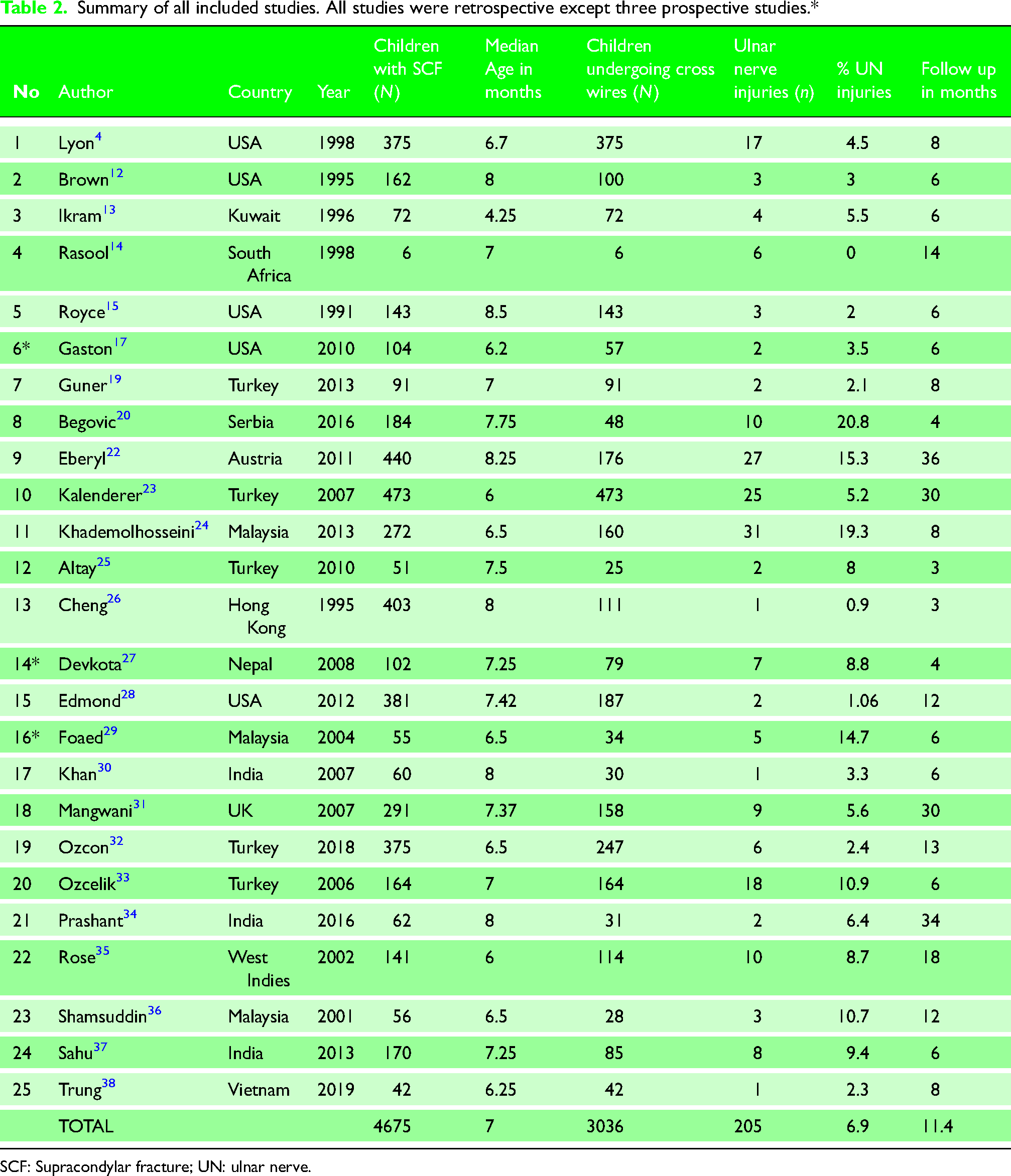
SCF: Supracondylar fracture; UN: ulnar nerve.
Types of management and outcomes (summary data).
Discussion
SCHFs are common paediatric elbow injuries. The management of displaced fractures is aimed at achieving anatomical reduction, maintenance of the reduction until fracture healing with avoidance of neurovascular complications, and achieving better functional and cosmetic results.2,10 The most acceptable treatment is a closed reduction with cross pinning as it provides excellent mechanical stability.2,7,39–41
Flexion types of SCHF are rare but there is a high prevalence of ulnar nerve palsy because the posterior shift of the fractured proximal humerus fragment and anterior displacement of the distal humerus leaves the posteriorly lying ulnar nerve susceptible to stretching.42,43 It is consistently linked to an increased likelihood of open reduction. 44 In extension type SCHF, the closed reduction can be achieved most of the time and the ulnar nerve is not commonly injured, but it is the most frequent nerve injured after treatment by the medial pin with an incidence as high as 30%.4,10,12,15,41,42,45–47 Conversely, the two lateral pin fixation avoids the danger of iatrogenic ulnar nerve injury but provides less biomechanical stability. 2
It is important for surgeons to be able to weigh the potential risks and benefits of this most accepted fixation technique. According to Slobogean et al., 48 the incidence of iatrogenic ulnar nerve injury is higher, approximately one for every 28 patients treated with the crossed pinning technique compared to the lateral pinning technique.
Royce et al. 15 suggest that iatrogenic ulnar nerve injury associated with the pinning of SCHF is more common than reported in the literature. Conversely, Lyons et al. 4 proposed that this iatrogenic nerve injury does not necessarily indicate that it occurred as a result of the intervention. It is possible that it had simply not been documented at the time of presentation or the injury may also occur during manipulation to achieve closed reduction.
Most of the studies have revealed that 86–100% of these nerve injuries are neurapraxias that settle without any active management within 6 months.12,21,48,49
The likely causes reported in the literature are contusion, constriction, or narrowing of the cubital tunnel and direct penetration of the nerve or its sheath by the medial pin.13,14,33,50 A number of authors have reported that the medial pin rarely pierces the nerve but commonly hampers it within the cubital tunnel by tethering adjacent soft tissue.14,51–54 Foead et al. 29 suggested that injury to the ulnar nerve could be due to local irritation or pressure from the medial pin during insertion. Kwok et al. 54 reported operated findings on 18 cases of iatrogenic nerve injuries related to percutaneous pins, either due to nerve penetration, epineural stripping, or tethering by fibrous scar tissue or K-wires.
Neurological examination in children demands skill and experience. 54 Post-operative signs of ulnar injury are early clawing and pain on extension of the ring and little fingers due to loss of flexor carpi ulnaris and weakness of long flexor function to the small and ring fingers. 4 The sensory function is assessed by touch sensation over the little finger and ulnar half of the ring finger while the motor function is checked by the movement of the flexor digitorum profundus of the little finger.
Clinical examination should also evaluate the sympathetic function that can be observed by swelling, discolouration, and anhidrosis in the hand. A warm dry digit denotes nerve dysfunction and indicates the need for exploration of the nerve. 50 Kwok et al. 54 recommended always documenting the presence or absence of Tinel's sign as it is a valuable clinical indicator in determining whether a nerve injury is degenerative or non-degenerative in nature. The presence of neuropathic pain and a positive Tinel sign also requires exploration of the nerve, but neuropathic pain is often difficult to recognize in children and may manifest as an exaggerated aversion to the light touch of the hand or forearm. 54
Many approaches are recommended to minimize the risk of injury to the ulnar nerve during the insertion of the medial pin including mini-incision, a greater extension of the elbow, or retrograde placement of the medial pin.28,53,55–57 The placement of the medial pin is one of the most important factors in the development of iatrogenic ulnar nerve lesion, 4 which can be prevented if proper care is taken. 50 Karim et al. 2 reported zero instances of iatrogenic ulnar injury by the medial pin in a series of 30 patients. They suggested inserting the first lateral pins in a hyperflexed elbow to achieve stability followed by the medial pin but in a 90° extension of the elbow. This minimizes the risk of nerve injury by reducing anterior subluxation of the nerve. To further protect the nerve, a mini-incision was placed over the medial condyle and the surgeon's thumb was placed over it and swept posteriorly over the cubital tunnel protecting the ulnar nerve. However, a divergence of the pins in different columns of the distal fragment is also important. Three lateral entry pins or two lateral entry pins that are divergent and are located in both the lateral and the central column provide torsional stability that is similar to that achieved with cross pins. 58 Green et al. 59 using the mini-incision technique reported only one case of transient neuropraxia in a series of 65 patients who underwent cross-pinning for displaced SCHF.
The result of this systematic review found that management is controversial as there is a lack of prospective controlled studies to propose the advantage of either method over the other.4,12,16,33,60 Although a total of 205 iatrogenic ulnar nerve injury children were pooled from 25 studies, there were no randomized or non-randomized controlled or comparative studies in the literature.
Three strategies are mentioned in the literature for the management of iatrogenic ulnar injury identified in the early postoperative period. These are the early surgical interventions of exploration of the ulnar nerve,12–16 removal of the medial pin13,15,17,18 or a wait and watch policy.4,18–21
The crucial question is whether to treat conservatively or surgically and at what point should exploration of the ulnar nerve be considered. How long should one wait for a nerve to show signs of recovery before considering exploration? In Table 3, 27 patients underwent exploration in comparison to 167 patients who had only observation but there was no significant difference in the time of full recovery. However, 11 children who had immediate removal of medial pin showed early signs of recovery. Oetgen et al. 18 reported three iatrogenic ulnar nerve injuries in a series of 137 patients and all had immediate removal of medial pin with ultimate resolution of the palsy. Kalenderer et al. 23 evaluated the results in 473 children, which is the largest series in the literature and reported a 5.2% incidence of iatrogenic ulnar nerve injury. They suggested following up patients for 7 months without any intervention.
Khademolhosseini et al. 24 have reported a high incidence (14.3%) of post-operative nerve injury but showed complete resolution in all patients and did not recommend immediate removal of the medial pin or exploration of the ulnar nerve. A review of 101 children with SCHF suggested an initial wait and watch policy and only considering surgical intervention if there is no clinical or electromyography evidence of recovery at 5 months after injury. 61 Royce et al. 15 recommended waiting up to 6 months before proceeding with an exploration of the nerve. Lyon et al. 4 reported a full return of normal function of the ulnar nerve in their 11 cases without intervention; even in the presence of an abnormal EMG, normal function returned within 4 months. Those who had removal of pins or exploration in the other six cases did no better than the rest. Ikram and Royce were in favour of early exploration of the ulnar nerve as delay can result in a longer period of return to normal function.13,15
Brown and Zinar, 12 reviewed162 fractures and reported four cases of iatrogenic lesions of ulnar nerve injury, all were explored, and the medial pin was found to be injuring the nerve in all cases and so they recommended urgent re-exploration in cases of post-operative nerve palsy. Ramachandran et al. 50 suggested considering the exploration of the nerve in the presence of neuropathic pain as it implies ongoing nerve compression.
In a recent systematic review of 179 patients, 85.5% of nerve injuries were managed expectantly, 11.7% had removal of the medial pin and 2.8% were managed with ulnar nerve exploration. 62
Limitation
There are several limitations to this systematic review that we wish to acknowledge. Overall, 22 out of 25 studies were retrospective with no randomized or non-randomized controlled trials giving the potential for bias and providing weaker realistic evidence. Some of the studies have short follow-up. There is a marked degree of heterogeneity with studies differing in terms of interventions and outcomes which makes it difficult to draw definite conclusions. Many important data, including a distinction between sensory and/or motor deficit, findings at surgical exploration, and detailed recovery at final follow up are missing.
Graff et al. 62 mentioned lack of strong literature regarding the management of iatrogenic ulnar nerve injury in their systematic review.
Additional research is required to ensure percutaneous fixation remains safe. It will be beneficial to conduct randomized controlled trials to guide clinicians in the management of iatrogenic ulnar nerve palsies.
Conclusion
The risk of iatrogenic nerve injury in the management of SCHF can be prevented if adequate care is taken during the insertion of the medial pin.
Iatrogenic or fracture-related nerve injury is a benign condition that may settle naturally, and a wait and watch policy seems to be an effective method for the treatment of this complication. However, exploration may be considered if there is no sign of recovery of the ulnar palsy and EMG-documented injury to the nerve.
Footnotes
Acknowledgements
The study was approved by the International Medical University Joint Committee on Research and Ethics (IMU 473/2020) and the Medical Research & Ethics Committee (MREC) of the Malaysian Ministry of Health (MOH), under NMRR-20-2920-57510 (IIR).
Authors’ contributions
MAI: conception of idea, data collection, review of manuscript and editing of manuscript; HSZ: data collection and analysis; JVG: data analysis, data collection and preparation of manuscript; all authors read and approved the final manuscript. All authors contributed to drafting the work, revising critically, and final approval of the version to be published. They also agreed to be accountable for all aspects of the work in ensuring that questions related to the accuracy of any part of the work are appropriately investigated and resolved.
Declaration of conflicting interests
The authors declared no potential conflicts of interest with respect to the research, authorship, and/or publication of this article.
Funding
The authors received no financial support for the research, authorship, and/or publication of this article.
Ethical approval
Ethical approval was not sought for this article because it was not required for this study since it is a systematic review of published literature.
Informed consent
Informed consent was not sought because no patients or members of the public were involved.