
Abstract
Objective:
To assess and compare the validity of 2D modified Easy Box and measurement of the Beta angle on standard conventional orthopantomogram (OPG) versus 3D cone-beam computed tomography (CBCT) OPG-constructed view.
Design:
A retrospective agreement study.
Methods:
The aim of this study was to construct an Easy Box on a standard conventional OPG and to validate this novel method by comparing it with the Easy Box method on 3D CBCT. After approval from the Ethics Committee, OPG and CBCT radiographs were obtained for the study from departmental records and five private practices in the same location (Indore, India). The radiographs were selected based on record availability and with written consent from the participants before the commencement of the study. The records were analysed to enable a comparison and to assess the accuracy of Easy Box construction on both 3D CBCT and standard conventional OPG radiographs. The location of the impacted canine within the Easy Box boundaries and the measurement of the Beta angle were determined on both views.
Results:
A perfect agreement was obtained for the comparison of 3D Easy Box CBCT analysis with 2D modified Easy Box on OPG for impacted maxillary canines (Kappa = 1.0). A Bland–Altman (LoA) analysis showed no proportional bias in the comparison of the Beta angle on 3D and 2D OPG radiographs.
Conclusion:
Beta angle and 2D modified Easy Box on a conventional OPG yield similar results when compared to Easy Box on 3D CBCT OPG-constructed view. The standard OPG was valuable and cost-effective, particularly in the early stages of diagnosis and treatment planning, either as a substitute or when CBCT was unavailable.
Introduction
A delay in the natural eruption process of a tooth can lead to impaction. Impaction of a tooth is a pathological situation where the tooth fails to attain a normal functional position past its root formation. It may be due to a systemic condition, genetics, ankylosis, peg-shaped maxillary lateral incisors, missing maxillary lateral incisors (guidance theory), physical obstruction or a primary failure of eruption (Kharbanda, 2019). After third molars, the second most commonly impacted teeth are maxillary canines. Because they contribute to an aesthetically pleasing and stable functional occlusion, maxillary canine impactions need to be managed carefully (Benson et al., 2021; Dachi and Howell, 1961; Fernandez et al., 1998; Grover and Lorton, 1985; Kramer and Williams, 1970; Takahama and Aiyama, 1982).
An impacted maxillary canine’s prognosis for alignment depends on several variables, including the canine’s connection to nearby structures, the amount of space available, its location, angulation, and the patient’s age and cooperation (McSherry, 1996). Several authors have developed prognostic indices for the prediction of prime clinical components, such as duration of treatment and level of difficulty of disimpaction, for impacted canines (Ericson and Kurol, 1988a, 1988b; Lindauer et al., 1992; Pitt et al., 2006; Power and Short, 1993). Some of the drawbacks of these indices include distortion, superimposition and difficulties in understanding the third dimension because they were based on 2D radiographs (Alqerban et al., 2011, 2014; Elefteriadis and Athanasiou, 1996; Pitt et al., 2006; Xie et al., 1996).
With the advent of cone-beam computed tomography (CBCT) in 1982, it became feasible to determine the location of an impacted maxillary canine more accurately in all three planes of space. The surrounding anatomic relations, including hard and soft tissues with their thickness, density, distribution and the exact location in all spatial planes, could be determined (Haney et al., 2010; Peck et al., 2007).
A new prognostic tool called the ‘Easy Box CBCT’, developed recently by Puthalath et al. (2023), uses CBCT images to assess impacted canines. The Easy Box comprises four borders, namely mesial, distal, superior and inferior (Table 1). Their study considered two views: the axial view and the OPG-constructed view. The Easy Box was constructed in both of these views from CBCT data. When the impacted canine was within the box, it showed a good prognosis for orthodontic alignment. They also assessed the angulation of the impacted canine in relation to the mesial border of the box, referred to as the Beta angle. Canines with a Beta angle greater than 45° were shown to be harder to align orthodontically (Puthalath et al., 2023). Since this method has only been introduced recently, it is not yet in widespread use.
Construction of Easy Box on standard conventional (plain film) OPG and 3D OPG-constructed view (from Puthalath et al., 2023).
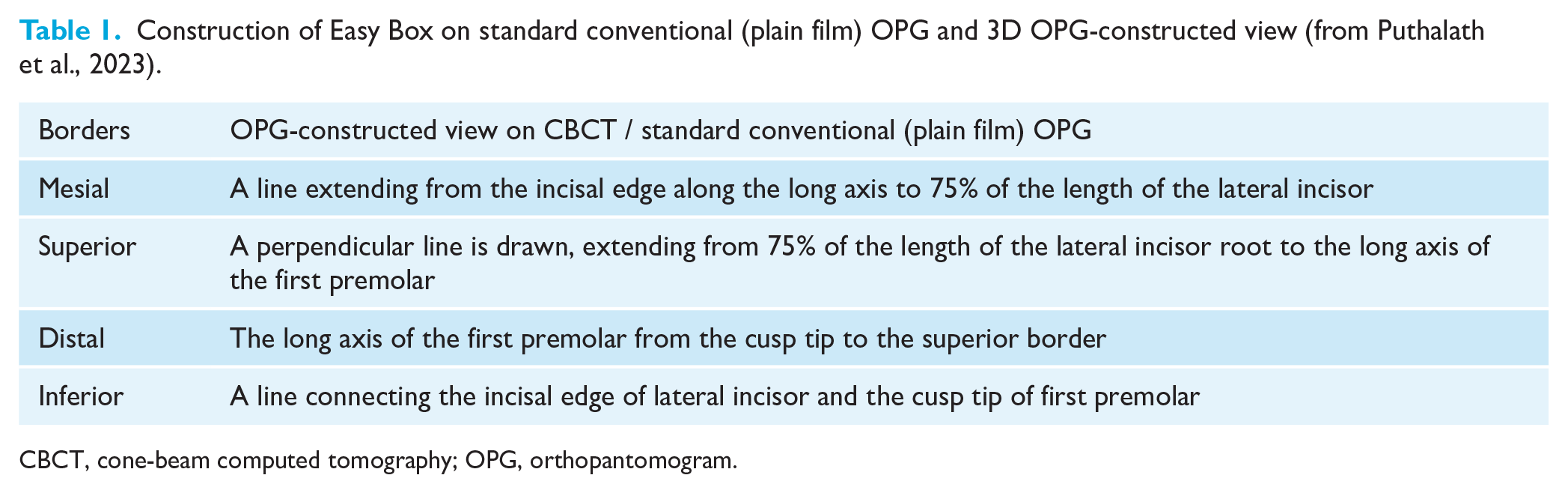
CBCT, cone-beam computed tomography; OPG, orthopantomogram.
Purpose of the study
The aim of the present study was to construct an Easy Box on a standard conventional OPG and to validate this novel method by comparing it with the Easy Box method on 3D CBCT. The overall rationale of the study was to validate the Easy Box on conventional (plain film) OPG radiographs for the assessment of impacted canines with the aim of limiting radiation dose to patients and possibly eliminating the need for CBCT. Therefore, we undertook this as a pilot study.
Objectives
The objectives of the study were to assess and compare the validity of a 2D modified Easy Box and to measure the Beta angle on standard conventional OPG versus 3D CBCT OPG-constructed view.
Sample size
The sample size for the pilot study was 25 participants.
Study design
A retrospective agreement study was conducted.
Selection criteria
The inclusion criteria were as follows: patients with unilateral/bilateral impacted maxillary canines; good-quality CBCT images with raw data; and good-quality routine OPG. The exclusion criteria were as follows: patients with a syndrome; patients with cleft lip and/or palate; history of trauma/surgery in the oral and maxillofacial region.
Material and methods
This retrospective agreement study was carried out after obtaining approval from the Ethical Committee of Index Institute of Dental Sciences, Indore (approval no. IIDS/IEC/2022/177(E)/ORTHO/04). Since the OPG is an essential diagnostic aid, it was the primary radiograph taken for the determination of the presence of impacted canines. The CBCT scan was supplementary, having been requested to determine the exact spatial relation, to adjacent anatomic structures, of the canines. The data for our study were collected retrospectively from a convenience sample, due to the limited availability of participants.
Standard conventional (plain film) OPG and CBCT data were obtained for the study from both departmental records and five private practices, in Indore, India. Participants were selected based on predefined inclusion and exclusion criteria, record availability and their willingness to provide consent. Written consent was obtained from all participants, before the commencement of the study, to allow the use of their images. Participant’s records (n = 25) included standard conventional (plain film) OPG radiographs and 3D CBCT data. The records were analysed to enable a comparison and to assess the accuracy of Easy Box construction across both 3D CBCT and standard conventional OPG radiographs.
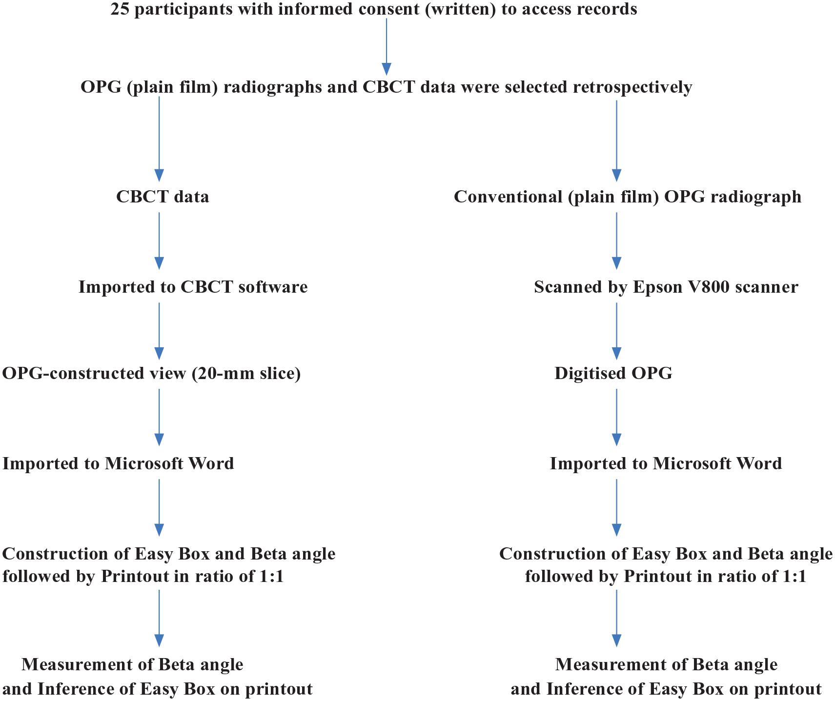
Puthalath et al. (2023) demonstrated the construction of an OPG-constructed view using CBCT data in their study and validated their findings. The thickness of the slice, of the OPG-constructed view from the CBCT was adjusted to 20 mm with the help of CBCT software. The standard conventional (plain film) OPG was scanned using an Epson V800 scanner and digitised. On the HP 15s series laptop, the OPG-constructed view from the CBCT and a digital copy of the conventional (plain film) OPG were imported to Microsoft Word 2019 software for the construction of Easy Box (Table 1).
The construction of the Easy Box, as demonstrated by Puthalath et al. (2023), was undertaken on the digital copy of the conventional (plain film) OPG. For the construction of the Easy Box on the OPG as well as the CBCT OPG-constructed view, four borders were drawn, namely the mesial, distal, superior and inferior (Table 1). Furthermore, the location of the impacted canine, whether it was inside or outside the Easy Box boundaries, was determined on both views. The Easy Box construction was carried out by the principal investigator (RD) to eliminate bias.
This was followed by the measurement of the Beta angle on both views (3D and 2D radiographs). The Beta angle was formed between the lateral incisor’s long axis and the long axis of the impacted canine. The Beta angle was measured on the printed copies (ratio 1:1) of the Word document containing the OPG-constructed view from the CBCT scan and a digital copy of standard conventional (plain film) OPG. In our study, we considered only the OPG-constructed view of the 3D CBCT Easy Box to compare with the 2D OPG radiographs since the axial view could not be determined from the 2D OPG radiographs (Table 1; Figures 1 and 2).
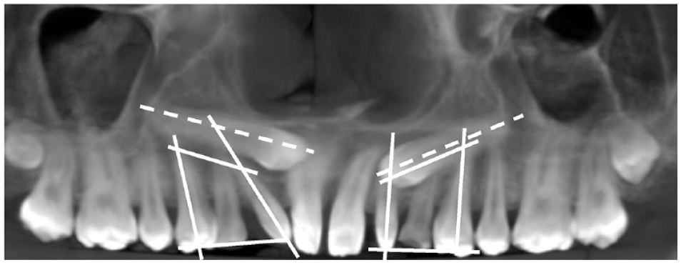
Construction of Easy Box on 3D cone-beam computed tomography data imported into a Word document of a participant.
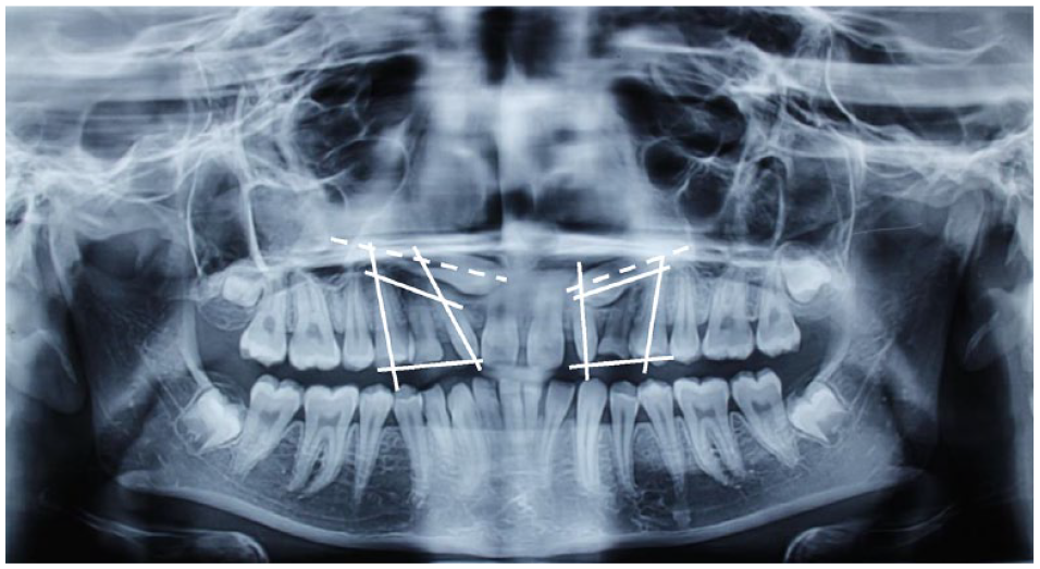
Construction of Easy Box on conventional/standard OPG imported into a Word document of the same participant as in Figure 1.
Assessment of reliability
The difference between all eight linear measurements (the borders of the Easy Box on 3D CBCT OPG-constructed view and 2D OPG) and the two measurements of the Beta angle of 10 randomly selected records were reassessed, with at least 15 days between assessments, to evaluate intra-observer reliability.
Statistical analysis
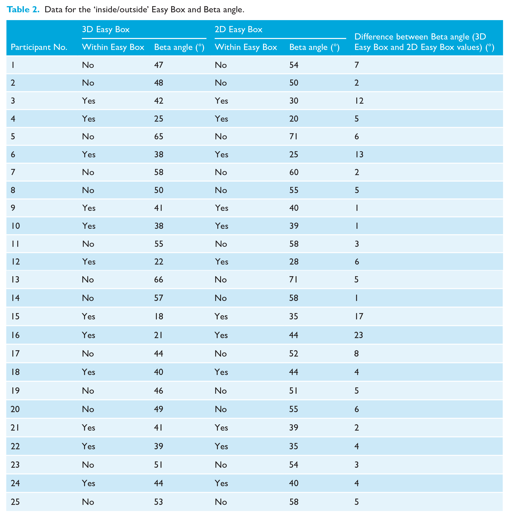
Descriptive and inferential statistical analyses were carried out on data from the images of the 25 participants (Table 2). SPSS Statistics version 20.0 (IBM Corp., Armonk, NY, USA) was used for the analysis of the data and Microsoft Word and Excel 2019 software were used to generate graphs and tables.
Data for the ‘inside/outside’ Easy Box and Beta angle.
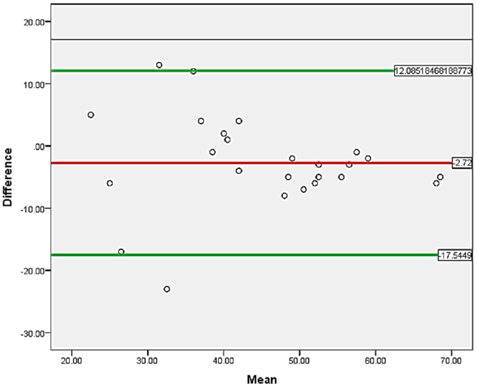
Kappa analysis was used to analyse the agreement between the assessment of the canine within the Easy Box, i.e. inside or outside (Table 3). A Bland–Altman (LoA) analysis was used to analyse the agreement between the measurements of the Beta angle (Table 4; Figure 3).
Comparison of 3D Easy Box CBCT analysis with 2D modified Easy Box on OPG for impacted maxillary canines using Kappa statistical analysis.
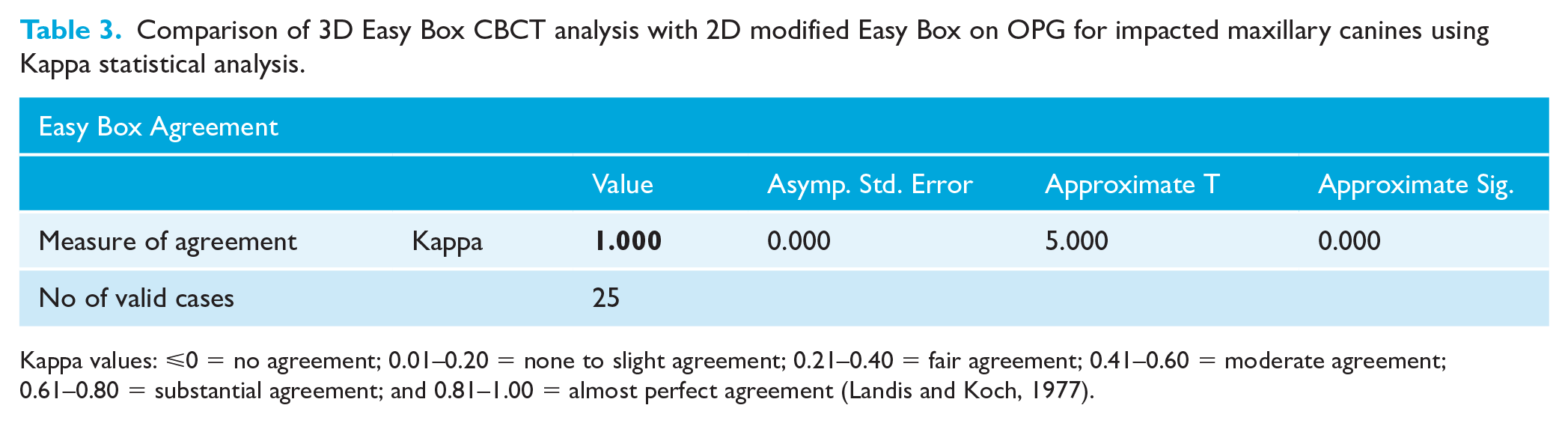
Kappa values: ⩽0 = no agreement; 0.01–0.20 = none to slight agreement; 0.21–0.40 = fair agreement; 0.41–0.60 = moderate agreement; 0.61–0.80 = substantial agreement; and 0.81–1.00 = almost perfect agreement (Landis and Koch, 1977).
Comparison of Beta angle among both groups using a Bland–Altman (LoA) analysis.
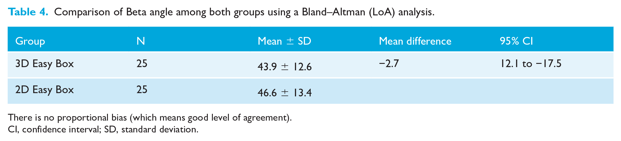
There is no proportional bias (which means good level of agreement).
CI, confidence interval; SD, standard deviation.
Comparison of Beta angle (mean ± SD) among both groups using a Bland–Altman (LoA) analysis.
A Bland–Altman (LoA) analysis was performed to assess intra-observer reliability (Table 5).
Intra-observer reliability of linear and angular measurements of the ‘Easy Box’ in 3D CBCT and 2D OPG radiographs using a Bland–Altman (LoA) analysis.
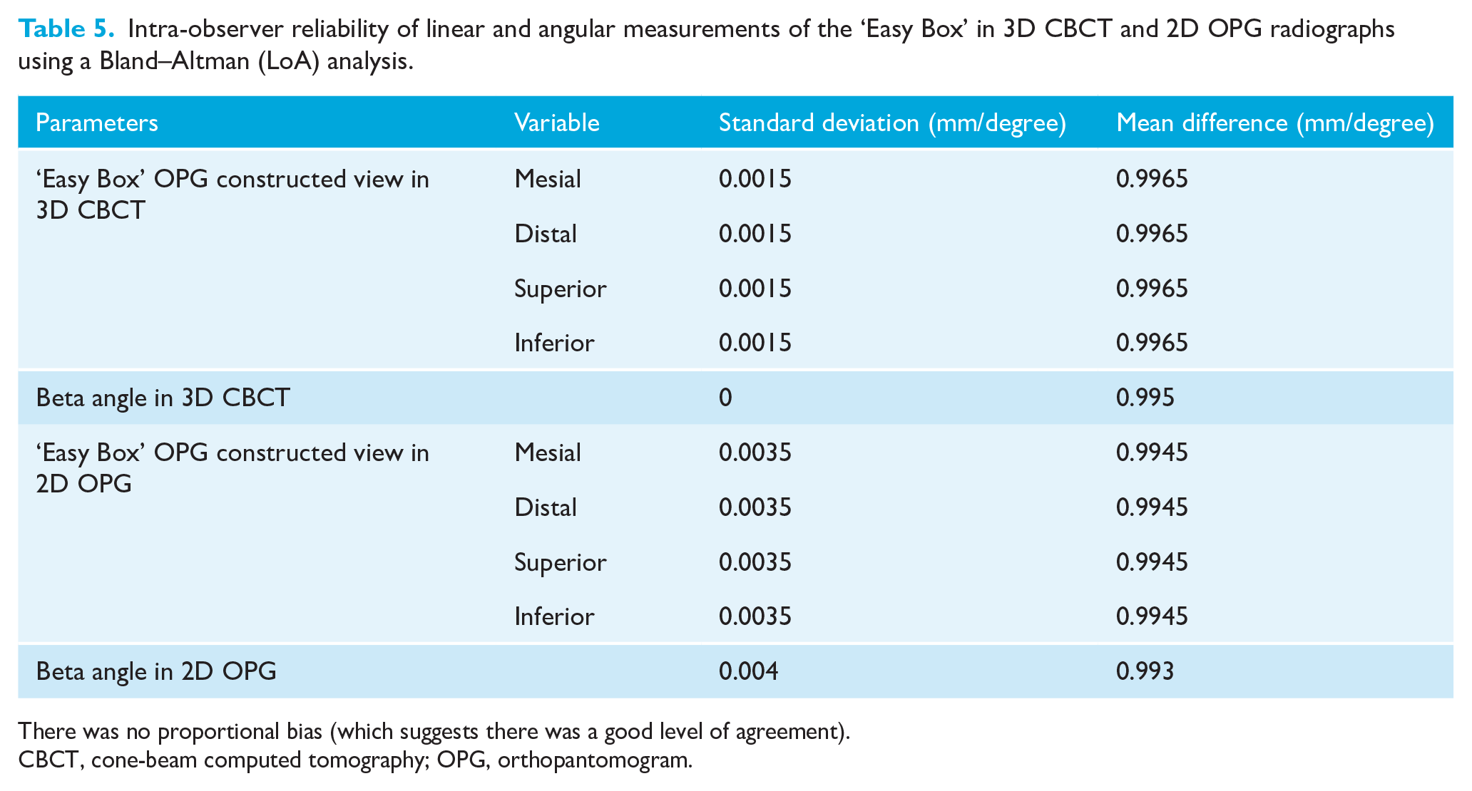
There was no proportional bias (which suggests there was a good level of agreement).
CBCT, cone-beam computed tomography; OPG, orthopantomogram.
Summary of the methodology
Results
Figures 1 and 2 depict the borders of the ‘Easy Box’, beyond which an impacted canine was rated to be harder to align orthodontically (Table 1). A Beta angle of 45° was found to be the angle greater than which it became harder to disimpact and align a maxillary canine with orthodontic traction (Puthalath et al., 2023).
A Bland–Altman (LoA) analysis showed good intra-observer reliability (Table 5). The data showed good agreement (low standard deviation) and minimal bias (mean difference close to mean value) between the measurements for all variables. The standard deviation for the Easy Box OPG-constructed view in 3D CBCT was 0.0015 and for Easy Box on 2D OPG was 0.0035.
Easy Box
A comparison of 3D Easy Box CBCT analysis with 2D modified Easy Box on OPG for impacted maxillary canines using the Kappa statistical analysis was found to be in perfect agreement as evidenced by a kappa value of 1.00 (Table 3). There was 100% agreement between the CBCT and OPG; 52% of canines were outside the Easy Box.
The following data are from Table 2:
For 3D Easy Box: • Inside Easy Box: 12 participants • Outside Easy Box: 13 participants
For 2D Easy Box: • Inside Easy Box: 12 participants • Outside Easy Box: 13 participants
Beta angle (°)
A Bland–Altman (LoA) analysis was used to compare the Beta angle on 3D and 2D OPG radiographs (Table 4; Figure 3). In terms of Beta angle, the 3D CBCT Easy Box in our study had a mean value of 43.9° ± 12.6° and the 2D Easy Box on standard conventional (plain film) OPG had a mean value of 46.6° ± 13.4°. The mean difference was −2.7 (95% confidence interval [CI] = 12.5–−17.5. There is no proportional bias, which means a good level of agreement between both groups. The mean difference between the Beta angle values obtained using the 3D Easy Box and 2D Easy Box methods was calculated to be 5.76° ± 6.01° (95% CI = 3.02–8.50) (Table 2).
Discussion
One of the most challenging orthodontic problems in clinical practice is the presence of impacted permanent maxillary canines (Bishara, 1992). Owing to their bulbous root and long tortuous eruption path, they have the second highest prevalence of impaction after mandibular third molars (Bishara, 1992). The major complications seen with impacted teeth include ankylosis, cyst formation, infection and resorption of the lateral incisor root, and, on occasion, resorption of the central incisor root (Shafer et al., 1963). Studies have been conducted to identify the causes and predictors of impacted maxillary canines with the aim of trying to prevent their impaction (Sacerdoti and Baccetti, 2004; Uribe et al., 2017).
It is important to start orthodontic treatment in a timely manner for these cases and in doing so it will potentially lead to reduced cost, duration and complexity of treatment (Lindauer et al., 1992). It is a challenging task to diagnose such cases due to the complex interplay of diverse factors. However, in 1988, Ericsson and Kurol introduced the sector classification, which was a major milestone for the identification of the severity of impaction and potential difficulty of orthodontic alignment of impacted maxillary canines (Ericson and Kurol, 1988a, 1988b; Lindauer et al., 1992). This was followed by the description of the prediction angle given in 1993 (Power and Short, 1993).
The level of an impacted permanent maxillary canine from the occlusal plane was identified as a prognostic factor for their alignment. A depth of 14 mm or more, away from the plane of occlusion, indicates an extensive period of orthodontic treatment (Stewart et al., 2001). This concurs with the study done by McSherry (1996), which reported that impacted canines, with their cusp tip against the apical third of the lateral incisor roots, had a poor prognosis.
Another factor that predicts the difficulty of orthodontic alignment of an impacted canine is the Beta angle, as proposed by Ericson and Kurol (1988b). Studies by Kuftinec and Shapira (1984) and Stivaros and Mandall (2000) agree that as the angulation increases, it becomes more complex to treat and manage the impacted maxillary canine.
With the advent of CBCT in 1982, the identification of an impacted tooth, in all three planes of space, became easier. Warford et al. (2003) identified an angle formed by the bi-condylar line and the long axis of the impacted canine as a prognosis predictor on an OPG. The mean angulation was found to be 63.2° for impacted canines. Pitt et al. (2006) formulated the treatment difficulty index for the prognosis of unerupted maxillary canines. Similarly, the KPG index (Kau et al., 2009) was developed using CBCT images of patients with canines that were impacted and scores were given (0–5) in three dimensions, according to its anatomical location, root tip and cusp tip.
Recent advancements have introduced two tools—the ‘Cone Beam Computed Tomography Maxillary Canine Impaction guide’ (CBCT-MCI) (Alhummayani and Mustafa, 2021) and the ‘Difficulty Index’ (Chauhan et al., 2022)—designed to assess the location and severity of maxillary canine impactions using CBCT images. However, the practical use of both indices is hampered by their complexity, attributed to the inclusion of multiple prognostic factors which led to the development of ‘Easy Box’ (Puthalath et al., 2023).
‘Easy Box’ was introduced to facilitate the rapid assessment of impacted canines. The Easy Box comprises four borders, namely the mesial, distal, superior and inferior (Table 1). Puthalath et al. (2023) considered two views: the axial view and the OPG-constructed view. The degree of difficulty of disimpaction is greater if the canines were not within the ‘Easy Box’ provided that the Beta angle was less than 45°. Nevertheless, it was similar for those when the Beta angle was greater than 45° even with the canines lying within the ‘Easy Box’.
It is worth noting that the construction of the Easy Box has not been previously applied to standard conventional OPGs. The novel ‘Easy Box’ on a standard conventional (plain film) OPG radiograph overcomes certain limitations of 3D CBCT, which include that they are comparatively expensive, mass screening is not feasible in low-income countries, there is no access in remote areas and it has a high radiation exposure since the radiation dose of a CBCT is approximately 3–6 times that of an OPG (Signorelli et al., 2016).
Agreement between the identification of impacted maxillary canines using the 3D Easy Box CBCT analysis and the 2D modified Easy Box on an OPG was found to be perfect, as evidenced by a kappa value of 1.00. The Bland–Altman (LoA) analysis revealed a good level of agreement between the Beta angle determined from 3D CBCT and conventional OPG radiographs, indicating that the Beta angle determined on conventional OPG is similar to 3D CBCT.
With the result obtained, we can conclude that the comparison of the 3D Easy Box CBCT analysis with the 2D modified Easy Box on OPG as a prognostic tool for impacted maxillary canines showed no statistically significant difference. However, in light of our initial findings of ‘no difference’, it is advisable to consider conducting this study with increased statistical power and a larger sample size for further definitive studies.
Limitations
The present study has a technical limitation. It was difficult to replace the axial view as performed in the 3D CBCT Easy Box study conducted by Puthalath et al. (2023).
Since this was a pilot study and because our data were collected retrospectively as a convenience sample, we lacked the statistical power to detect smaller differences in diagnostic effects.
Our study does not apply to transmigrated and/or transposed canines.
Implications for clinical practice
As OPG radiographs are easily available to practitioners, the construction of the ‘Easy Box’ is a relatively straightforward and inexpensive tool to help determine the prognosis of impacted canines.
Implications for research
There is a necessity for prospective studies to encompass diverse racial populations and sexual dimorphism, incorporate larger sample sizes and validate the results of orthodontic treatment accordingly. Our current study, based on conventional/standard OPGs, suggests potential avenues for future research.
Merits
Perfect agreement was observed between the 2D modified Easy Box on an OPG and the 3D Easy Box CBCT analysis. Furthermore, a good level of agreement was found in terms of the Beta angle. This suggests that, for positional assessment, CBCT may not be required, especially when considering that the measurement of the Beta angle was originally proposed and routinely performed on conventional OPG radiographs (Ericson and Kurol, 1988b; Kuftinec and Shapira, 1984; Stivaros and Mandall, 2000).
Based on the findings of our study, OPG radiographs emerge as potentially advantageous and cost-effective, serving as an essential diagnostic aid when compared to CBCT, given that the radiation dose of a CBCT is approximately 3–6 times higher than that of an OPG (Signorelli et al., 2016).
Conclusion
The agreement between the identification of impacted maxillary canines using the 3D Easy Box CBCT analysis and the 2D modified Easy Box on an OPG, was found to be perfect as evidenced by a kappa value of 1.00.
The Bland–Altman (LoA) analysis showed a good level of agreement between the measurements of the Beta angle on 3D and 2D OPG radiographs.
The innovative ‘Easy Box’, for standard conventional (plain film) OPGs, can potentially be useful, especially during early diagnosis and treatment planning for impacted canines, as OPGs are used routinely in orthodontic practice and have a reduced radiation dose when compared to CBCT.
Footnotes
Declaration of conflicting interests
The author(s) declared no potential conflicts of interest with respect to the research, authorship, and/or publication of this article.
Funding
The author(s) received no financial support for the research, authorship, and/or publication of this article.