
Abstract
Objective:
To evaluate the chemical and dimensional changes of metallic and nickel-free metallic brackets in vitro and ex vivo, regarding oxidation, tie-wing deformation and surface roughness.
Design:
A split-mouth, in vitro and ex vivo controlled clinical trial.
Methods:
A total of 34 adult participants, aged 20–35 years, underwent conventional orthodontic treatment. The right upper central incisor carried the metallic bracket and the left upper central incisor carried the nickel-free metallic bracket. At the 120th day of treatment with a 0.018-inch steel alignment wire, the upper central incisor brackets were removed (ex vivo) and compared with unused brackets (in vitro), distributed into four groups (n = 34): group 1 = in vitro metallic brackets; group 2 = ex vivo metallic brackets; group 3 = in vitro nickel-free metallic brackets; and group 4 = ex vivo nickel-free metallic brackets. Analyses were performed using scanning electron microscopy (SEM) and confocal laser scanning microscopy (CLSM) to assess slot dimensions and surface irregularity (roughness), respectively, and by energy dispersive spectroscopy (EDS) for oxidation. Analyses were performed with a significance level of 5%.
Results:
Evaluation of composition showed that ex vivo nickel-free and metallic brackets had higher percentages of oxygen (O) and carbon (C), lower percentages of niobium (Nb) and chromium (Cr) (P <0.05), and no significant difference in the percentage of iron (Fe) (P >0.05). Brackets showed no dimensional changes when comparing in vitro and ex vivo conditions. Ex vivo metallic brackets had increased roughness compared to in vitro metallic brackets.
Conclusions:
The oral environment promoted corrosion in both metallic and nickel-free brackets. Nickel-free brackets did not have rougher surfaces than metallic brackets, and there were no dimensional changes after use in participants.
Plain language summary
Introduction
Metallic orthodontic brackets are widely used due to their biocompatibility and their physical and mechanical characteristics, such as a lower risk of fracture, a lower modulus of elasticity and a lower friction coefficient when compared to ceramic brackets (Assad-Loss et al., 2008; Kusy and Whitley, 2001). Metallic brackets can be made from a variety of alloys, such as chromium-cobalt alloy (Cr-Co), stainless steel, titanium and nickel-free alloys (Ni) (Eliades and Athanasiou, 2002; Houb-Dine et al., 2017).
The fact that orthodontic devices corrode is well established, and it is time-dependent in the oral environment, while short-term use of archwires is generally considered safe (Stoyanova-Ivanova et al., 2025). However, brackets are more subject to corrosion—loss of metal or conversion to oxide—when in contact with the oral environment. The oral environment in which they are inserted can interfere with the ion-release process (Di Spirito et al., 2024; Menezes et al., 2010; Shintcovsk et al., 2015). In addition, the dimensional changes that can occur in the bracket slot and tie-wings during orthodontic treatment modify the relationship between the orthodontic wires and their slot, influencing the magnitude of friction, which causes a loss of applied force in orthodontic mechanics and interferes with the tooth movement process (Eliades and Bourauel, 2005; Regis et al., 2011; Sarul et al., 2022).
One of the most commonly used metals in the manufacture of brackets is nickel because of its stabilisation (Shintcovsk et al., 2015). Orthodontic brackets are commonly produced from AISI 304L stainless steel (18%–20% Cr, 8%–10% Ni, <0.03% C), whose chromium-derived passive film ensures high corrosion resistance and mechanical stability during orthodontic loading (Oh et al., 2005). However, the structure of metallic brackets has a rough surface, which makes them more susceptible to the corrosive process and, consequently, to the release of ions, usually Ni and Cr, into the oral cavity (Amini et al., 2012; Zigante et al., 2020). This can lead to Ni sensitivity through cytotoxic, allergic and inflammatory reactions, promoting the recruitment of immune cells to the exposed area (Pazzini et al., 2016; Tramontana et al., 2020; Zigante et al., 2022). A nickel concentration of around 30 ppm may already be sufficient to provoke an allergic response (Petoumeno et al., 2008; Sfondrini et al., 2009).
To avoid contact dermatitis, some manufacturers produce nickel-free metallic brackets, which may present changes during their use that differ from those of metallic brackets, being of great relevance for orthodontic treatment (Menezes et al., 2010; Silver et al., 2018). Nickel-free brackets must be able to preserve their structural characteristics—surface, dimensions and chemical composition—so that there is no interference in orthodontic mechanics.
Aims and hypotheses
The aim of this study was to evaluate the physicochemical and dimensional changes of nickel-free and metallic brackets in vitro and ex vivo, regarding oxidation, tie-wing deformation and surface roughness. The null hypotheses in this study were that nickel-free metallic brackets do not oxidise more than metallic brackets, that the absence of nickel does not influence slot dimensions during use and that nickel-free brackets do not have rougher surfaces than metallic brackets.
Material and methods
Participants
This study was carried out in accordance with the protocol of the Research Ethics Committee (CAAE: 79481417.7.0000.5385) and the CONSORT checklist of items for reporting in vitro studies of dental materials (Faggion, 2012). The orthodontic treatment was performed in adult participants of both sexes, aged 20–35 years, who agreed to participate in the study and signed the informed consent form.
Outcomes and sample size
The sample was collected from participants treated at a graduate clinic in orthodontics. The sample size was determined based on the number of brackets assessed in the in vitro and ex vivo tests, employing a split-mouth design with repeated measurements over time and assuming a test power of 0.80, a significance level of 0.05 and a medium effect size (f = 0.25), resulting in 34 brackets per group.
Metallic and nickel-free metallic maxillary central incisor brackets (Roth Max, Dental Morelli Ltda., Sorocaba, Brazil – Lots: 2270634, 2254182, 2150080, 2228032) were used to analyse oxidation, tie-wing deformation (primary outcome) and surface roughness in vitro and after the use of these brackets in participants undergoing orthodontic treatment.
Inclusion and exclusion criteria
Patients with no previous orthodontic treatment, Angle Class I malocclusion and permanent dentition with crowding of no more than 4 mm in the upper arch were included in the study. Patients with agenesis, anterior or posterior crossbite, open bite or overbite were excluded from the study. In addition, smokers, pregnant patients, patients with periodontal disease, systemic diseases, syndromes or those using medications that could interfere with bone remodelling were excluded.
Allocation
The ex vivo brackets were assigned in patients using a split-mouth study design, with the metallic bracket on the right central incisor and the nickel-free bracket on the left central incisor. The unused brackets (in vitro) and removed brackets (ex vivo) were randomised for the chemical and physical assessments. Therefore, the study was divided into four groups (n = 34): group 1 = in vitro metallic brackets; group 2 = ex vivo metallic brackets; group 3 = in vitro nickel-free metallic brackets; and group 4 = ex vivo nickel-free metallic brackets (Figure 1).
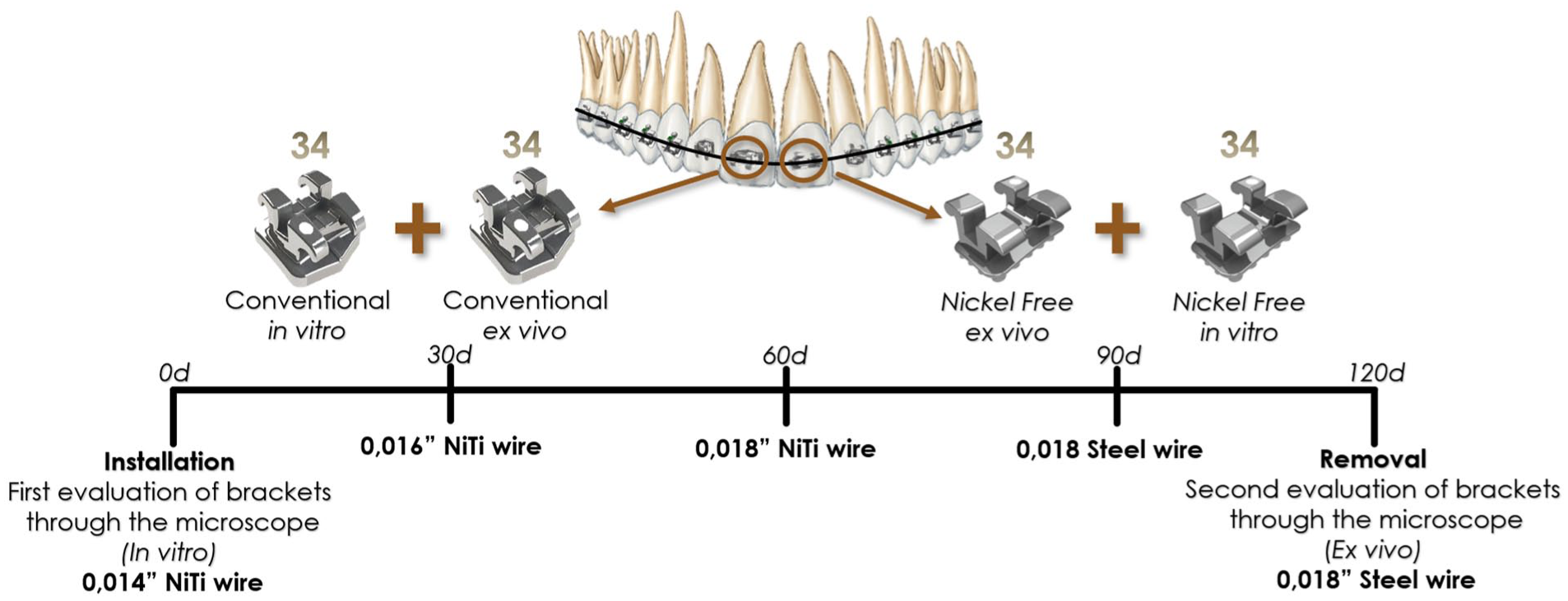
Flowchart of sample characterisation.
Intervention
Orthodontic treatment was carried out using the preadjusted appliance technique. Each participant was treated according to the treatment plan indicated for the malocclusion present through the established diagnosis. All participants received the same dentifrice and toothbrush (Colgate Triple Action Original Mint Toothpaste, Extra Clean Toothbrush, Colgate-Palmolive Company, New York, NY, USA).
Brackets were bonded in the centre of the clinical crown of the participants' upper central incisors following the protocol of the resin manufacturer used (Transbond XT adhesive, 3M Unitek, Monrovia, CA, USA). The right central incisor carried the metallic bracket and the left central incisor carried the nickel-free metallic bracket at the same time. After alignment with 0.014-inch NiTi, 0.016-inch NiTi, 0.018-inch NiTi and 0.018-inch steel wires—an average of 120 days of treatment—the brackets were carefully removed with shear force at the base, using bracket remover pliers 346 (Quinelato Quality/Schobell Industrial Ltda., Rio Claro, Brazil) (Regis et al., 2011). After collection, the metallic brackets were replaced to complete the participants’ treatment.
After removal, the brackets were placed in a container containing 100 mL of distilled water for 10 min and subsequently dried at room temperature and packaged separately in plastic envelopes. The brackets were washed with detergent and distilled water at a ratio of 10:1 for 30 min in a Gnatus ultrasonic vat (Gnatus Ltda., Ribeirão Preto, Brazil). The same protocol for washing and drying the brackets was applied to the in vitro groups, and the chemical and physical assessments were identical to those for the ex vivo brackets (Regis et al., 2011).
Chemical assessment
Brackets were analysed using a scanning electron microscope (SEM; JEOL SM-6390LV, Tokyo, Japan) with respect to oxidation and dimensions. Double-sided carbon tape was applied to the stub (sample holder), and using clinical forceps, four brackets from the same group were glued to each stub, arranged in an acrylic holder.
Oxidation was evaluated using energy dispersive spectroscopy (EDS). EDS was performed on all brackets to analyse the chemical composition, allowing the quantification and comparison of the release of iron ions (Fe), chromium (Cr), niobium (Nb), oxygen (O), carbon (C), silicon (Si), calcium (Ca), manganese (Mn), molybdenum (Mo), nickel (Ni), lead (Pb) and tungsten (W) in vitro and ex vivo (Menezes et al., 2010).
Physical assessment
To assess the dimensional changes of the tie-wings, ImageJ software (National Institutes of Health, Bethesda, MD, USA) was used on in vitro and ex vivo SEM images. The measurement between the left and right tie-wings (horizontal dimension) was assessed on the lower edge of the upper tie-wings and the upper edge of the lower tie-wings, parallel to the long axis of the slot. The vertical dimension was assessed at the centre between the upper and lower tie-wings on the left and right sides, parallel to the long axis of the bracket, in a 40× magnified image (Menezes et al., 2010; Regis et al., 2011) (Figure 2).
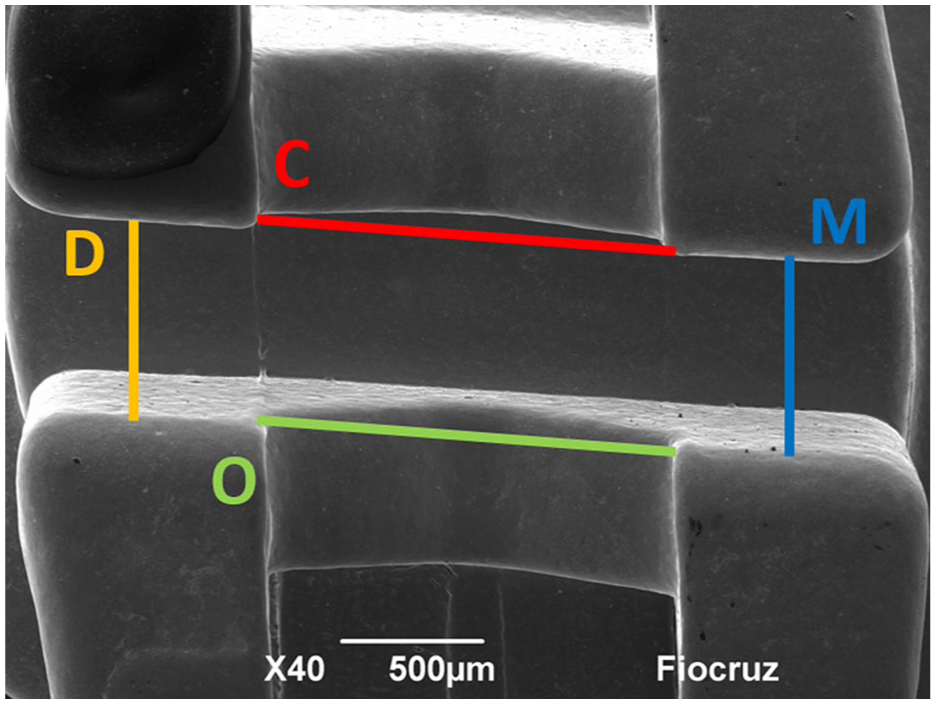
Representative SEM image showing measurements taken in the bracket slots. Tie-wing measurements: horizontal assessment. (O) Horizontal occlusal tie-wings dimension; (C) horizontal cervical tie-wings dimension; (D) vertical left tie-wings dimension; (M) vertical right tie-wings dimension. 40× magnification.
To analyse surface irregularity—roughness—of the slot and 3D images, the brackets were analysed using a Confocal Laser Scanning Microscope (CLSM) (LEXT OLS4000, Olympus, Tokyo, Japan), coupled with OLS4000 software (Olympus, Japan) (Bayguinov et al., 2018; Kuskonmaz et al., 2020). The central topographic areas of the brackets’ surfaces, measured in µm, were used in an area of approximately 0.5 mm². The 3D images were obtained in the central region of each sample using a 10× objective, producing images with 214× magnification, which were saved in TIFF format.
Statistical methods
Intra-examiner reproducibility of tie-wing dimensional changes was assessed using descriptive statistics (mean, SD, median), the intraclass correlation coefficient (ICC) and the Bland–Altman method. Analyses were conducted in two stages, with a 30-day interval between them, using a significance level of 5%.
The exploratory analysis indicated that the percentage data of the Fe, Cr, O, C, Si, Mn and Mo elements, as well as deformation, were analysed using generalised linear models according to the split-mouth design with repeated measures over time. In addition, the percentage of impurities and/or other metals present in very low amounts was analysed using generalised linear models considering the split-mouth design. Nb data were analysed using a paired t-test. The Ni, Pb and W data did not fit a normal distribution and were analysed using the non-parametric Wilcoxon test.
The normality of the roughness data was tested using the Kolmogorov–Smirnov and Shapiro–Wilk normality tests and was found to be normally distributed. Repeated measures multivariate analysis was used, followed by the Bonferroni post-hoc test (P <0.05). The analyses were performed using SAS (SAS Studio 3.8), R (R Core Team) and SPSS Statistics software (IBM), with a significance level of 5%, on in vitro and ex vivo brackets.
Results
A reliability test for dimensional changes of the slot measurements was performed, and the first and second measurements were very close, with no statistically significant differences (P >0.05), confidence limits in the range of −0.01–0.01 cm and ICC ⩾0.9 (Figure 3).
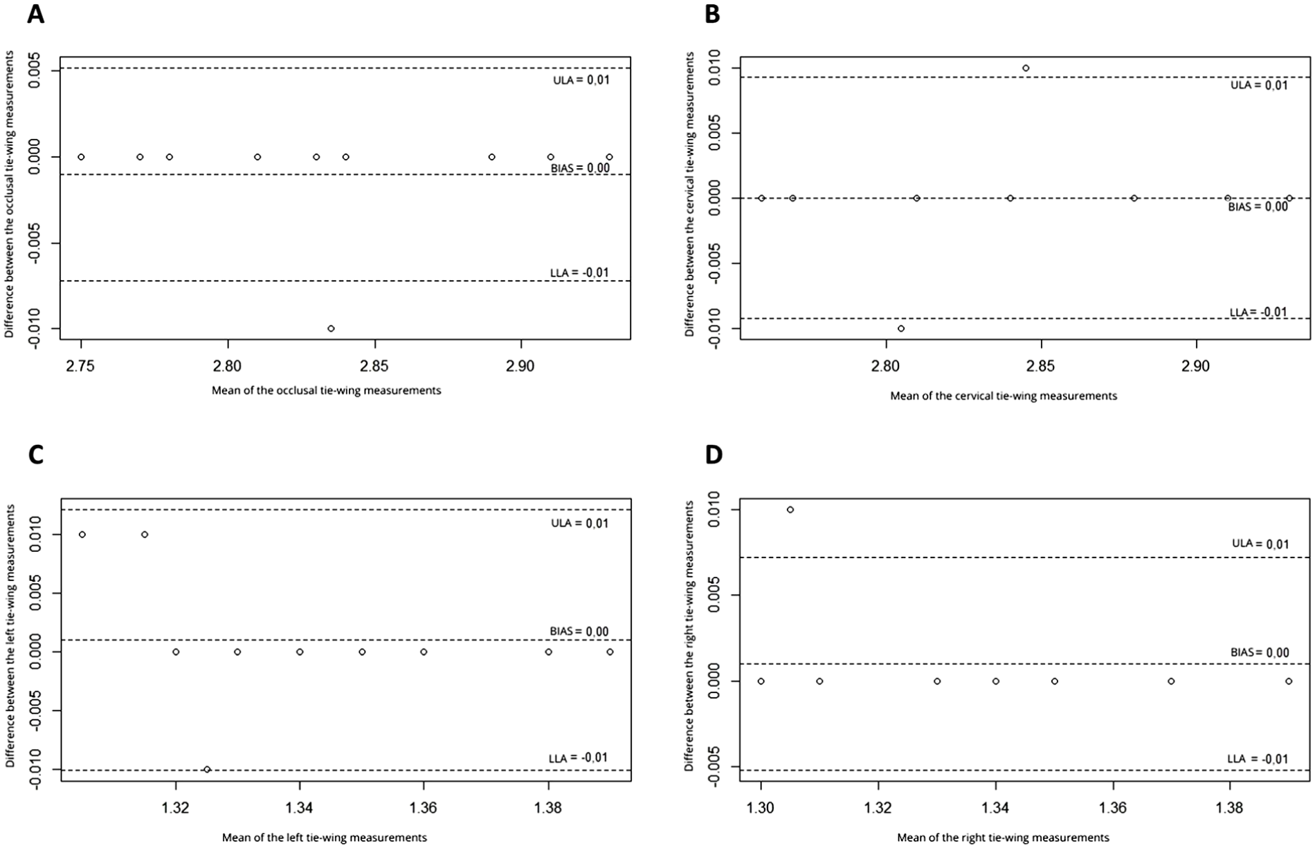
Scatter plot of differences vs. means of tie-wing measurements between the first and second assessments. (a) Horizontal occlusal tie-wings dimension; (b) Horizontal cervical tie-wings dimension; (c) Vertical left tie-wings dimension; (d) Vertical right tie-wings dimension. LLA, lower limit of agreement; ULA, upper limit of agreement.
Table 1 shows that there was no significant difference between the types of brackets and between the groups (in vitro and ex vivo) regarding the percentage of Fe (P >0.05). The percentage of Cr in vitro was significantly lower in nickel-free brackets (P <0.05), and in ex vivo, it was significantly lower for both types of brackets (P <0.05). In metallic brackets, the percentage of Nb was significantly lower in the ex vivo brackets (P <0.05), whereas in the nickel-free type, Nb was not detected. The percentage of O was significantly higher in vitro metallic brackets (P <0.05) and significantly higher in ex vivo for both types of brackets. The percentage of C was also significantly higher in ex vivo for both types of brackets. The percentage of Si was significantly lower in ex vivo for both types. Mn and Mo were not detected in metallic brackets, and in nickel-free brackets, they presented a lower percentage in the ex vivo brackets (P <0.05). There was no significant difference between the two types regarding the percentage of impurities and/or other metals present in very low amounts in the ex vivo brackets.
The percentage of elements (oxidation evaluation), according bracket type and group.
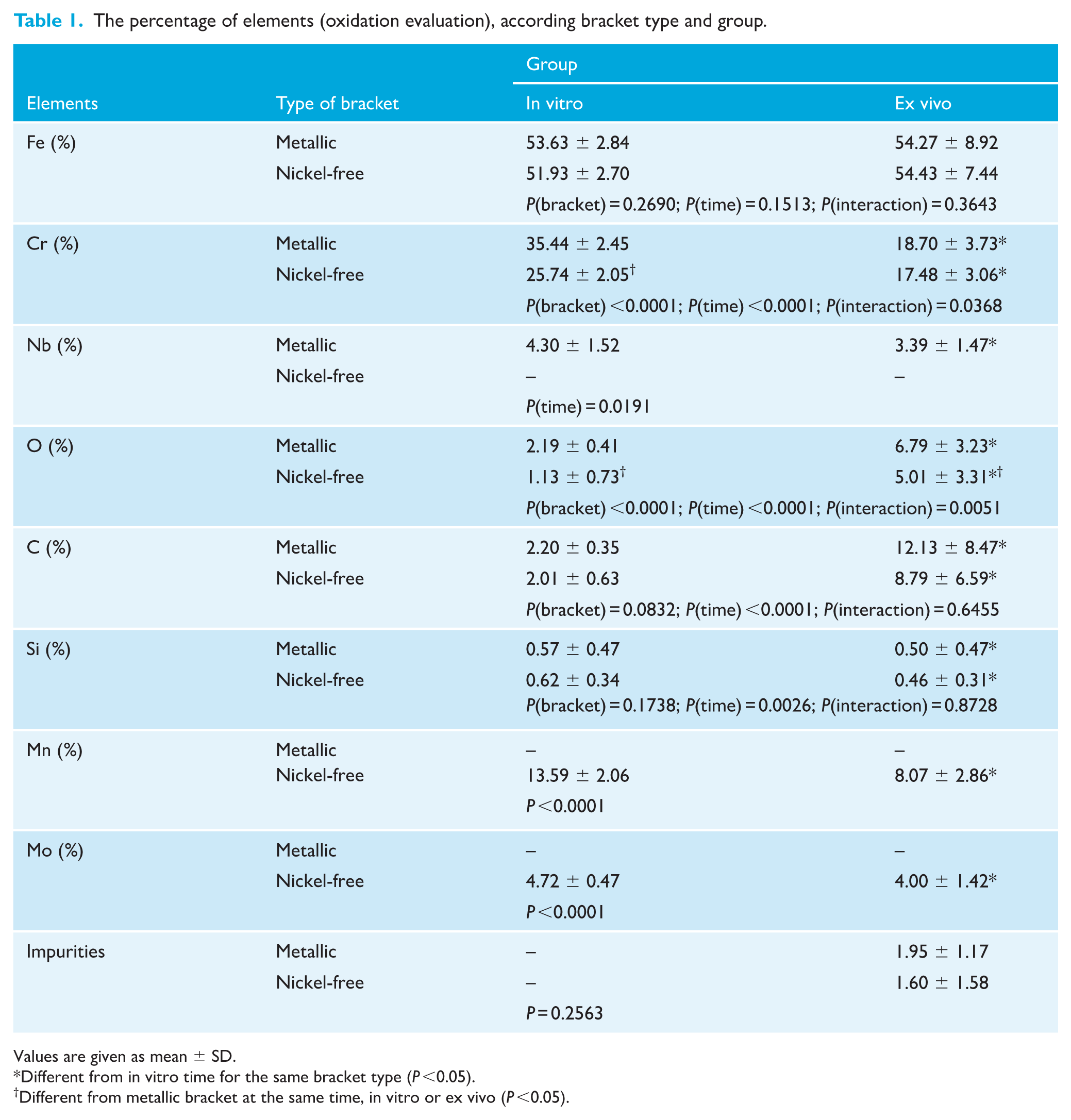
Values are given as mean ± SD.
Different from in vitro time for the same bracket type (P <0.05).
Different from metallic bracket at the same time, in vitro or ex vivo (P <0.05).
Table 2 presents the results of the elements whose data do not fit a normal distribution (Ni, Pb and W), summarised by median and interquartile range (IQR) values. Ni was not detected in nickel-free brackets, and there was no significant difference between in vitro and ex vivo for metallic brackets (P >0.05). Pb and W were not detected in either metallic or nickel-free brackets.
The percentage of elements (oxidation evaluation), according bracket type and group.
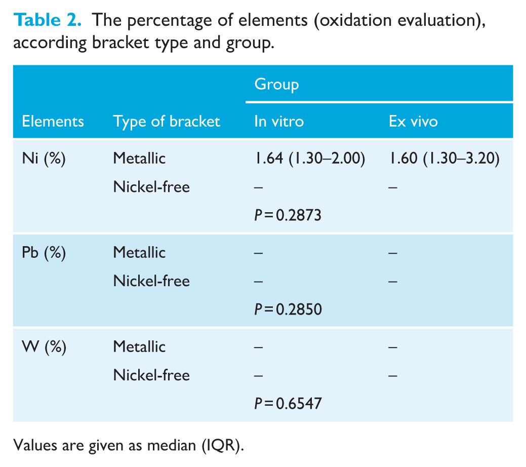
Values are given as median (IQR).
Table 3 shows that there was no deformation of metallic or nickel-free brackets between the in vitro and ex vivo analyses. Horizontal tie-wing dimensions were larger in nickel-free brackets (P <0.05). Vertical tie-wing dimensions were smaller in nickel-free brackets (P <0.05) in both in vitro and ex vivo analyses.
Slot deformation measurements (in mm), according bracket type and group.
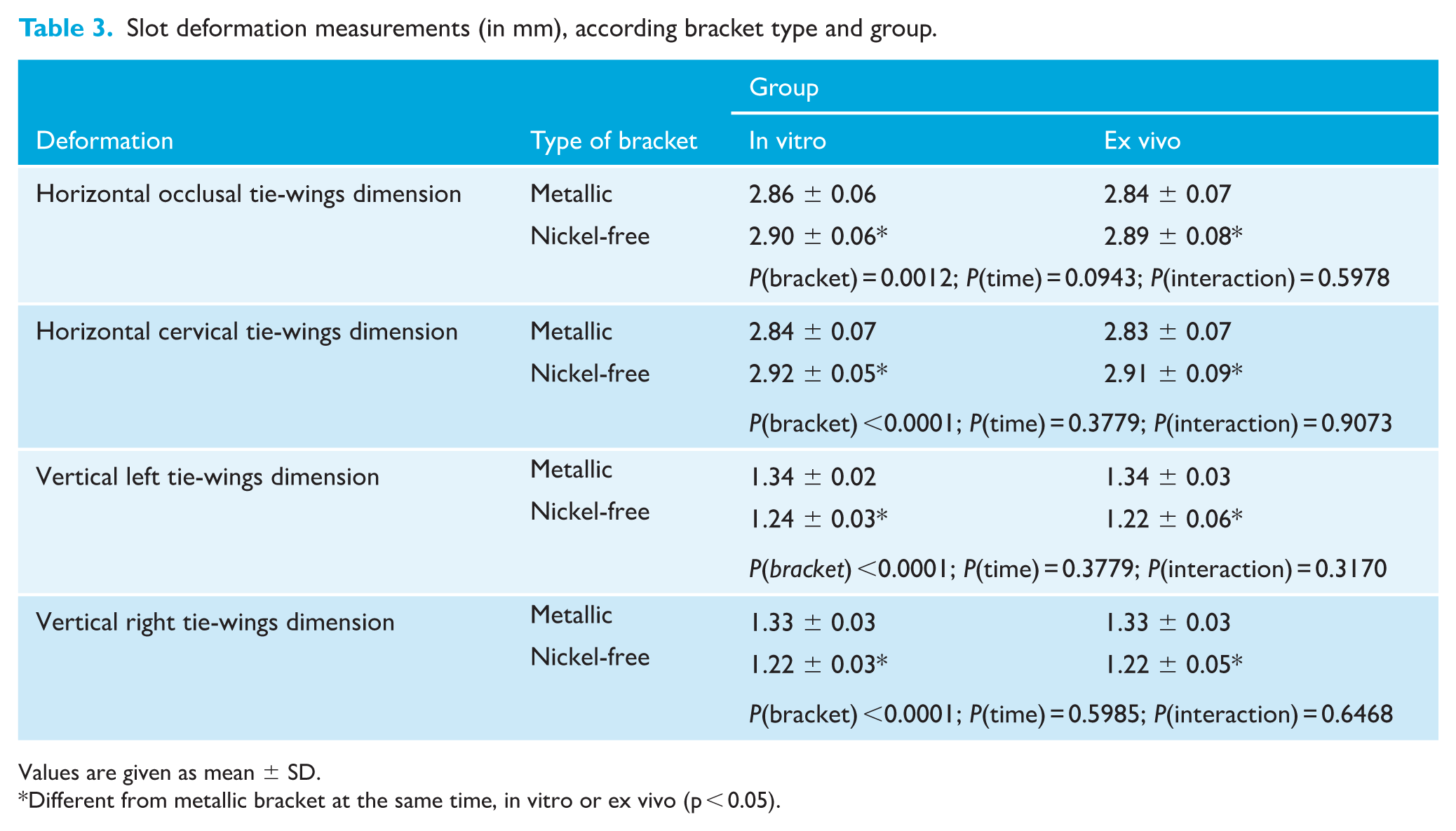
Values are given as mean ± SD.
Different from metallic bracket at the same time, in vitro or ex vivo (p < 0.05).
Table 4 shows that no statistically significant differences were found in the mean roughness values between metallic and nickel-free brackets (P >0.05). Metallic brackets showed a statistically significant difference in roughness values between in vitro and ex vivo groups (P <0.05). No differences in roughness were observed for nickel-free brackets between in vitro and ex vivo groups (P >0.05) (Figure 4).
Slot roughness (µm), according bracket type and group.
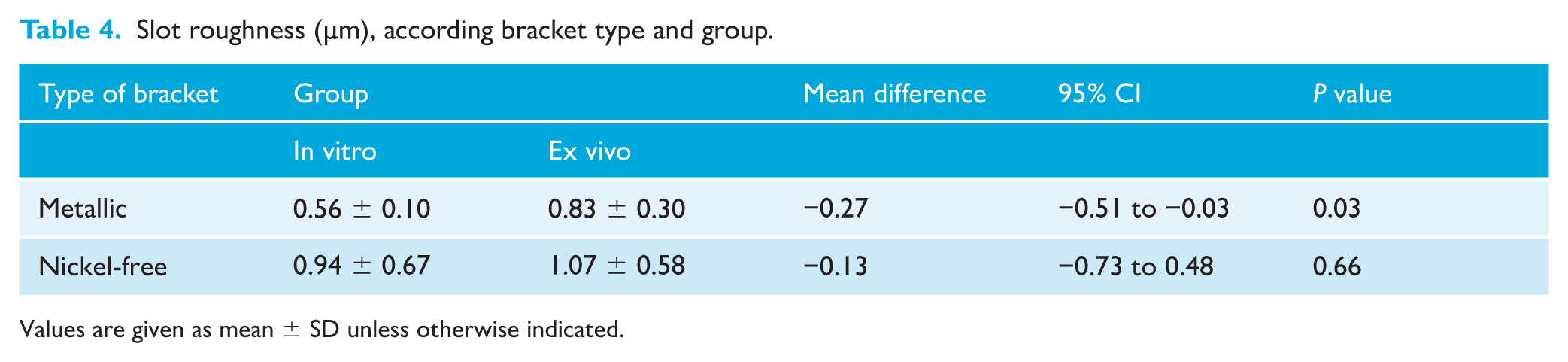
Values are given as mean ± SD unless otherwise indicated.
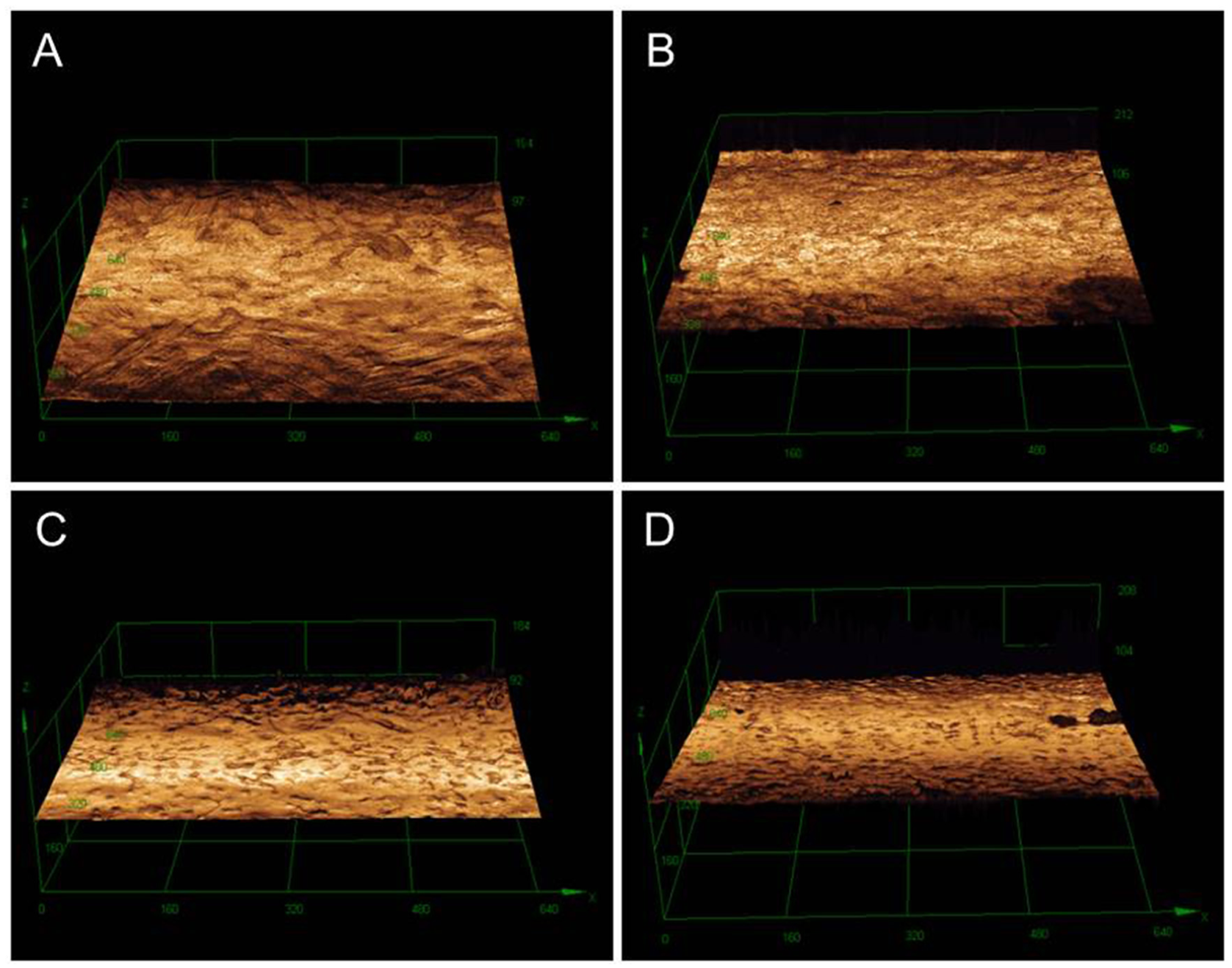
Bracket slot surface roughness (µm) measured by confocal laser scanning microscopy. (a, b) Metallic brackets in vitro and ex vivo, respectively; (c, d) nickel-free brackets in vitro and ex vivo, respectively. 214× magnification.
Discussion
Summary of findings
Using the methodologies described above, the following null hypotheses were confirmed: (1) nickel-free brackets do not undergo further oxidation; (2) the absence of nickel does not influence dimensional changes in the tie-wings during use; (3) nickel-free brackets do not have rougher surfaces than metallic brackets.
Comparison with other studies and clinical implication
There is a predominance of Fe, Cr and Ni in the composition of metallic brackets (Menezes et al., 2010; Shintcovsk et al., 2015). In the present study, a high percentage of Fe and Cr was observed in these brackets, with Nb having a higher percentage than Ni. Nb is a hypoallergenic, corrosion-resistant metal that confers mechanical resistance to steel and lower toxicity compared to Ni, which may contribute to the lower allergenic effect of metallic brackets (Genchi et al., 2020; Sufarnap et al., 2023). The presence of Nb may explain the reduction in Ni concentration found in this study.
Although Ni was not found in large proportions in metallic brackets in the present study, it is known that even a small percentage of Ni present in alloys is not considered safe for patients with a history of allergy to this metal (Sfondrini et al., 2009; Zigante et al., 2020). The corrosion process, which results in the release of Ni, has been demonstrated (Shintcovsk et al., 2015), and hypersensitivity reactions are the most common problems related to this metal, being considered cytotoxic, carcinogenic, allergenic and mutagenic (Petoumeno et al., 2008; Suryawanshi et al., 2024).
In this study, there was no significant difference between the types of brackets or between the groups—in vitro and ex vivo—regarding the percentage of Fe. This is because Fe has a moderate reactivity potential on the reactivity scale, or the electrolytic voltage of metals, which is the metal’s ability to donate electrons, that is, to undergo oxidation (Kusy et al., 1998). Above iron (Fe) on the oxidation scale are Mn and Cr, so even after a relatively short time in the mouth in this study (120 days), these metals were oxidised before Fe, as demonstrated in the results of the present study.
Stainless-steel brackets have a layer consisting of chromium oxide (Cr2O3) to provide resistance to discoloration and corrosion; if this layer breaks, corrosion will occur (Kusy et al., 1998). The consumption of acidic foods and beverages by patients may contribute to an accelerated corrosion process (House et al., 2008; Kao and Huang, 2010). In this study, the in vitro chromium percentage was significantly lower in nickel-free brackets, which may be related to a greater propensity for corrosion. However, in the ex vivo condition, both types of brackets showed a lower percentage compared to the in vitro measurements over time in the mouth, which may indicate corrosion.
The percentages of O and C in this study were higher in ex vivo conditions for both types of brackets, indicating the presence of by-products from the microbial biofilm formed on the surface of these brackets. Consequently, carbon deposits on bracket surfaces can lead to biological consequences, including cytotoxicity. Notably, studies have shown that nickel-free brackets exhibit superior biocompatibility compared to other materials (Retamoso et al., 2012). Moreover, the retention of oral biofilm around orthodontic brackets significantly elevates the risk of developing white spot lesions (Khoroushi and Kachuie, 2017). In addition, the accumulation of carbon deposits on brackets increases friction, potentially compromising the efficacy of orthodontic treatment (Zhang et al., 2023). Precipitated biofilm shows a dark phase in the images, suggesting the presence of elements with low atomic numbers, with C, O and Ca being the main elements detected by energy dispersive spectrometry (Regis et al., 2011). Furthermore, high concentrations of O, as shown in this study, suggest the formation of oxides on the surface of the accessory (Saporeti et al., 2012).
The dimensions of the in vitro tie-wings varied between the types, demonstrating slightly different slot sizes; in nickel-free brackets, the occlusal and cervical measurements were greater, while the measurements of the right and left tie-wings were smaller than those of metallic brackets. When evaluating dimensions, other studies have also found differences between brands; however, the brackets evaluated showed dimensional stability during orthodontic treatment (Oh et al., 2005; Regis et al., 2011). A critical factor that affects the expression of a bracket prescription is the accuracy of its slot, as it directly interferes with alignment and even torque strength during orthodontic mechanics (Flores et al., 1994; Sarul et al., 2022; Silver et al., 2018). In the present study, there was no significant difference in tie-wing dimensions between in vitro and ex vivo analyses. For metallic brackets, the mean difference was −0.27 µm, and for nickel-free brackets, −0.13 µm, indicating stability of the slots.
The stainless-steel characteristics of metallic brackets, evaluated through CLSM, were correlated with surface flaws related to the manufacturing process (Kao and Huang, 2010). The slot surface evaluation of titanium alloy and stainless-steel brackets and orthodontic wires showed higher frictional values for rougher and more irregular surfaces (Magesh et al., 2021; Krishnan and Kumar, 2004; Kusy and Whitley, 1997). The final polishing process in manufacturing is related to changes in the characteristics of the slot surface, where less polished brackets are more susceptible to the release of ions in the oral cavity and, therefore, corrosion, in addition to interfering with sliding mechanical properties, increasing friction (Assad-Loss et al., 2008; Camporesi et al., 2016). These data were corroborated in this study, as metallic brackets showed an increase in roughness in ex vivo conditions, indicating corrosion.
Nickel-free brackets must have better polishing compared to metallic brackets, in an attempt to hinder or minimise corrosion of the alloy, a finding confirmed in the present study, where there was no statistically significant difference between the two conditions (Menezes et al., 2010; Shintcovsk et al., 2015). In addition, a more polished surface reduces the risk of mechanical trauma due to friction with the oral mucosa, as reduced contact of Ni ions with deeper tissues makes the onset of allergic reactions less likely (Rahilly and Price, 2003). Thus, nickel-free brackets with more polished surfaces may cause fewer allergic reactions (Pantuzo et al., 2007).
Limitations
A 4-month period allows for the observation of potential early corrosion or wear of the brackets but remains relatively short compared to the total duration of orthodontic treatment, which limits the assessment of long-term effects.
Implications for research
Considering the peculiarities of the oral environment and the mechanical requirements during orthodontic treatment (Belasic et al., 2021; Quadras et al., 2019; Stoyanova-Ivanova et al., 2025), new studies should be conducted to assess the biocompatibility, biodegradation and anti-corrosive properties of metallic brackets in clinical trials—analysing, for example, salivary or blood nickel levels in patients wearing brackets—in order to make orthodontic treatment safer.
Conclusion
Within the study design, it can be concluded that:
The release of ions in both types of brackets showed that oxidation occurred in the oral environment.
The absence of Ni does not influence dimensional changes in the slots during use.
Nickel-free brackets do not have rougher surfaces than metallic brackets.
Footnotes
ORCID iDs
Author contributions
LCF: conceptualisation, project administration, investigation, data curation, writing – original draft and writing – review & editing. LKS: conceptualisation, supervision, investigation, methodology, writing – original draft. GFC: conceptualisation, writing – original draft. JGN: conceptualisation, methodology, formal analysis, writing – original draft. BC: conceptualisation, methodology, formal analysis, writing – original draft. MSJ: conceptualisation, supervision, project administration, methodology, formal analysis, resource, writing – original draft and writing – review & editing.
Funding
The authors received no financial support for the research, authorship, and/or publication of this article.
Declaration of conflicting interests
The authors declared no potential conflicts of interest with respect to the research, authorship, and/or publication of this article.