
Abstract
The goal of this study was to establish reactivity of lignin-derived synthetic polyphenolic material under irradiation by ultraviolet (254 nm) and visible (460 and 525 nm) light in order to deeper examine relationships between the optical properties of this complex mixture and its individual constituents. In all photoirradiation experiments, blue shift of the fluorescence spectrum was observed. We aimed at understanding whether these changes could be explained on the basis of the chromophore interactions hypothesis, which implies destruction of electron-acceptor pairs via free radical transformations to be responsible for the alteration of optical properties. For this, changes in molecular composition were explored by Fourier transform ion cyclotron resonance mass spectrometry. Irradiation with UV resulted in a pronounced oxidation of polyphenols, which was manifested in the van Krevelen diagram by the formation of components with higher O/C ratio. At the same time, irradiation by visible light had led to the appearance of more condensed molecules depleted of oxygen. Consideration of changes in relative contribution of 500 most abundant components in polyphenol materials revealed higher transformation yields under UV light as compared to the visible light. Further studies using deuteromethylation followed by Fourier transform ion cyclotron resonance mass spectrometry enabled to enumerate the number of carboxylic groups in individual components of the parent polyphenol material. It was shown that at all wavelengths irradiation mainly impacted carboxylic-rich unsaturated and aromatic compounds, which can be considered as strong electron-acceptors. We suggest that their transformation is responsible for the blue shift of fluorescence spectrum, thus emphasizing the role of chromophore interaction mechanism of the optical properties formation.
Keywords
Introduction
Natural humic substances (HS) are ubiquitous polyphenolic mixtures formed by a combination of abiotic and microbial transformations of bio-polymers, such as lignin, tannins, cellulose, etc.1,2 Photochemical processes involving HS are particularly important for global carbon cycle studies due to the continuous release of HS fractions in aquatic systems followed by intensive solar irradiation.3,4 Understanding of HS photochemistry and evaluation of its role in HS transformation in the environment requires thorough exploration of their optical properties.
HS are the extended systems of chromophores characterized by rather specific yet similar optical properties. Firstly, absorption spectra of HS in the 350–1000 nm range can be described by a decaying exponential function of wavelength (with the slope varying in between 50 and 150 nm−1). 5 Secondly, fluorescence spectra are broad and bell-shaped without any pronounced vibronic structure with a position of maximum monotonically increasing with excitation wavelength, while the fluorescence quantum yield continuously decreases with excitation wavelength. Collectively, these properties are responsible for a characteristic “envelope” of fluorescence spectra of HS. 6 The abovementioned optical properties, which are not inherent for individual molecular fluorophores, have stimulated discussion of two opposite hypotheses about the formation of the optical properties of HS. Namely, two different models were suggested: the superposition model, when optical properties of HS are considered to be a sum of impacts of its constituents, 7 and the “charge-transfer model”, 8 which implies an important role of electronic interactions between HS components in the formation of optical properties. Despite an extensive research in the field, the validity of these two models is still under discussion, as there are contradictory experimental data, which support either of them. 9 For instance, some of our recent results are more supportive of the interaction model. That is, consideration of optical properties and molecular composition of HS and dissolved organic matter (DOM) from various environments showed that high contribution of bulky aliphatic species, which impact the distance between chromophores (thus lowering the degree of interaction between them) leads to a significant decrease of the long-wave absorbance. 10 Further, fractionation of coal HS by acidity and aromaticity of their components revealed a red-shift of fluorescence emission band asymmetry, which accompanied the enrichment of the sample in acidic aromatic components. 11
Initially, Del Vecchio and Blough 8 suggested the importance of charge transfer interactions in the formation of HS optical properties based on the results of photobleaching experiments. In their work, the appearance of holes in the absorption spectra of HS was observed following irradiation with intense light at several wavelengths. The authors showed that the observed effect could hardly be explained by means of the superposition model. In this work, we aimed at the analysis of alterations of molecular composition caused by UV and visible irradiation of samples. The main idea was to verify whether molecular transformations accompanying photoirradiation-induced changes of optical spectra could be indeed interpreted in the frames of the charge transfer model.
The method of choice for examination of light-induced transformations in complex systems is the Fourier transform ion cyclotron resonance mass spectrometry (FTICR MS), which due to its powerful analytical capabilities allows to resolve thousands of molecular ions in a single HS sample.12,13 This gives a chance to connect specific changes in the optical properties with individual constituents of HS and to determine, which components undergo transformations. 14 However, extreme molecular heterogeneity of HS hampers even FTICR MS analysis. It was shown that charge competition during direct infusion into the most powerful 21 T FTICR MS instrument results in underestimation of a number of molecules in crude petroleum, which were determined after fractionation of samples. 15 This aggravates results of HS molecular study using FTICR MS.
In order to overcome this limitation, in this work we applied photoirradiation to the previously described synthetic water-soluble polyphenolic material obtained by oxidation of hydrolyzed lignin—BP-Cx-1. 16 This sample resembles HS in terms of optical properties, and at the same time it is characterized by relative simplicity of molecular composition with the dominance of nitrogen- and sulfur-depleted aromatic components as compared to natural HS, which also include residues of peptides, organic sulfur, and carbohydrates. 17 The objective of this study was to establish interconnection between photoreactivity of BP-Cx-1 and light-induced changes in its optical and molecular properties. To add structural information on the individual components of BP-Cx-1, enumeration of carboxylic groups was performed by selective deuteromethylation coupled to FTICR MS.
Materials and methods
Solvents and other reagents used in this study were commercially available. Methanol of HPLC grade (Lab-Scan) was used for elution and dissolution of BP-Cx-1 components. High-purity distilled water (18.2 MΩ) was prepared using a Millipore Simplicity 185 system. D-enrichment of deuterated methanol CD3OD was 99.8% (AstraChem). Bond Elut PPL (Agilent Technologies) cartridges (100 mg, 3 mL) were used for isolation and purification of the labeled BP-Cx-1. Parent BP-Cx-1 was provided by Nobel Ltd as a sterile 0.42% ammonia solution (batch X112K14A1) described elsewhere. 16 The carbon distribution obtained by qualitative 13C NMR is provided in Supporting Information file.
Photoirradiation protocol
Three irradiation wavelengths were selected for photobleaching experiments: 254, 460, and 525 nm. A low-pressure mercury lamp with a 100 mW power near the sample was used as a source of UV radiation at 254 nm. The second light source was a light-emitting diode with emission at 460 ± 15 nm and radiation power of 3 W. Finally, the radiation source at 525 nm—the second harmonic of an Yb-doped fs-laser with an average power of 5 W (repetition frequency 80 MHz, pulse duration 100 fs, TEMA Avesta, Russia)—was used. For photobleaching experiments at the wavelengths of 460 and 525 nm, the BP-Cx-1 samples were diluted with Millipore distilled water to a concentration of 1 mg/mL, while in the case of photobleaching at 254 nm, the samples were diluted to the concentration of 0.1 mg/mL so that the optical density at the used wavelengths was close to unity. Samples were placed in a 3 mL quartz cuvette and irradiated for 12 and 2 h for 254 and 460, 525 nm, respectively, leading to the similar irradiation dose of about 30 kJ per sample for different wavelengths. Photoirradiation experiments at all wavelengths were reproduced in triplicates. A nonirradiated control sample with BP-Cx-1 concentration of 1 mg/mL was stored at 4°C.
Optical measurements
Absorption spectra were measured using Lambda 25 spectrophotometer (Perkin-Elmer) in the 250–900 nm wavelength region. The bandwidth of both slits was set to 1 nm, the increment was 1 nm, and the scanning rate was 480 nm/min.
Fluorescence spectra were recorded using the FluoroMax-4 fluorometer (Horiba Jobin Yvon). All samples were measured in the regime of excitation emission matrices measurements, excitation wavelength was varied in the range of 280–580 nm with a step of 20 nm, and the detection range was set to 300–700 nm. To avoid reabsorption and inner filter effects, optical density was set to < 0.1 at 280 nm when measuring fluorescence spectra by dilution with distilled water (Millipore). All measurements were performed in a quartz cuvette with 1 cm optical path at ambient temperature (25 ± 2°C).
Fourier transform ion cyclotron resonance mass spectrometry
All experiments were run on a FT MS Bruker Apex Ultra mass spectrometer equipped with a harmonized cell18,19 (Bruker Daltonics), 7 T superconducting magnet, and electrospray ion source (ESI) in negative ionization mode located at the facilities of the Institute of Biomedical Chemistry. Prior to analysis, parent and treated BP-Cx-1 samples were diluted with water–methanol mixture (1:1) to concentration of 100 mg/L. They were directly injected into the ESI source using a microliter pump at a flow rate of 90 µL/h with a nebulizer and drying gases flows. A source heater temperature of 200°C was maintained to ensure rapid desolvation in the ionized droplets. The mass spectra were both externally and internally calibrated: the former was performed using synthetic carboxylated polystyrene standard, 20 and the latter—by the known residual peaks of fatty acids 21 reaching accuracy value of <200 ppb. The spectra were acquired in triplicates within a time domain of 4 megawords in ESI(−) and 150 scans were accumulated for each spectrum. Resolving power was 530000 at m/z = 400. The FTICR MS data were processed using the open source browser-based application UltraMassExplorer created by Leefmann et al. 22 (http://dockersrv1.awi.de:3838/ume). The generated CHONS formulas were validated by setting sensible chemical constraints typical for DOM23,24 (O/C ratio ≤ 1, 0.3<H/C ratio ≤ 2.2, element counts (C ≤ 120, H ≤ 200, 0<O ≤ 60, N≤ 2, S ≤ 1) and mass accuracy window < 0.5 ppm). Additionally, only formulae presenting in each replicate within group (control and irradiated by 254 nm, 460 nm, and 525 nm) were considered. The assigned CHNOS formulae were further plotted into van Krevelen diagrams, which represent relationships of H/C ratio versus O/C ratio. 25
Deuteromethylation procedure and carboxylic group enumeration
Carboxylic groups in the parent sample were selectively deuteromethylated following the previously developed method. 26 Briefly, thionyl chloride (60 μL) was added dropwise to the solution of 0.5 mg of BP-Cx-1 fractions in 1.5 mL of CD3OD under continued stirring and ice-cooling. The reaction mixture was then refluxed for 4 h and dried under vacuum. The residue was redissolved in 1.5 mL of cold 1 M NaOH, diluted by water and isolated using SPE Bond Elute PPL cartridge according to the procedure described for DOM samples. 27 The data treatment of the labeling experiments included a search and extraction of peaks related to deuteromethylation series of individual BP-Cx-1 components from the full mass spectrum.26,28 These series are produced by peaks with the m/z difference of 17.03448, which corresponds to the substitution of a carboxylic proton with a CD3 group. These deuteromethylation series were manually determined for 500 abundant peaks in the parent BP-Cx-1 sample to identify the number of carboxyl groups in each parent compound. A number of analyzed peaks was limited to 500 out of labor- and time-saving reasons: they were represented by peaks with S/N ratio >10. Error constraint was set to 0.0005 m/z.
Data treatment
Data treatment was conducted using the R software (https://www.r-project.org) and custom-made Python scripts using NumPy, Pandas, Matplotlib, and SciPy modules. Firstly, we created a pivot table of molecular formulae with their relative abundance according to the corresponding mass spectra. Obtained data frame was used for calculation of Tanimoto similarity score (T score), which enables the comparison of intensity distributions within all mass spectra. 29 Changes of relative intensity of most abundant ions in mass spectrum of parent sample was used to evaluate the degree of transformation after photoirradiation at different wavelengths. During the fluorescence spectra processing, the line of Raman scattering was subtracted from the emission spectra by linear interpolation procedure in the region of Raman scattering.
Results
Changes in optical properties of BP-Cx-1 induced by photoirradiation
As a result of irradiation of BP-Cx-1 samples at different wavelengths, changes in the fluorescence and absorption spectra were observed. Photoirradiation at all the wavelengths used (254, 460, and 525 nm) was accompanied by a decrease in the red edge of fluorescence emission upon excitation at 300 nm (Figure 1(a)) and at longer wavelength excitation (Figure 1(b)). Moreover, for irradiation at 254 nm, changes in fluorescence spectrum were accompanied not only by a decrease of the red edge of fluorescence spectra, but also by a significant shift of the fluorescence maximum to the blue region (∼30 nm for excitation at 300 nm and ∼20 nm for excitation at 380 nm). The changes in the shape of the fluorescence band, estimated as the maximum difference between the normalized fluorescence signal of the irradiated samples and the fluorescence spectrum of the control sample at the same emission wavelength, were in the order of 5% for irradiation at 460 nm, 10% for 525 nm, and 25% for irradiation at 254 nm. Despite typical solar simulations are conducted by longer wavelengths (280–360 nm), 30 due to similar photon energies irradiation with 254 nm should lead to the same molecular changes but with more pronounced trends toward oxidized products. 31
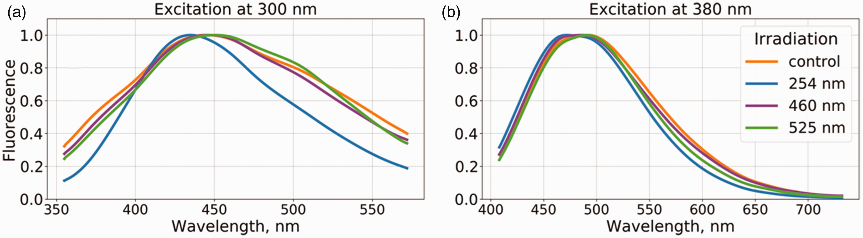
Normalized fluorescence spectra of BP-Cx-1 samples upon excitation at: (a) 300 and (b) 380 nm.
At the same time, the absolute values of the optical density of BP-Cx-1 changed less than 5% for irradiation at 460 and 525 nm, and only at 254 nm irradiation a significant decrease in the optical density was observed (Figure S1). We did not observe significant changes in the band shape of the optical density spectra of irradiated samples, which is consistent with other studies where the effect of photobleaching on the optical properties of HS was investigated. 32 In order to shed light on which molecular formulas degrade and form during irradiation at different wavelengths, photoirradiated samples were measured using FTICR MS.
Molecular compositions of samples as measured by FTICR MS
All mass spectra were represented by singly charged ions in the mass-window of 200-1000 Da. Collectively, 2278 formulae present in each replicate were resolved with a dominance of CHO-composition, which is typical for lignin-derived polyphenols. 33 For visual inspection of molecular composition changes upon irradiation, van Krevelen diagrams were plotted as shown in Figure 2. It is clearly seen that irradiation at 254 nm resulted in the disappearance of the maximum amount of ions – 967, while in the cases of 460 and 525 nm full degradation of only 68 and 164 ions was determined, respectively. Low photolability of BP-Cx-1 is in agreement with the previously reported results on the photobleaching of CHO-components of porewater organic matter under sunlight irradiation. 34 It should be mentioned that according to 13C NMR the sample likely contains cellulosic components (Table S1), which also may undergo photobleaching. However, carbohydrates possess lower ionization efficiency as compared to oxy-acids.
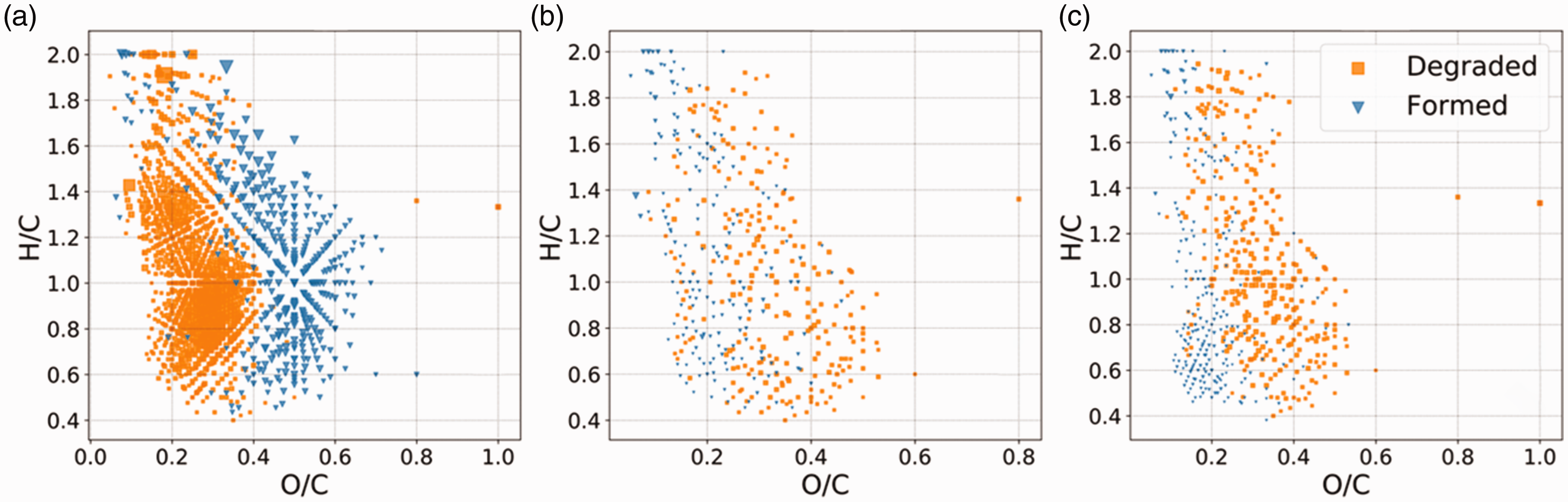
Van Krevelen diagrams representing molecular composition of the samples obtained by FTICR MS with designated reactive and newly formed species. The samples were irradiated at (a) 254 nm, (b) 460 nm, (c) 525 nm. The blue and orange dots correspond to totally degraded and newly formed species, respectively.
According to the previous reports on transformation of polyphenols, photoirradiation may lead to the formation of unsaturated and aliphatic species. Likely, this is due to the cleavage of aromatic systems followed by an addition of unsaturated fragments to the existent aromatic systems or their polymerization as it was previously proposed during the synthesis of humic-like models 33 and oxidation of natural lignin-derived HS. 35 Surprisingly, in the case of visible-light irradiation the formation of less oxidized and more condensed species was observed: 213 and 253 new ions for 525 nm and 460 nm, respectively. In the case of 254 nm, the formation of 362 new ions was observed. In agreement with the literature, 32 higher O/C ratios were characteristic for new ions. At the same time, no significant increase in H/C ratios was observed suggesting the presence of hydroxylation processes during irradiation rather than the aromatic ring cleavage.
Changes in the relative intensity distribution of major components may also affect optical properties. In order to evaluate the degree of BP-Cx-1 tranformation under different conditions, T-scores were calculated (Figure 3(a)) to evaluate the differences between the irradiated samples. It is seen that the sample irradiated at 254 nm is characterized by the lowest similarity to the control sample as compared to 460 nm and 525 nm. In order to prove that irradiation affects molecular content of the samples to a larger extent as compared to deviations due to reproducibility of FTICR MS results, full Euclidean distances were calculated pairwise for all individual samples (5861 molecular formulae). From Figure S2 it is clearly seen that except for one of the replicates for the 525 nm irradiation, the samples were grouped according to the experimental conditions.
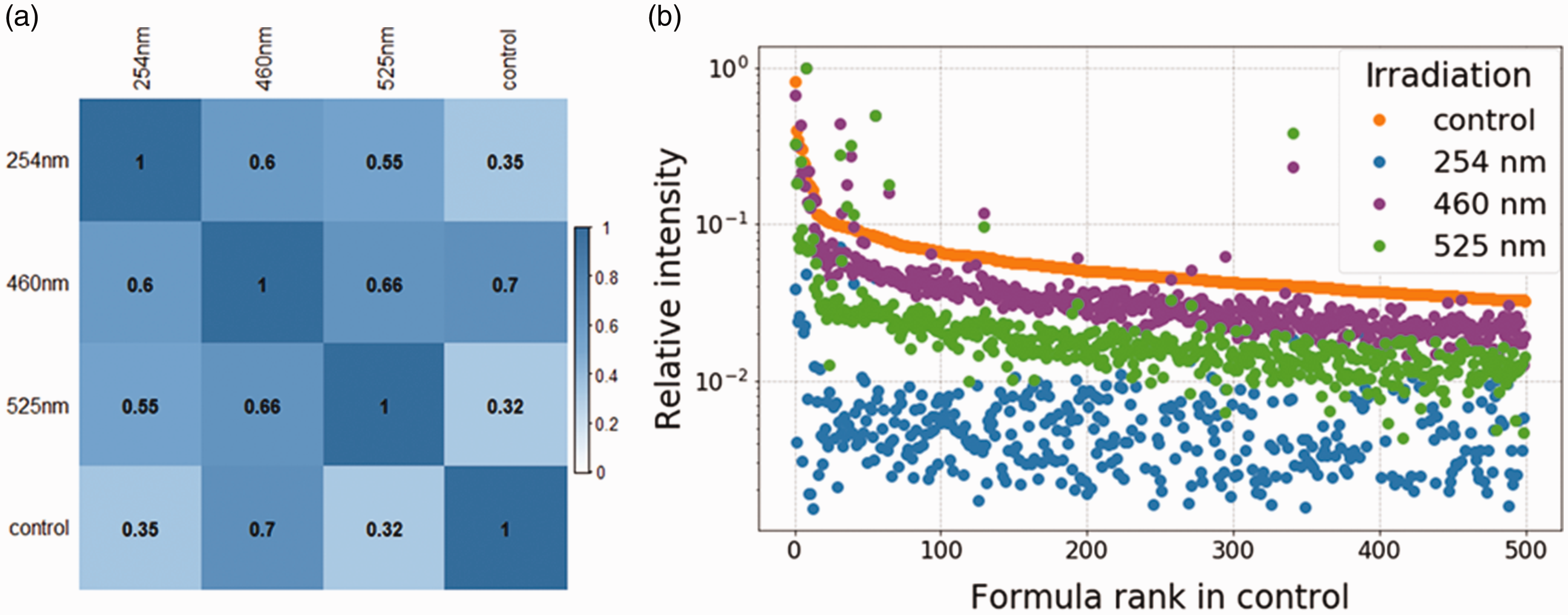
(a) Pairwise similarity matrix based on T-score calculated for ion abundance according to FTICR MS data in control and irradiated samples; (b) relative intensities of the 500 most abundant ions in the control sample and their changes after irradiation.
Despite the T-scores were higher for samples within each group, Figure 2 revealed that BP-Cx-1 reactivity is not structure-specific and a wide range of components undergo transformation during irradiation. Irradiation by different wavelengths led to different transformation degrees. To examine this, relative intensities of the 500 most abundant ions ranking from 1 to 500 were plotted in Figure 3(b). In this respect, transformation degree decreases in the range 254 nm > 525 nm – 460 nm. Plotting relative intensity of these species against double dond equivalent (DBE) and molecular mass showed the same trend and similar transformation degree, which did not depend on m/z and DBE values (Figure S3).
Determination of structural features of reactive compounds
To deeper understand the observed trends in reactivity and changes of optical properties of the sample after irradiation, it is necessary to explore structural features of BP-Cx-1 components, which undergo transformation upon light exposure. This was performed by selective deuteromethylation, which allows to enumerate carboxylic groups in individual components of complex mixtures.26,36 The color-coded van Krevelen diagram is presented in Figure 4.
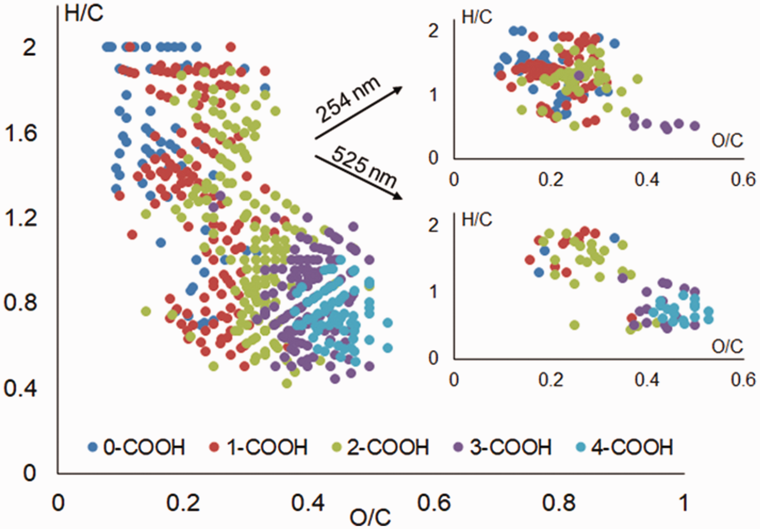
Van Krevelen diagram with color-coded amount of carboxylic groups in individual BP-Cx-1 components. Insets show components disappeared upon photo-irradiation.
Previously by deuteromethylation we have shown that in case of natural HS components with O/C < 0.5 include up to two carboxylic groups, 26 while BP-Cx-1 is enriched in carboxylic functionality. Higher numbers of carboxylic groups were also proposed for condensed compounds of natural HS extracted from soils by analysis of Kendrick mass defect series. 37 These components are acceptors of electrons and their presence may facilitate charge-transfer with donor compartments. 10 Therefore, photoirradiation led to the disapearance of acceptors, which well fits the blue shift of fluoresence emission spectra. Namely, consideration of most abundant ions, which disappear after photobleaching, revealed that 254 nm irradiation affected a wide range of carboxylic acids from lignin-like components with one carboxylic group up to aromatic polycarboxylic acids. At the same time, 525 nm irradiation affected mostly aromatic polycarboxylic acids. Moreover, only long-wave irradiation affected most oxidized species with up to four carboxylic groups.
Possible mechanisms responsible for optical properties alterations after photoirradiation with UV and visible light
The observed changes in fluorescence spectra can be explained by structural transformations of the samples caused by irradiation wavelengths. Previsouly, it was shown that continious blue light irradtiation leads to photobleaching of long-wave absorbing species. 38 Irradiation of carboxylic acids, especially aromatic acids, results in the formation of reactive oxygen species, 39 which further initiates radical-condensation of phenols and quinones. The possible reaciton pathway summarizing photo-induced transformations of carboxylic acids in BP-Cx-1 under UV and visible light is presented in Figure 5. Hydroxylated and condensed species are frequently reported compounds obtained a the result of these reactions. For instance, the impact of reactive oxygen species on lignin-derived organic matter led to the formation of condensed “black-carbon” species depleted of oxygen. 35 The similar results were obtained in our study after visible-light irradiation of the sample as it is seen in Figure 2(b) and (c). UV light efficiently transforms all sample’s components. In case of riverine and marine DOM formation of unsaturated and aliphatic species was reported. 40 In our work, the formation of more oxidized compounds with the same H/C ratios was observed. This evidences the hydroxylation of aromatic rings, which is inherent to phenols. 33 If optical properties of the sample under study resulted from superposition of individual fluorophores, the formation of more oxidized aromatic species would result in a red shift of fluorescence emission similarly to a “yellowing” model mixture of phenols by UV irradiation as it was previously reported. 38 Otherwise, we explored the molecular damage of carboxyls and a blue-shifted fluoresence. Hence, we suggest that photobleaching of BP-Cx-1 in our study transformed and destroyed donor–acceptor pairs, which are necessary for the implementation of the charge-transpher mechanism.
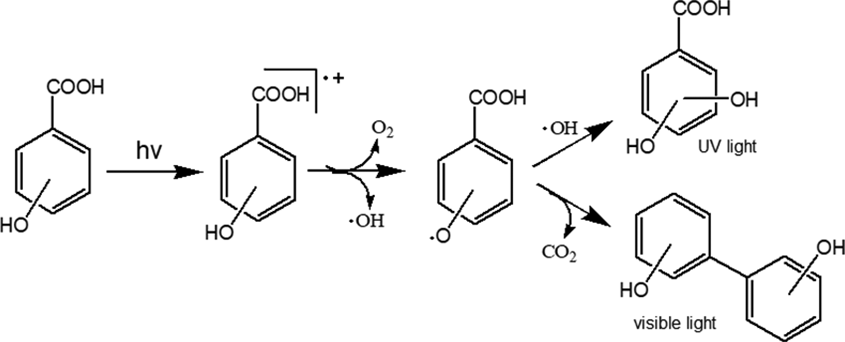
Possible reaction pathway of aromatic hydroxyacids induced by light exposure based on molecular compositions of newly formed species in BP-Cx-1 as determined by FTICR MS.
Conclusions
Photoirradiation of model polyphenolic mixture by different wavelengths showed the lack of selectivity in reactive species as it was determined by FTICR MS. In all cases, a wide range of aromatic and unsaturated compounds underwent partial and full degradation. It was found that UV irradiation at 254 nm impacted parent material to a larger extent compared to visible light irradiation at 460 nm and 525 nm. This was shown by the examination of changes in the relative intensity of abundant ions from parent material and by the enumeration of fully degraded components. Further application of deuteromethylation showed that irradiation at different wavelengths affected aromatic polycarboxylic acids, which are electron-acceptors. This was in agreement with the blue shift of fluorescence emission spectra upon photobleaching and supports the hypothesis about the role of donor–acceptors pairs in the formation of optical properties of polyphenols.
Supplemental Material
EMS917067 Supplemental Material - Supplemental material for Photoreactivity of humic-like polyphenol material under irradiation with different wavelengths explored by FTICR MS and deuteromethylation
Supplemental material, EMS917067 Supplemental Material for Photoreactivity of humic-like polyphenol material under irradiation with different wavelengths explored by FTICR MS and deuteromethylation by Alexander Zherebker, Boris Yakimov, Anna Rubekina, Oleg Kharybin, Elena I Fedoros, IV Perminova, Evgeny Shirshin and Evgeny N Nikolaev in European Journal of Mass Spectrometry
Footnotes
Declaration of conflicting interests
The author(s) declared no potential conflicts of interest with respect to the research, authorship, and/or publication of this article.
Funding
The author(s) disclosed receipt of the following financial support for the research, authorship, and/or publication of this article: This work was supported by Russian Science Foundation grant 19-75-00092. Optical measurements were supported by IHSS (Young investigator research grant to ES) and Russian Foundation for Basic Research (grant no. 18-32-20116). Research contribution of BY was supported by “Basis” foundation stipendium for PhD students (scholarship no. 18-2-6-182-1).
Supplemental material
Supplemental material for this article is available online.
References
Supplementary Material
Please find the following supplemental material available below.
For Open Access articles published under a Creative Commons License, all supplemental material carries the same license as the article it is associated with.
For non-Open Access articles published, all supplemental material carries a non-exclusive license, and permission requests for re-use of supplemental material or any part of supplemental material shall be sent directly to the copyright owner as specified in the copyright notice associated with the article.