
Abstract
PVA used in packaging applications has been faced with a UV light degradation challenge, which often reduces its durability while in use. The UV light stability enhancement effect of nanocrystalline cellulose (NCC) reinforcement in PVA was studied. Polyvinyl alcohol composite film was reinforced with NCC from palm oil waste (PVA-NCC film) and exposed to UV light (22 W, SUV-16 254 nm) for different time duration to study the material durability enhancement. The percentage weight loss of the samples was measured to observe the UV light degradation effect. Furthermore, the samples’ structural, morphological, and tensile properties were studied before and after exposure to UV light with FT-IR, scanning electron microscopy (SEM), and tensile test. The results showed physical degradation, morphological and tensile properties enhancement of PVA with NCC’s addition. The addition of NCC to the PVA matrix reduced the degradation rate under UV light significantly. Also, the percentage of weight loss was observed to change with the exposure time to UV light.
Introduction
The functional properties of polymeric materials in packaging applications are often affected by environmental factors such as UV light (from the sun). 1 The potential polymer film’s UV stability for packaging application is significant to manufacturers and end-users for product preservation. Polymer composites reinforced with biomass have attracted researchers’ attention to producing materials with durability against environmental influences when used.2,3 Environmental degradation of polymer composites can occur by chemical, physical, biological processes or combination.4,5 Several factors, such as sunlight (UV), temperature, moisture, air, microorganism, chemical agent, high energy radiation, and mechanical stress, have contributed to the degradation process. 6 Many methods have been used to determine the resistance of polymer composites to these environmental factors. 4 These methods include soil burial, natural weathering, accelerated weathering, and resistance in the seawater/chemical solutions. 7 However, only a few studies have been dedicated to the resistance of polymeric material to UV light. Evaluation of the UV stability of polymer composite film used for packaging application is very significant to its functional properties because of exposure to sunlight (UV).
One of the methods for evaluating the UV resistance of polymer composites films used for packaging application is the study of degradation of the composite with direct exposure to UV light. The exposure of the films under UV light has been proposed to affect polymer composites’ physicochemical properties. UV light has been reported to destroy the covalent bonds in materials, which result in a series of undesirable degradation effects in polymer composites. 8 The exposure of polymer films to UV light is critical for estimating their shelf-life in multi-purpose applications. 9 This can be transferred from the internal or external environment through a polymer composite wall, resulting in continuous changes in product quality and reduced shelf-life. 10
Furthermore, UV light has also been reported to affect lipid oxidation. The oxidation of polymer composites reduces its shelf-life due to the impacts on the organoleptic qualities for packaging applications. This process is often called photooxidative degradation. Photooxidative degradation has been reported to break down polymer chains, reduce molecular weight, and decrease mechanical properties deterioration with time. 11 Therefore, polymer composite films with a UV light protective component can prevent this type of oxidation.
The degradation process of polymer composite after exposure to UV light is dependent on the type of material. Photodegradable molecules are caused by photon absorption. The polymer degradation is also dependent on the exposure time of the visible or UV light. Photodegradation may occur either in the absence or presence of oxygen. Photodegradation can break cross-linked polymers without oxygen, whereas, with oxygen, photooxidative degradation occurs. 12 The degradation of polymer composites commonly occurs in three steps. The macromolecule chains are first depolymerised into monomers and oligomers. 13 After which, the monomers and oligomers are taken up as biomass. Lastly, the biomass is seen to break down into CO2 and H2O because of oxygen (O2).
Cellulose with nanosized crystals called crystallite is classified as crystalline. Nanocellulose is commonly used as reinforcements in polymer matrices to enhance their properties for specific applications such as packaging, biomedical, etc. 14 Hydrogen bonds between hydroxyl groups connect the cellulose chains. Nanocellulose produced high rigidity and the structural strength in polymer composites. Cellulose materials are abundantly available and easy to process than other comparable options. 15 Cellulose is biodegradable, biocompatible, and requires lesser energy to isolate from precursor materials.16,17 Nanocellulose reinforced in polymers increases their tensile strength and tensile modulus.18–20 Previous studies have investigated the enhancement of nanocellulose in polyvinyl alcohol (PVA) for polymer composites. 21 The composite is soluble in water, non-toxic, semi-crystalline, chemical resistant, with excellent film-forming ability.22–24 It has high tensile strength, flexibility, high optical clarity, and is biocompatible.24–26 The hydroxyl group in the partially hydrolysed PVA has been reported to interact with the hydrophilic surface of cellulose, leading to strong hydrogen bonds. Furthermore, the amount of cellulose loading as reinforcement in a polymer matrix significantly affects hydrogen bonding strength between cellulose and polymer matrix. 22 Therefore, the incorporation of nanocellulose in polymer composites usually has an indispensable contribution to their service life and determines the type of application. 27
Studies on the durability and performance evaluation, under UV light, of the polymeric-nanocellulose composite are limited. Few researchers have studied polymer composites’ degradation and analysed the changes in their physical, chemical, and morphological properties. Karvanis et al. 28 studied UV light’s effect on cellulose nanocrystal filled in epoxy. It was stated that the degradation process of polymer composites was dependent on the exposure time of the UV light. The UV light exposure was also reported to increase the polymer composite’s brittleness with micro-cracks forming on its surface. Epoxy polymers filled with cellulose nanocrystal have been used as a UV filter functional nanofiller material. It displayed remarkable structural stability and minor discolouration over a prolonged period of exposure. Liu et al. 29 investigated Vectran fibre after exposure to UV light and reported that the film surface was damaged, rough, and delaminated. Benedetto et al. 30 also confirmed the UV light effect on a composite fibre. Their FT-IR analysis results indicated that the composite fibre changed the structure’s characteristic bands of transmittance. This change occurred due to the formation of aromatic structures via cyclisation caused by breaking its bonds. Furthermore, a change in the physical properties has been reported after exposure to UV light for polymer composites reinforced with biomass. 31 The UV light was reported to cause discolouration of dyes and pigments; yellowing of film, among other problems. 11
In this study, analyses of the impact of UV stability enhancement by NCC reinforcement on PVA was studied. NCC’s degradation and morphological properties reinforced in PVA composites film (PVA-NCC) were studied when exposed to UV light. FT-IR and SEM analysis were used to investigate the morphological properties of the structural changes in the functional group and fracture surface of PVA-NCC before and after exposure time. The density, initial weight, and final weight after exposure to UV light were determined. Furthermore, the colour changes during the exposure time were observed with visual photographs.
Materials and methods
Materials
PVA (Mw 60000) and sodium hydroxide (NaOH) were purchased from Merck KGaA, made in Germany. Formic acid (98–100%) and sodium hypochlorite (NaOCl) was purchased from PT (Smart LAB, Indonesia). Oil palm waste (OPW) extracted NCC and was provided from smallholder plantations on Nagan Raya, Kabupaten Nagan Raya, Aceh Province, Indonesia.
Preparation and characterisation of Nanocrystalline cellulose (NCC)
NCC was prepared with formic acid hydrolysis from cellulose that was isolated from OPW fibre. The pulping process was done with NaOH (1 M) and continuous bleaching with 5% NaOCl. NCC was isolated with sulphuric acid (56% concentration) hydrolysis of OPW fibre pulp using a modified method by ref. 32 at 45°C for 100 min. The acid to cellulose ratio was 12:1, and the hydrolysis took place for 100 min. The reaction mixture was diluted with ice water to terminate the hydrolysis process. The mixture was centrifuged under flowing water until a turbid mix and neutral pH were obtained. The hydrolysis experiments were conducted at a fixed temperature of 45°C and cellulose to acid ratio of 1:12 under vigorous and constant stirring. The obtained suspension was sonicated (Branson Sonifier 450) for 5 min with an 800 W and freeze-dried to solid NCC.
The isolated NCC was characterised by transmission electron microscopy and FT-IR functional group analysis. The TEM samples were prepared in water, stained with acetone on a copper grid, and placed in the TEM Perkin-Elmer, PC1600, Winter Street Waltham, MA, USA, for observation. The FT-IR samples were also prepared after 24 h of drying in an oven at 40°C. The powdered samples were mixed with potassium bromide (KBr) and pressed into a circular film. The circular film was observed with transmittance, and the functional group graph obtained from the FT-IR EFTEM Libra – Carl Zeiss, UK were analysed. PVA-NCC films were prepared via the solution casting method on a glass plate with dissolved PVA (100%) in distillate water and different NCC loading (1, 3, 5, 7, and 9 by weight%), respectively.
UV light exposure experiment
PVA-NCC composite films were subjected to UV light and put in a UV-irradiation lightbox with UV lamp 22 W, SUV-16 254 nm, Merck AS ONE, Japan. 33 The box was closed and sealed so that no light escaped from it. Exposure time was 12 days, and samples were weighed every 2 days. After the exposure time, films were taken out for subsequent characterisation.
Physical properties
PVA-NCC composite films were characterised for density, which prepared the sample to be cut (2.5 × 2.5 cm). Samples were weighed and measured for thickness. PVA-NCC composite film density was measured using ASTM D 1895-2003 standard. The PVA and PVA-NCC composite films were cut into 2 cm × 2 cm dimension, and the mass was measured. Films were calculated with equation (1). Five random measurements for each composite film were taken.

where ρ is density (g/cm3), m is the mass of PVA-NCC composite films (gr), and v is the volume of the PVA-NCC composite film (cm3).
The percentage average mass loss for both the PVA-NCC composite films and neat PVA films before and after exposure was calculated using equation (2). The percentages were compared to the initial weight at 12-day intervals.

Ml is the average mass loss percentage, mi is the mass of the PVA-NCC composite films before exposure time, and md is the weight of the PVA-NCC composite films after 12 days of exposure time.
Furthermore, each sample’s weight was measured at 2 days of exposure time intervals, and the weight value in mg with time was plotted to show the weight reduction with time synonymous to the thermal degradation plot.
The change in the appearance of PVA-NCC films and neat PVA during exposure was taken by Rax Vision optical microscope (model S44207, Florida, USA) at low magnification of 4× lens at 2 days.
Tensile and morphology properties
FTIR spectra analysis was used to characterise the functional group structural analysis of neat PVA and PVA-NCC composite films in absorption spectra. The FT-IR analysis was done with FTIR spectroscopy Perkin Elmer 1600 Infrared Spectrometer, and information about the organic structure of composites was obtained. The film sample was put in the spectrometer, and spectra were recorded at wavenumbers ranging from 400 to 4000 cm−1.
The tensile properties of the neat PVA and PVA-NCC composite film were measured with Instron Universal testing machine MT1175 (Dia-Stron Instruments, Andover, UK) at ASTM 3039 standard testing. The samples were cut to dumbbell shape and standard sizes of 25 mm wide and 250 mm long, and the tensile properties were measured with a standard force of 50 kN, 2 mm/min. The result of tensile strength, tensile modulus, and percentage elongation of 5 replicates was recorded. The average values of the five (5) replicated were calculated and the standard error for neat PVA and PVA-NCC films determined. The tensile fracture surface of neat PVA and PVA-NCC composite films were analysed for morphology using a scanning electron microscope (SEM model EVO MA10, Carl-ZEISS SMT, Germany). Composite film samples before and after UV light irradiation used for fracture surface were mounted in an SEM holder using a double-sided electrically conducting adhesive tape to prevent surface charge on the specimens when exposed to an electron beam. SEM was prepared and retrieved under conventional secondary electron imaging conditions with an acceleration voltage of 5 kV.
Results and discussion
Characterisation and properties of Isolated NCC
The transmission electron microscopy (TEM) and the FT-IR functional group analysis of the isolated NCC are shown in Figure 1. The TEM images showed the NCC’s nano sizes at 500 nm and confirmed nanoparticles’ formation with this method. Furthermore, the FT-IR images showed typical bonds present in NCC. The - OH stretch is observed between 3300 and 3600, reflecting the crystal structure’s hydrogen bonding. Bands were observed at 2800 cm−1, 1600 cm−1, 1400 cm−1, 250 cm−1, and 800 cm−1, typical representations of stretching vibration of C–H, C=O, symmetric bending mode of the CH2 group, and stretching vibration of the C–C groups, respectively. Previous research work on the isolation of NCC confirmed these identified functional groups’ presence to indicate the successful isolation of NCC.34,35
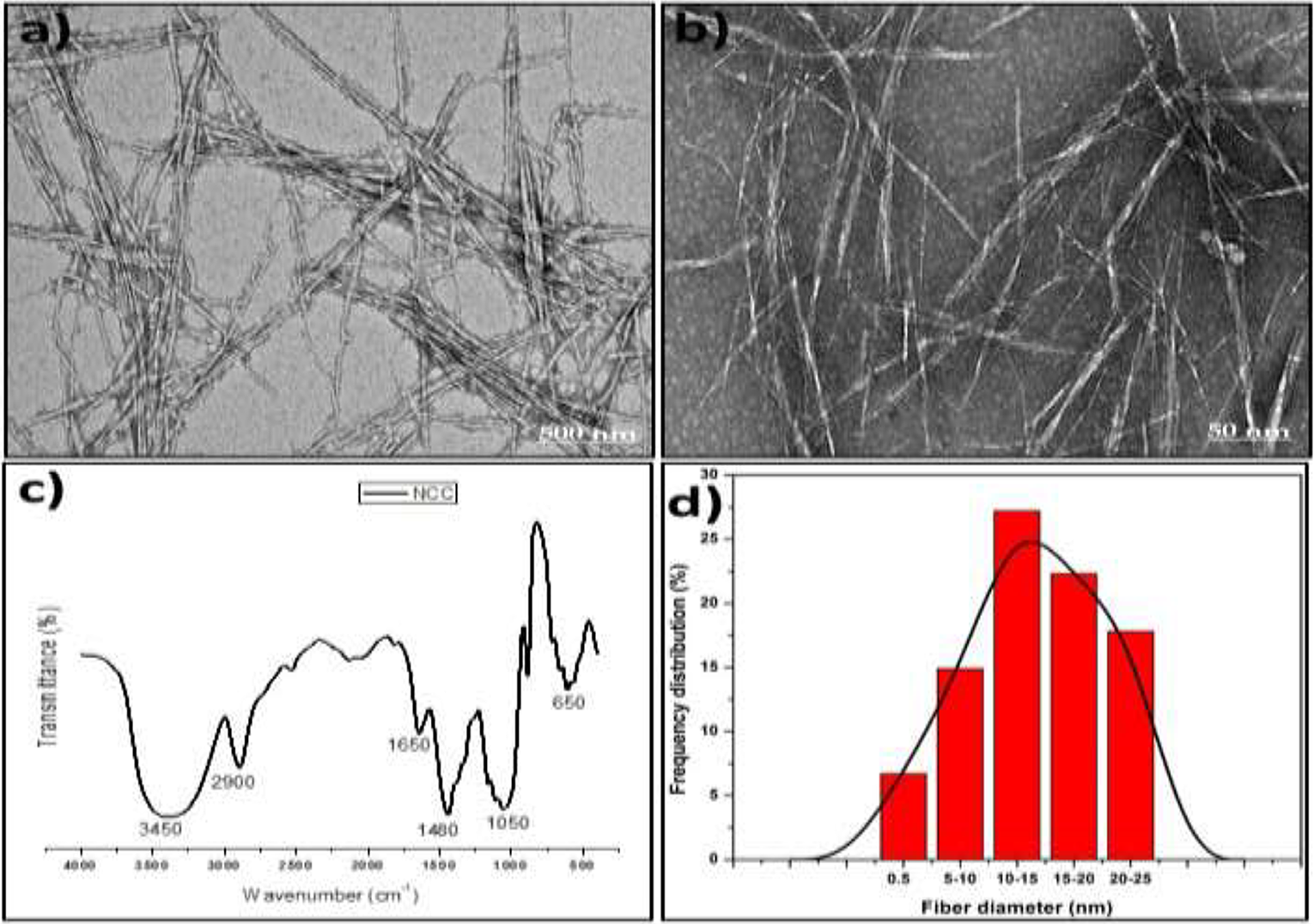
Transmission electron microscope (TEM), FT-IR and fibre size analysis of isolated NCC from OPW fibre.
Physical properties and percentage mass loss
Figure 2 showed the density and percentage of mass-loss properties of PVA-NCC composite films. Generally, the density of PVA-NCC composite films significantly increased with the addition of NCC loading. A slight close density value was observed with some samples due to the differences in the film’s thickness, which affected the section’s volume in the density calculation. Furthermore, the percentage mass loss for PVA-NCC1%, PVA-NCC3%, PVA-NCC5% PVA-NCC7%, and PVA-NCC9% were 17.65%, 13.46%, 8.00%, 7.69%, and 5.55%, respectively. The average percentage of mass loss after exposure for 12 days for all PVA-NCC composite films was about 11.88%, and the percentage of the value reduces with the addition of NCC. The composite density is expected to increase with NCC’s addition due to a reduction in the distance between molecules, which results in increased mass content. 36 Also, the reduction in percentage mass loss with NCC’s addition is probably due to interaction (chemical bonding and physical dispersion) between PVA and NCC. The internal bonding between PVA and NCC is probably from a chemical or physical bond (dispersion/miscibility effect). 37 Previous studies explained that the bonding between PVA and NCC is a chemical interaction between PVA and NCC and good dispersion between the PVA matrix and NCC filler. 21
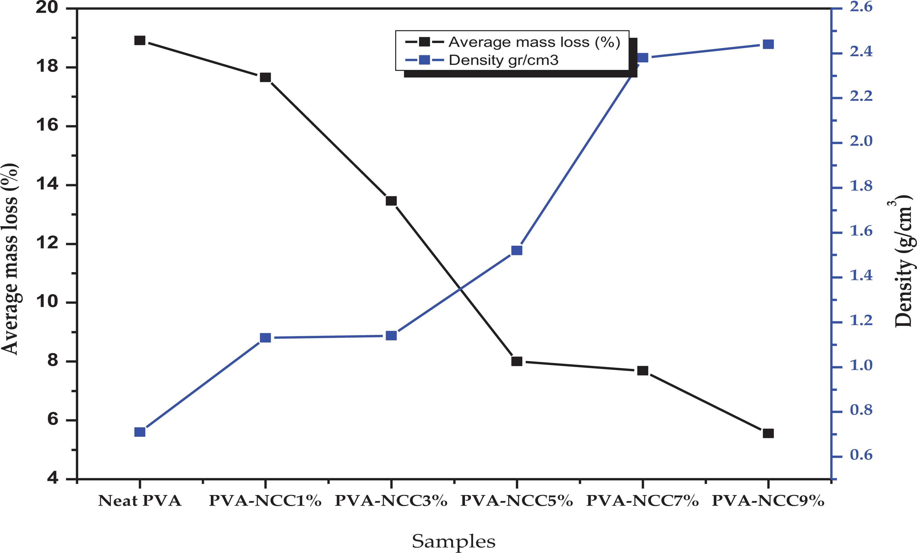
Percentage average mass loss after 12 days of exposure time and density before UV exposure of neat PVA and PVA-NCC composite films.
Figure 3 showed the graph of each sample’s weight degradation value (i.e., Actual weight value with time) of neat PVA and PVA-NCC composite films with the exposure time. The composite films’ weight loss (mg) with the duration of UV light exposure could indicate the degradation process. 38 The weight of PVA-NCC composite films and neat PVA were measured at a time interval of 2 days until 12 days. The weight measurement was recorded at 2 days intervals in milligrams to obtain the degradation. The weight at day ‘0’ is the sample’s initial weight before exposure to UV light, and the sample weight was measured after 2, 4, 6, 8, 10, 12 days. Each of the graphs showed degradation steps, which is probably due to the UV light’s degradation at different locations on the samples’ surface. The weight degradation values of all composite films were very significant with the exposure time. The mass change data indicated that the UV light of the PVA-NCC composite films led to bond disruption and produced volatile products, such as water, from breaking the hydrogen bond. 39 Furthermore, in correlation with the percentage average mass loss (Figure 3), the mass change reduced with NCC addition as more bond is formed between PVA and NCC, which require more energy to break. The neat PVA is seen with a high-value mass loss because lesser bond energy is required for UV degradation. Therefore, it can be inferred that NCC’s addition to the PVA results in a change in the composite’s thermal properties. A similar result on PVA’s thermal properties by NCC has been reported in previous studies. 21 Their study reported a lower onset temperature because of water content; however, the degradation temperature of PVA-NCC was enhanced more than the neat PVA.
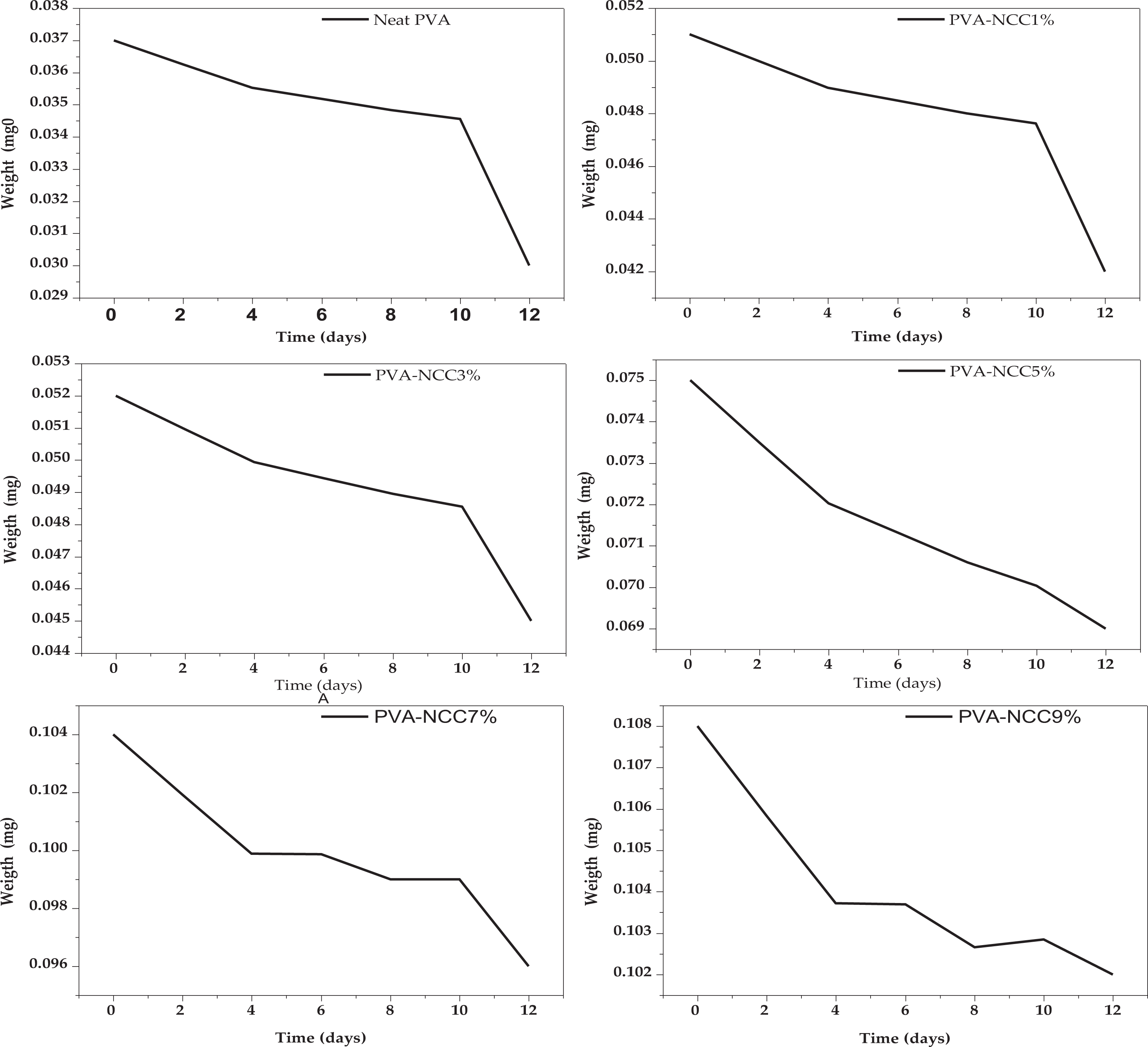
Weight degradation rate (days) of neat PVA and PVA-NCC composite films under UV light exposure.
The optical colour changes of the PVA-NCC composite films and neat PVA before and after UV light are shown in Table 1. Table 1 showed the change in PVA-NCC composite films’ appearance and neat PVA between a time interval of 2 to 12 days. The composite films were observed to roll upon exposure to the UV light due to the heat generated. The films changed colour with increased exposure time, and the colour changes of PVA-NCC was more significant between 10 and 12 days. The colour change was also observed to increase with NCC loading at PVA-NCC3%, PVA-NCC5%, PVA-NCC7%, and PVA-NCC9%.
Optical colour analysis of neat PVA and PVA-NCC composite films after UV light exposure.
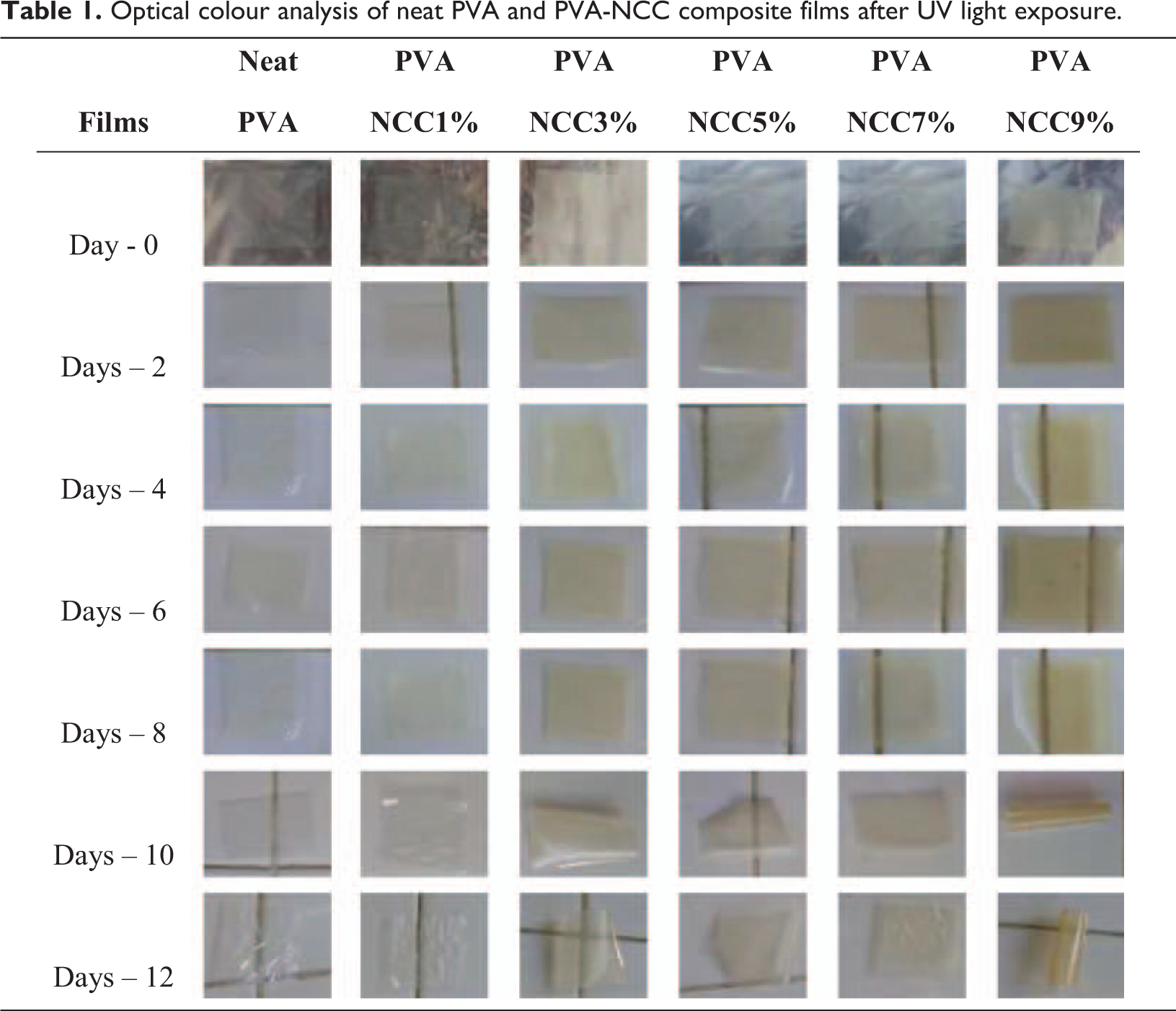
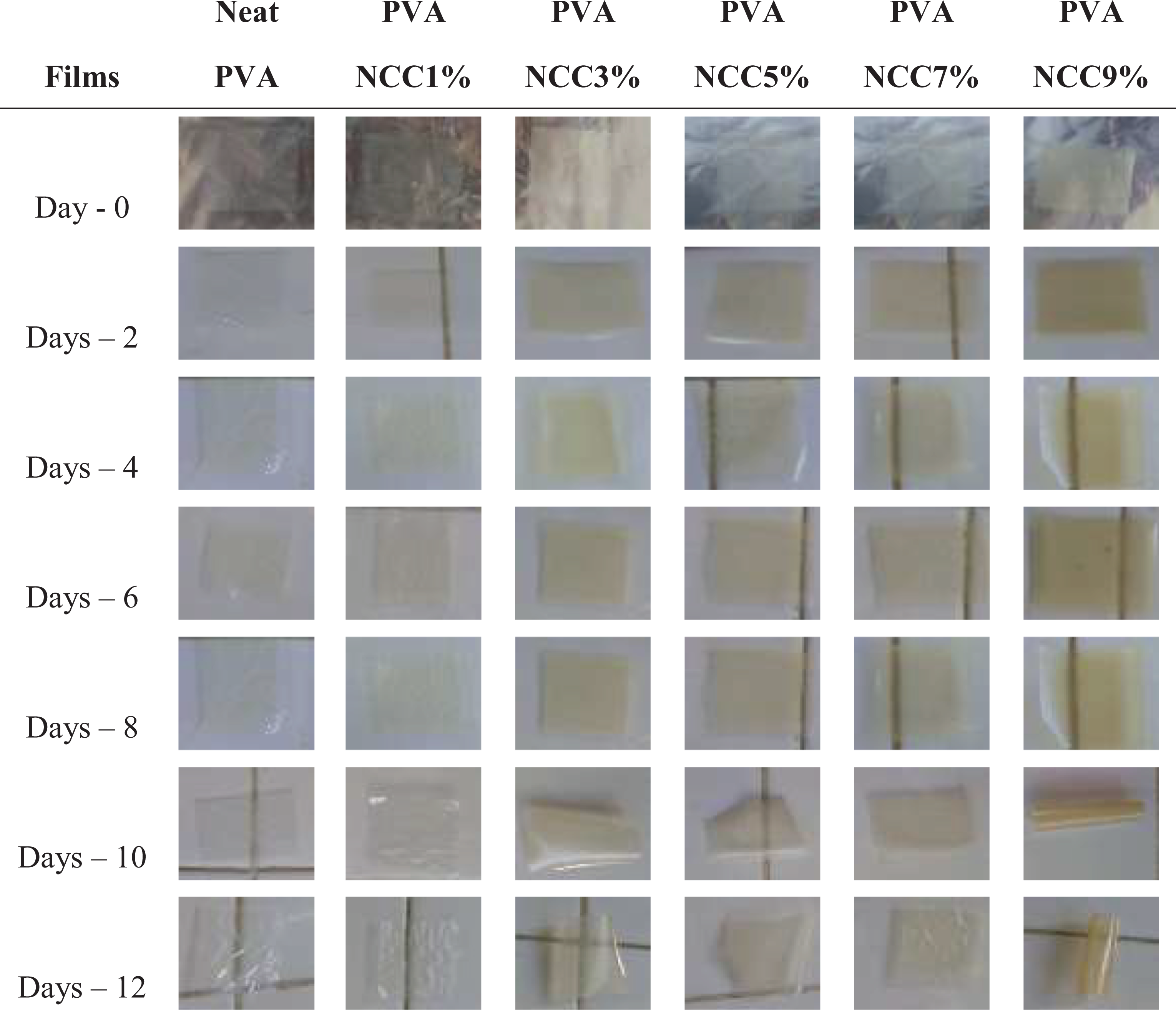
On the other hand, neat PVA and PVA-NCC1% did not significantly change in colour after 12 days of UV light exposure. The neat PVA and PVA-NCC1% colour did not change; rather, instead of the composite with higher NCC, reinforcement became more yellow. This showed that the colour change was because of heat on the NCC loaded in the PVA matrix.
Furthermore, it was also observed that there was a significant weight loss in the early days of exposure, making the PVA-NCC lose its flexibility. Therefore, it was reasonable to presume that the films’ surface was changed after accelerated ageing due to UV light. 40 The colour change was presumably a result of the surface oxidation of the composite films. A similar result by Chandrappa et al. 41 explained that the neat PVA film had very low absorption in the UV region (200–400 nm), while the PVA-NCC composite films showed sharp absorption in the UV region.
FT-IR analysis of PVA-NCC composite films
The FT-IR analysis was performed to observe composite film degradation and chemical structural changes. Figure 4 showed the band’s evaluation from FT-IR spectra on the degradation of the neat PVA and PVA-NCC composite films before and after UV light exposure. The spectra of the neat PVA showed the same results as the PVA-NCC composite films.
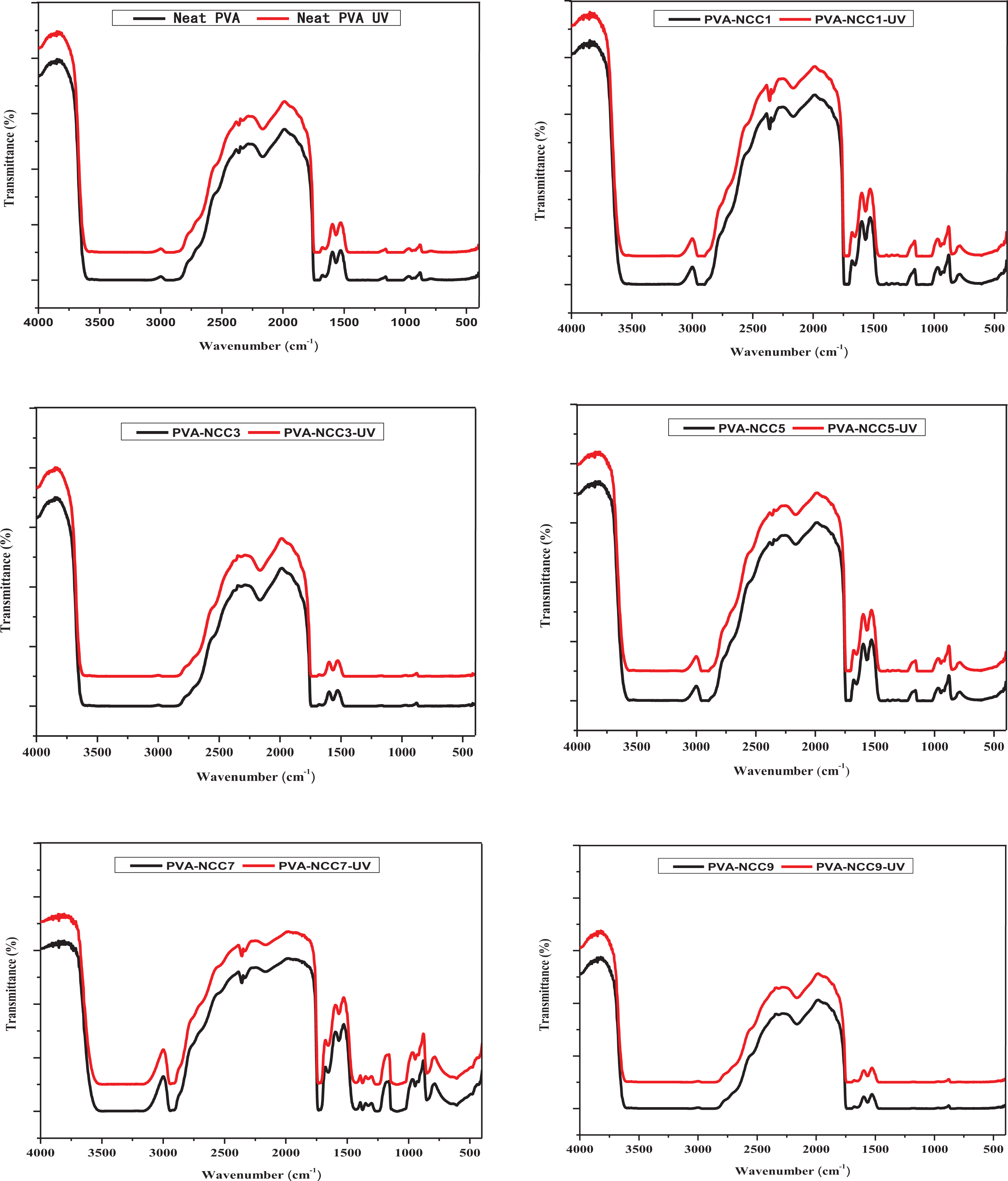
FT-IR analysis of neat PVA and PVA-NCC composite films before and after UV light exposure.
The FT-IR results showed bands at 3350 cm−1, which indicated a stretching vibration for O-H groups in intermolecular hydrogen bonds. 42 These spectra confirmed the presence of NCC is in the composite films. 20 The band at 2933 cm−1 indicated the stretching vibration of C–H, which showed methyl groups’ presence. The band around 1720 cm−1 was due to the O=C–O bonds due to the ester group’s formation between NCC and PVA. In the band of 1430 cm−1, it was observed that the symmetric bending mode of the CH2 group was present in the film. Also, the C–O group deformation was observed with the band at 1330 and 1040 cm−1. The bands at 928 cm−1 and 820 cm−1 indicated a syndiotactic structure for CH2 and stretching vibration of the C–C groups.18,43 The OH stretching spectra in the NCC was observed to be broad and strong. This indicates the formation of intermolecular hydrogen bonds between cellulose molecular chains and PVA. 44 Previous studies have established the formation of a hydrogen bond between nanocellulose and PVA. 45 The 1095 cm−1 band was sensitive to hydrogen bonding in PVA. A shift was observed in the spectra due to loading NCC. The plane aromatic rings C–H at the 873 cm−1 bands indicated that aromatisation occurred, and the C–H deformation of benzene rings was identified in a band at 870 cm−1. 30
According to the FT-IR spectra, the difference between the before and after UV light showed a similar functional group. This showed that no new bond was formed with exposure to UV light. Regarding the FT-IR result with increased NCC percentage, it is also expected that the functional group should be similar for all the composite except the neat PVA. However, the intensity (y-axis) difference may be due to the increase in the NCC percentage in the polymer mix. The change of spectra in the FT-IR analysis (Figure 5) indicates a polar group’s development in the polymer composites. The spectra of the neat PVA and PVA-NCC composite films were almost unchanged during ultraviolet light exposure. However, the intensity of spectra slightly increased after exposure time. The difference in intensity of neat PVA and PVA-NCC composite films indicated NCC’s presence on the weathered surface. Ching et al. 22 and Kord et al. 46 stated that nanocellulose in the PVA became a barrier for UV light penetration. The change of intensity composite films was due to absorption caused by new defects generated within PVA by UV light. This increased the light scattering and resulted in UV-induced imperfection in the PVA matrix. 36 Commonly, UV light facilitates the degradation of polymer molecules. 47
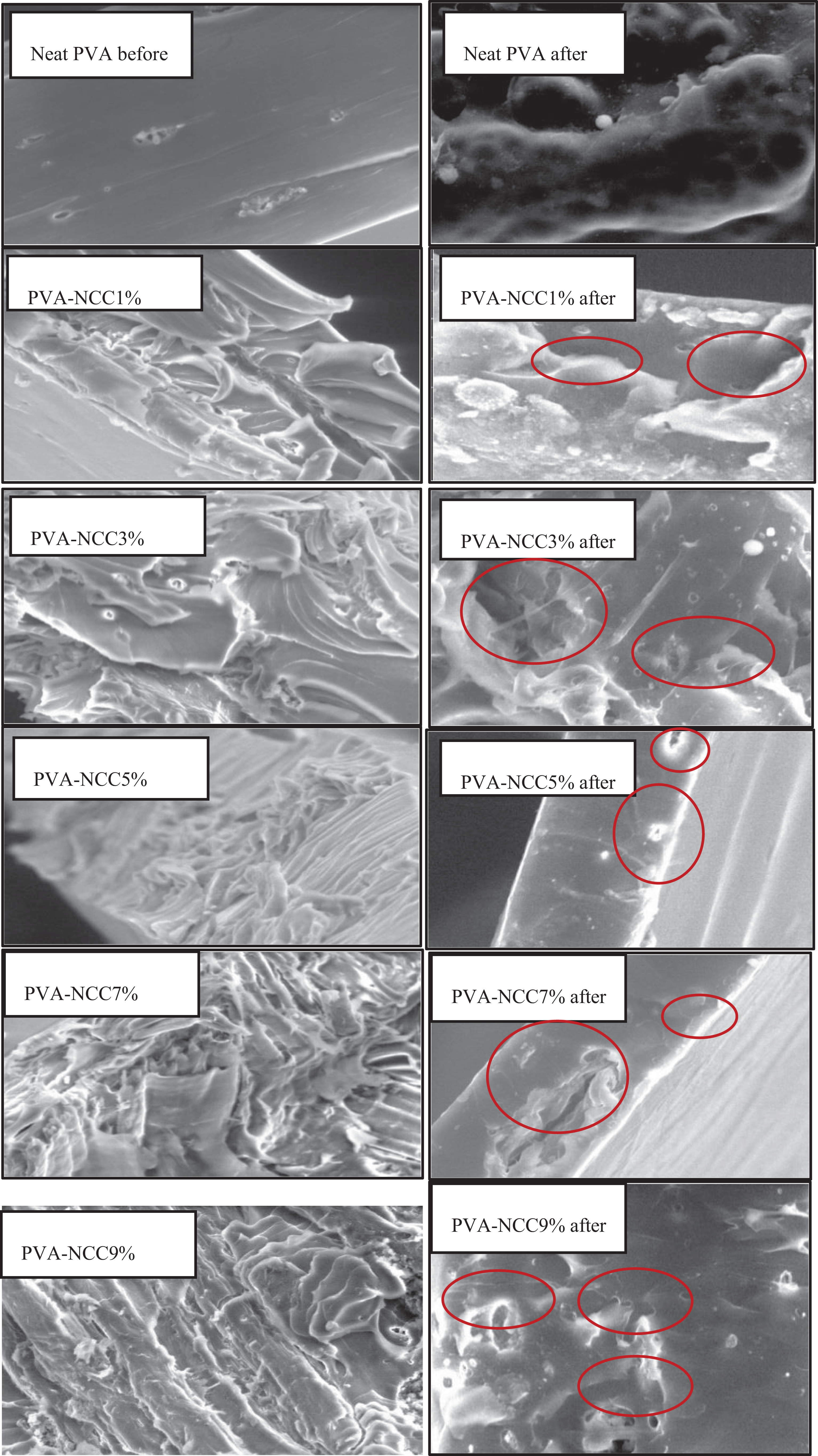
SEM analysis of PVA-NCC composite films before and after UV light exposure.
Tensile and morphological properties of PVA-NCC composite films
The tensile properties of neat PVA and PVA-NCC composite films before and after exposure to UV light were presented in Table 2. The result showed a significant increase in the tensile properties with the addition of NCC. The highest tensile properties were observed with PVA-NCC5%. A similar result was observed in the tensile modulus result with the highest modulus of 210.04 MPA at PVA-NCC5%. However, the elongation of the film reduced with the addition of NCC. Generally, NCC’s addition is seen to reduce the percentage elongation of the composite, probably due to the increased brittleness of the film with NCC’s addition. This was expected because of the crystalline nature of NCC. The increase in the tensile strength and modulus results from the NCC’s filler effect and a similar report has been documented by Wei et al. 21 and Pereira et al. 37
Tensile properties of neat PVA and PVA-NCC composite films before UV light without UV light exposure.
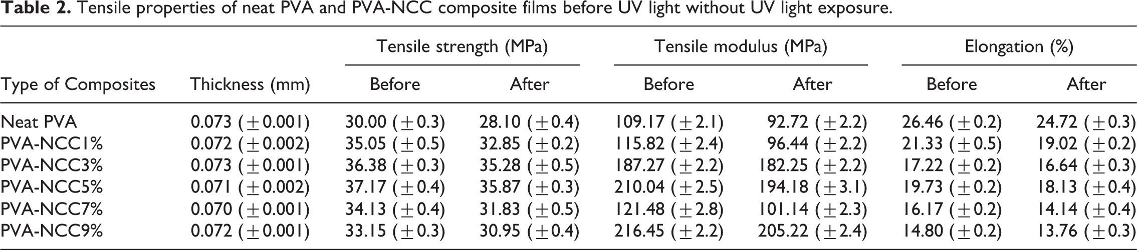
Furthermore, the reduction in the elongation value is probably due to the increased bonding from the filler, which reduced the composite film’s stretching ability. A similar reduction in the percentage of elongation has been reported by ching et al. 22 Furthermore, the reduction in the neat PVA and PVA-NCC composite film’s tensile properties showed UV light degradation on the material. The tensile result corroborated the previous observation reported in the visual analysis and the average percentage mass loss of the material.
The SEM analysis of the fractured tensile surface of neat PVA and PVA-NCC composite films before and after UV light exposure are shown in Figure 5. When comparing neat PVA and PVA-NCC composite films with different NCC loadings, it was revealed that PVA-NCC composite films had fractured surfaces with an appreciable amount of phase-separated reinforcements that mostly appear in the aggregate forms. Although the surface of the neat PVA was still smooth after UV light exposure, it was shown that the light did not impact the neat PVA because of the lack of NCC interaction. On the other hand, the PVA-NCC composite film showed a significant impact of the UV light on the material’s morphology, as shown in the SEM images. The chalking phenomenon took place after UV light exposure. The SEM images showed cavities and a few cracks on the composite film surface after UV light exposure. Also, PVA-NCC5, PVA-NCC7, and PVA-NCC9 before exposure showed hole-like layers due to the NCC’s uneven miscibility and the PVA matrix. Cavities formed after exposure time are indicated in Figure 5 in the red circles. The cavities may have been induced by the escape of volatile products from the PVA matrix. Beneddetto et al. 30 stated that NCC morphology could help mechanical and chemical anchoring adhesion between NCC and PVA by increasing the effective surface area of interfibrillar regions. Structural changes resulted from an active photochemical process during the exposure time.
Figure 5 showed the extent of textural degradation in the cross-section (fracture surface) of PVA-NCC composite films after UV light exposure. The neat PVA film’s fractured surface was still smooth after UV light exposure, whereas many small uniform cavities were observed in the PVA-NCC composite films. These indicated exposure time on the composite films, which resulted in NCC’s degradation in the PVA matrix. In other words, the adhesion between the NCC and PVA matrix became weak with time. Several articles have stated that the degradation caused by UV light only occurred on the surface of the composite film and hardly occurred inside the composite. The cavities became very large, with increased NCC loading and UV light exposure time. 48 Furthermore, PVA-NCC composite films’ surface became rough, and cavity sizes increased after 12 days of UV light exposure. 48
Conclusions
PVA-NCC was successfully produced and exposed to UV light for photodegradation enhancement. The film’s UV stability was enhanced with the addition of NCC compared with the neat PVA. This study has established that nanocrystalline cellulose (NCC) reinforcement is for strength enhancement and UV stability resistance. The UV light was observed to cause changes in the neat PVA and PVA-NCC composite films’ physical and morphological properties. The degradation of PVA-based NCC was observed in the accelerated ageing, weight loss percentage, and yellowing of the exposed surfaces.
Furthermore, the FTIR characterisation showed that the composite films before and after exposure are similar, indicating no new functional groups were added due to light exposure. That is, there were no new chemical reactions between NCC and PVA with the exposure. However, when the exposure time increased, cavity size and surface cracks increased. This is the physical indication that degradation occurs in the composite film.
Footnotes
Acknowledgements
The authors would like to acknowledge the financial support received by the Research Institute and Community Service of Universitas Syiah Kuala, Ministry of Research, Technology, and Higher Education in accordance with the Research Contract No. 21/UN11.2/PD/SP3/2019.
Author contributions
Conceptualisation, NA Sri Aprilia and Abdul Khalil HPS; Data curation, NG Olaiya, Suraiya Kamaruzaman and Fitriani Fitriani; Formal analysis, Khairul Rahmah; Funding acquisition, Samsul Rizal; Methodology, Zuhra Zuhra; Project administration, Abdul Khalil HPS; Resources, Suraiya Kamaruzaman, Zuhra Zuhra and Samsul Rizal; Supervision, Abdul Khalil HPS; Validation, NG Olaiya; Visualization, CK Abdullah; Writing – review & editing, NG Olaiya and CK Abdullah.
Declaration of conflicting interests
The author(s) declared no potential conflicts of interest with respect to the research, authorship, and/or publication of this article.
Funding
The author(s) disclosed receipt of the following financial support for the research, authorship, and/or publication of this article: This research was funded by the Universiti Sains Malaysia research fund RUI 1001/PTEKIND 8014119 and Research Institute & Community Service of Universitas Syiah Kuala, Ministry of Research, Technology, and Higher Education in accordance with the Research Contract No. 21/UN11.2/PD/SP3/2019.