
Abstract
Purpose:
To investigate the relationship between postprocedure intravascular ultrasound (IVUS) findings and restenosis after placement of drug-eluting stents (DES) for femoropopliteal lesions.
Methods:
Between July 2012 and May 2013, DES were placed in 64 patients with 88 de novo femoropopliteal lesions. In 40 patients (mean age 74.2±9.4 years; 27 men), DES were placed in 50 lesions under IVUS guidance, and restenosis was monitored for 1 year. All patients were symptomatic (Rutherford 2–6), and 17 patients (43%) suffered from critical limb ischemia. IVUS findings after stenting were compared for patients with vs without restenosis, which was defined as a peak systolic velocity ratio >2.4 on duplex ultrasonography or >50% diameter stenosis on angiography.
Results:
Ten patients (14 lesions) developed restenosis, while 30 patients (36 lesions) did not. There were no significant differences in the frequency of diabetes or dialysis between the 2 groups. Female patients were predominant in the restenosis group (p<0.003). There were no significant differences of the percentage of TransAtlantic Inter-Society Consensus C/D lesions or stent edge dissection. Multivariate analysis indicated that cilostazol use [odds ratio (OR) 0.13; p=0.046], distal lumen cross-sectional area (CSA) (OR 0.86; p=0.035), and axial symmetry index (OR 0.60; p=0.045) were independent predictors of restenosis. Using receiver operator characteristic analysis, the best cutoff values of the distal lumen CSA and axial symmetry index for predicting restenosis were 17.1 cm2 and 0.6, respectively.
Conclusion:
IVUS guidance of DES placement in femoropopliteal lesions can offer useful predictors of restenosis at 1 year. The utility of distal lumen CSA and the axial symmetry index in the prediction of restenosis after femoropopliteal DES placement should be confirmed in a larger cohort.
Keywords
Introduction
Endovascular treatment (EVT) is minimally invasive and safe, so it is regarded as first-line therapy for femoropopliteal lesions.1,2 The primary patency rate at 12 months after placement of a self-expanding nitinol stent was reported to be 63% to 81% vs 37% to 61% at 12 months after balloon angioplasty,3–5 suggesting that nitinol stent placement is superior to balloon angioplasty. However, the primary patency rate at 12 months after autologous vein bypass grafting is 81% to 88%,6–9 so the outcome after stent placement is still relatively unsatisfactory, owed largely to in-stent restenosis (ISR).
Drug-eluting stents (DES) were developed to lower the incidence of ISR in the coronary arteries, 10 and a paclitaxel-eluting stent has also been developed for the femoral and popliteal arteries. The primary patency rate at 12 months after placement of a paclitaxel-eluting stent for femoropopliteal stenosis was reported to be 70% to 90%,11–14 which is comparable with rates for bare metal stents (BMS) reported recently (80% to 88%).15–17 Although DES are superior to BMS for preventing ISR in the coronary arteries (DES were reported to reduce target vessel revascularization at 12 months by 52% to 74% 18 ), the paclitaxel-eluting stent does not seem to have such a strong preventive effect on restenosis in the femoropopliteal segment.
Intravascular ultrasound (IVUS) is widely used during coronary stenting. Witzenbichler et al 19 reported that stent placement under IVUS guidance improved the 12-month outcome by preventing in-stent thrombosis, myocardial infarction, and target lesion/vessel revascularization. It was also reported that the minimum stent area after placement was related to the occurrence of in-stent restenosis.20,21 Cheneau et al 22 concluded that subacute in-stent thrombosis was caused by the combination of an inadequate postprocedure lumen dimension (final lumen <80% reference lumen area), dissection, thrombus, and tissue protrusion. Moreover, the outcome of coronary stenting was reported to be related to postprocedure IVUS findings.
In contrast, there have only been a few reports about the usefulness of IVUS in the lower limb arteries. Iida et al 23 reported that IVUS guidance improved long-term primary patency after placement of BMS for femoropopliteal lesions. In addition, Miki et al 24 stated that postprocedure IVUS findings had an influence on target lesion revascularization (TLR) after BMS placement for femoropopliteal disease. However, the role of IVUS in patients receiving DES for femoropopliteal lesions has not been assessed. Therefore, the present study was conducted to investigate the relationship between postprocedure IVUS findings and restenosis at 1 year after placement of paclitaxel-eluting stents in femoropopliteal lesions.
Methods
Patients
This was a retrospective, single-center, nonrandomized study. Between July 2012 and May 2013, 89 patients underwent stenting of de novo femoropopliteal lesions at our hospital (Figure 1), excluding those with acute/subacute lower limb ischemia or inflow aortoiliac lesions. DES were placed in 64 patients, among whom 40 patients (mean age 74.2±9.4 years; 27 men) were treated under IVUS guidance for 50 lesions and followed for 12 months after stenting. Baseline characteristics of the two groups are shown in Table 1. All patients were on exercise and drug therapy and had symptoms corresponding to categories 2 to 6 of the Rutherford classification. 1 Baseline lesion characteristics are shown in Table 2. When angiography revealed at least 50% diameter stenosis of the femoropopliteal arteries, vascular specialists (including vascular surgeons and interventional radiologists) decided whether or not EVT was applicable. The protocol (number 2014056) was approved by the ethics committee of our hospital, and all patients gave informed consent.
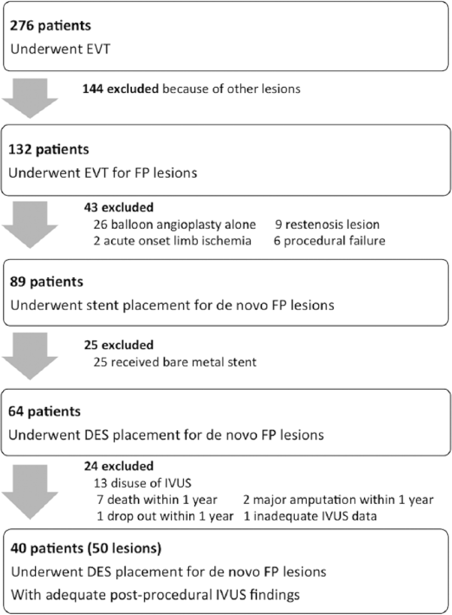
Study flowchart. DES, drug-eluting stent; EVT, endovascular therapy; FP, femoropopliteal; IVUS, intravascular ultrasound.
Baseline Patient Characteristics. a
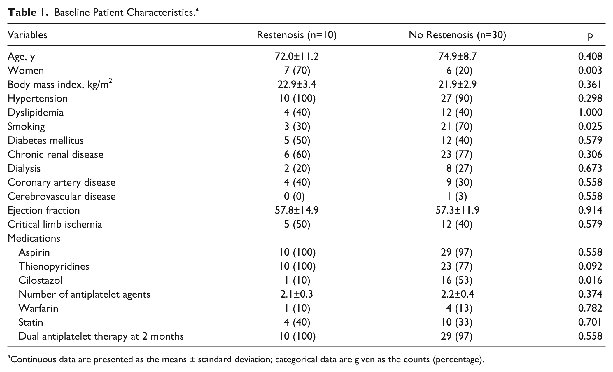
Continuous data are presented as the means ± standard deviation; categorical data are given as the counts (percentage).
Lesion and Procedure Characteristics. a
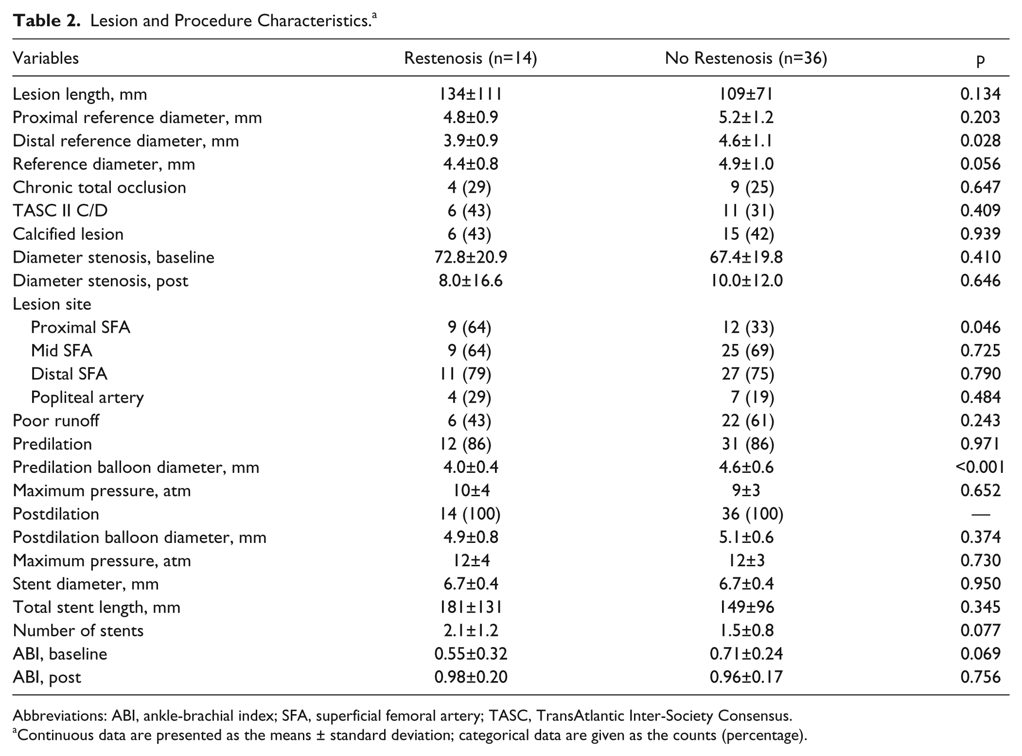
Abbreviations: ABI, ankle-brachial index; SFA, superficial femoral artery; TASC, TransAtlantic Inter-Society Consensus.
Continuous data are presented as the means ± standard deviation; categorical data are given as the counts (percentage).
Interventions
Dual antiplatelet therapy (DAPT) with aspirin (100 mg/d) plus clopidogrel (75 mg/d), ticlopidine (200 mg/d), or cilostazol (200 mg/d) was started at least 3 days before stenting. The crossover approach was used for EVT. A 6-F crossover sheath was inserted and unfractionated heparin (5000 units) was injected intra-arterially. A 0.014-inch guidewire was passed through the target lesion, and the operator decided whether predilation should be performed. A paclitaxel-eluting stent (Zilver PTX; Cook Medical, Bloomington, IN, USA) with a diameter 1 to 2 mm larger than the reference vessel diameter proximal to the target lesion was selected and deployed so that it fully covered the lesion, terminating in angiographically normal segments. When two or more stents were used for a long lesion, the overlap was ≤10 mm. The lowest point of the popliteal artery that was stented was marked by the proximal edge of the patella. All lesions were routinely postdilated. At the end of the stenting procedure, IVUS images were recorded with a commercially available IVUS console (s5 Imaging System; Volcano, Rancho Cordova, CA, USA) and a phased-array 20-MHz IVUS catheter (Eagle Eye Gold; Volcano) using manual pullback through the stented segment at a uniform speed. DAPT was continued for at least 2 months after the procedure.
Intravascular Ultrasound Analysis
Two experienced observers who were unaware of the clinical and angiographic findings analyzed the IVUS scans. The IVUS parameters measured or calculated were the maximum and minimum cross-sectional area (CSA) of the stent, proximal and distal CSA of the external elastic membrane (EEM), lumen CSA, plaque burden, reference lumen CSA, reference diameter, stent/reference diameter ratio, stent expansion ratio, radial stent symmetry index, and axial stent symmetry index (Table 3).24,25 The proximal and distal reference segments selected for analysis were the most normal-looking cross sections within 10 mm of the proximal and distal margins of the stent before the origin of any large side branches. 22 Stent edge dissection, tissue protrusion, and stent malapposition were also investigated.
Definitions of Intravascular Ultrasound Measurements.
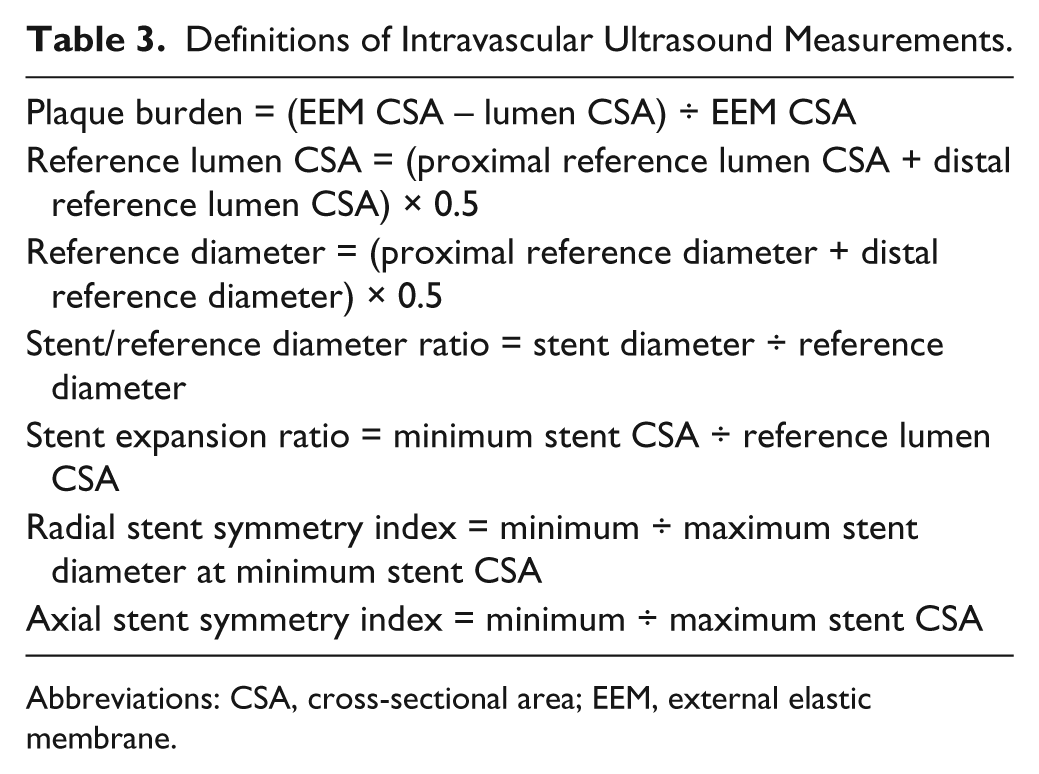
Abbreviations: CSA, cross-sectional area; EEM, external elastic membrane.
Definitions
Definitions of hypertension, dyslipidemia, diabetes mellitus, chronic kidney disease, dialysis, smoking, coronary artery disease, and cerebrovascular disease followed those previously reported. 24 Below-the-knee (BTK) runoff was assessed by angiography after stent placement. Poor runoff was defined as one or no BTK vessel runoff. 26 Stenting was classified as successful if angiography demonstrated that residual stenosis was ≤30% without flow-limiting dissection. 24 Restenosis was defined as a peak systolic velocity ratio >2.4 on duplex ultrasonography 27 or >50% diameter stenosis or occlusion on quantitative vascular angiography.
Stent edge dissection meant dissection at a stent edge (<5 mm proximal or distal to the stent margin) and was classified as intimal or medial dissection. Intimal dissection was defined as a disruption of the lumen surface that extended within the intimal plaque, while medial dissection was a disruption extending to the internal elastic membrane.24,25 Tissue protrusion referred to tissue between the stent struts extending inside a circular arc connecting adjacent struts on IVUS images. Tissue protrusion area was calculated as stent CSA minus lumen CSA at the segment with maximum protrusion. 24 Stent malapposition was defined as lack of contact between stent struts and the arterial wall. 22
Clinical Follow-up
Postprocedure evaluation of symptoms, measurement of the ankle-brachial index (ABI), and duplex ultrasonography were performed at 1, 3, 6, 9, and 12 months after stenting. When duplex ultrasonography suggested restenosis, angiography was performed for confirmation.
Statistical Analysis
Continuous variables with normal distributions are presented as mean ± standard deviation, and variables without normal distribution are presented as median and interquartile range (IQR). Categorical data are presented as frequencies. Continuous variables were compared by using the unpaired Student t test or the Mann-Whitney U test. Categorical data were compared with the chi-square test or Fisher exact test.
Univariate and multivariate logistic regression analysis were performed to determine predictors of restenosis; outcomes are reported as the odds ratio (OR) with 95% confidence intervals (CIs). Variables that achieved p<0.05 on univariate analysis were entered into the multivariate regression models.
Receiver operating characteristic (ROC) analysis was performed to determine the optimal cutoff value of distal lumen CSA and axial symmetry index for predicting restenosis at 12 months after stent placement, with each cutoff point being selected to give the highest value for the sum of sensitivity and specificity. The area under the ROC curve (AUC) was used as a measure of the accuracy of each parameter. In all analyses, p<0.05 was considered statistically significant. Statistical analysis was performed using JMP software (version 10.0.0; SAS Institute, Cary, NC, USA).
Results
In the present study, 50 lesions in 40 patients were investigated. Restenosis was detected in 14 lesions (28%, 10 patients) at 12 months after stenting. There was no significant difference of age between the restenosis group and the no restenosis group (Table 1). The percentage of female patients was significantly higher in the restenosis group (70% vs 20%, p=0.003), but diabetes mellitus, dialysis, and critical limb ischemia were comparable between the groups. While treatment with cilostazol was significantly less frequent in the restenosis group (10% vs 53%, p=0.016), there were no significant differences in the number of antiplatelet drugs or the percentage of patients receiving DAPT at 2 months after stenting. At 12 months, 35 (88%) patients continued DAPT and 5 (13%) patients took only aspirin.
There were no significant differences between patients with or without restenosis regarding the incidence of chronic total occlusion (CTO) or TASC II C/D lesions (Table 2). There was also no significant difference in lesion length (134±111 vs 109±71 mm, p=0.134), but the distal reference diameter was significantly smaller in the restenosis group (3.9±1.0 vs 4.6±1.1 mm, p=0.028). Minimum and maximum reference vessel diameters were 3.0 and 7.9 mm, respectively. Furthermore, there were no significant differences in the percentage of patients having poor runoff or predilation, the mean stent diameter, or the total stent length.
Intravascular Ultrasound Findings
Postprocedure IVUS findings are detailed in Table 4. The maximum stent CSA showed no significant difference between the 2 groups; the minimum stent CSA tended to be smaller in the restenosis group but was not statistically different. There were no significant differences of the proximal EEM CSA or proximal lumen CSA, but the distal EEM CSA and distal lumen CSA were significantly smaller in the restenosis group [26.7±7.7 vs 33.9±9.3 mm2 (p=0.012) and 14.1±5.0 vs 20.3±6.1 mm2 (p<0.001), respectively]. Both the radial stent symmetry index and axial stent symmetry index showed significant differences between the two groups [0.85±0.04 vs 0.80±0.09, (p=0.040) and 0.55±0.13 vs 0.69±0.15 (p=0.003), respectively]. In addition, the stent/reference diameter ratio was significantly larger in the restenosis group (1.49±0.20 vs 1.33±0.19, p=0.009), but there was no significant difference of the stent expansion ratio. While there was no significant difference in the frequency of stent edge dissection, tissue protrusion was significantly more common in the restenosis group (21% vs. 3%, p=0.029).
Postprocedure Intravascular Ultrasound Findings.
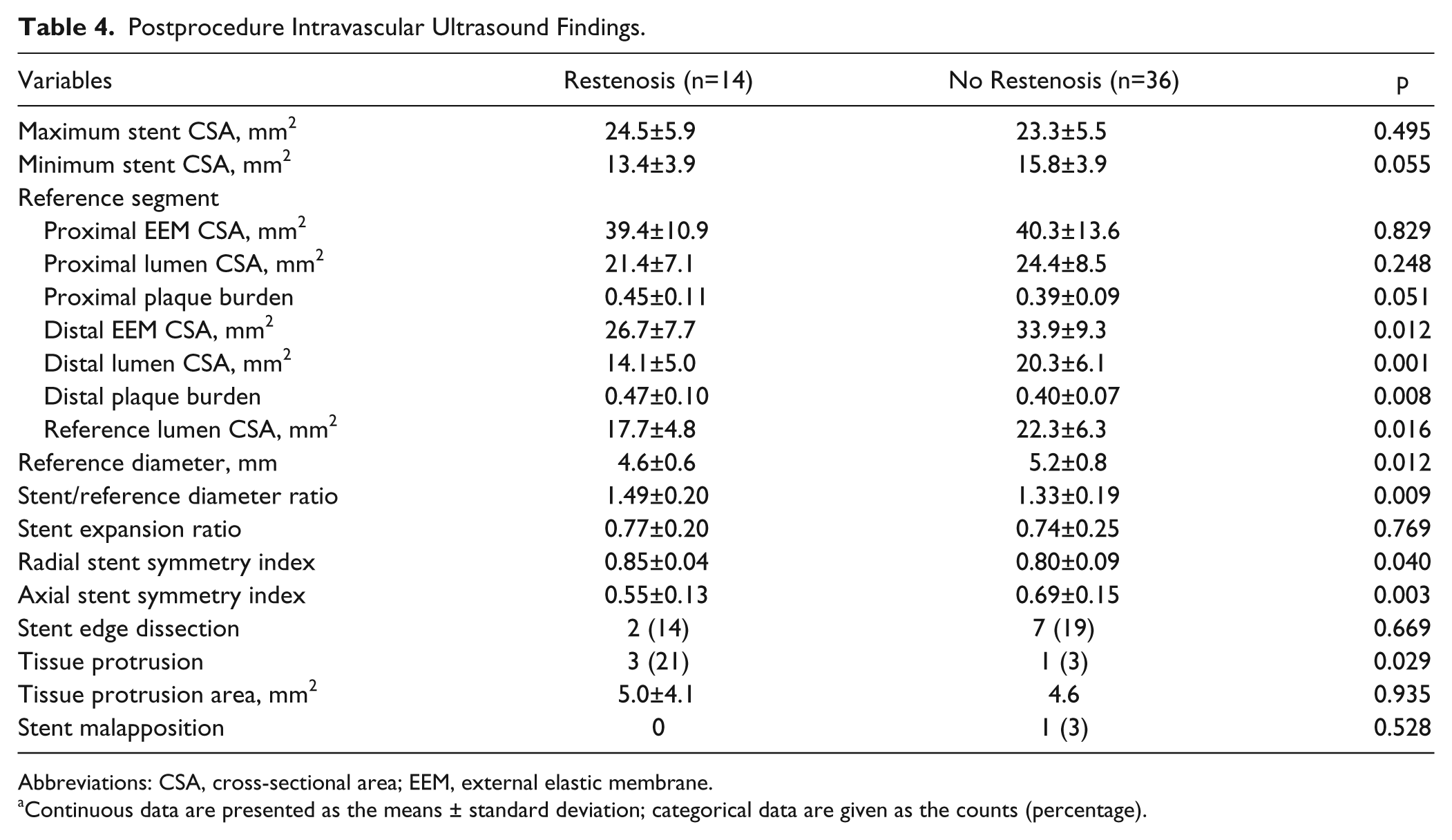
Abbreviations: CSA, cross-sectional area; EEM, external elastic membrane.
Continuous data are presented as the means ± standard deviation; categorical data are given as the counts (percentage).
Regression Analyses
Cilostazol, distal lumen CSA, axial symmetry index, and tissue protrusion were entered into the multivariate model. Female sex, distal EEM CSA, distal plaque burden, stent/reference diameter, reference diameter, reference lumen CSA were not entered into the logistic regression model because they were associated with distal lumen CSA. On multivariate analysis (Table 5), cilostazol use (OR 0.13, 95% CI 0.01 to 0.97, p=0.046), distal lumen CSA (OR 0.86, 95% CI 0.73 to 0.99, p=0.035), and axial symmetry index (OR 0.60, 95% CI 0.32 to 0.99, p=0.045) were independent predictors of restenosis. Tissue protrusion was not an independent predictor of restenosis.
Logistic Regression Analysis of Restenosis.
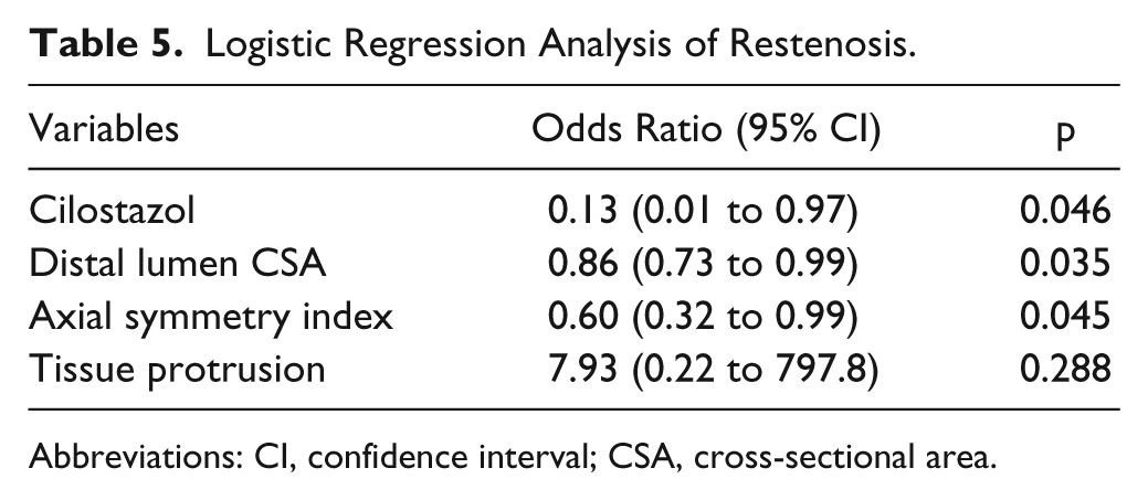
Abbreviations: CI, confidence interval; CSA, cross-sectional area.
Receiver Operating Characteristic Analysis
The outcome of the ROC analysis of the distal lumen CSA and the axial symmetry index is displayed in Figure 2. The AUC for the distal lumen CSA was 0.783 and the best cutoff value of distal lumen CSA for predicting restenosis within 12 months after stenting was 17.1 mm2, which had a sensitivity of 78.6% and a specificity of 72.2%. In the present series, the distal lumen CSA was <17.1 mm2 in 40.0% of all lesions. On the other hand, the AUC for the axial symmetry index was 0.769; the best cutoff value of axial symmetry index for predicting restenosis was 0.60, which had a sensitivity of 85.7% and a specificity of 69.5%. The axial symmetry index was <0.60 in 40.0% of all lesions.
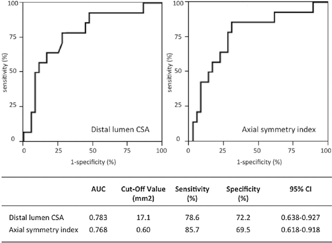
Prognostic values for distal lumen cross-sectional area and axial symmetry index in predicting restenosis within 12 months after stent placement were derived from the receiver operating characteristics curves and are shown in the table. AUC, area under the curve; CI, confidence interval; CSA, cross-sectional area.
Clinical Outcomes
The ABI values and Rutherford classification were significantly improved from before treatment to 12 months. Clinical improvement of at least one Rutherford class was achieved in 31 (78%) of 40 patients. The mean ABI value was 0.66±0.27 at baseline, 0.88±0.14 at 6 months, and 0.90±0.18 at 12 months. The median PSVR was 1.27 (IQR 0.95–1.67) at 6 months and 1.17 (IQR 0.93–1.61) at 12 months.
Discussion
Although there have been many reports regarding IVUS measurements predicting in-stent restenosis after coronary stenting,20-22,28 there have been few investigations after self-expanding stent placement in femoropopliteal lesions. In fact, as far as we could determine, there has been only one study on the relationship between clinical outcome and IVUS findings after placement of self-expanding BMS for femoropopliteal disease. 24 The present study takes this work a step further by assessing the relationship between restenosis and IVUS findings after placement of self-expanding DES for femoropopliteal lesions.
Relationships Between IVUS Variables and Restenosis
Lumen CSA
The distal EEM CSA, distal lumen CSA, reference lumen CSA, and reference diameter were all significantly smaller in the restenosis group. Miki et al 24 reported that the distal reference lumen CSA is useful for predicting TLR after placement of BMS for femoropopliteal lesions, which is consistent with the results of our study. However, Dake et al 29 reported that the angiographic reference diameter was not useful for predicting patency after placement of a Zilver PTX stent (p=0.07). This discrepancy may be attributable to the difference between measurement of the vessel diameter by IVUS and angiography. With angiography, the measured vessel diameter varies according to the calibration point and X-ray source. For example, the left circumflex artery is near the X-ray source during coronary angiography and its diameter differs the most from that measured by IVUS. 30 The same applies to femoropopliteal diameters. Because the proximal and distal ends of the superficial femoral artery are not in the same horizontal plane, the arterial diameter varies according to the calibration point. Because IVUS measures the reference vessel diameter more accurately, this diameter becomes a useful predictor of restenosis. The results of the present study suggest that the risk of restenosis after DES placement in small vessels is high. There is no clear definition of a “small” vessel, but the ROC analysis showed that 17.1 mm2 for the distal lumen CSA could predict restenosis at 12 months after stenting.
Drug-coated balloons (DCBs) are commonly used today, and good outcomes have been reported after employing DCB to treat complex, long, and restenotic lesions.11,31,32 However, the usefulness of these balloons for lesions in small vessels has not been investigated. Werk et al 33 reported that TLR was required in only 7.1% of patients at 1 year after DCB treatment for lesions of small vessels (mean reference diameter <5 mm). This report suggested that DCB may improve the outcome for small vessel lesions, but a large-scale prospective study would be needed for confirmation.
Asymmetrical Stenting
It has been reported that highly asymmetrical stent expansion (radial symmetry index <0.7) does not affect chronic neointimal tissue growth or the restenosis rate of coronary lesions. 34 In the present study, the radial symmetry index was 0.85±0.04 in the restenosis group and 0.80±0.09 in the nonrestenosis group, suggesting that the stents expanded properly in most subjects. However, the radial symmetry index was higher in the restenosis group (p=0.040). While the reasons for this difference are unknown, it seems that a low radial symmetry index is not related to restenosis.
The axial symmetry index was significantly lower in the restenosis group and was one of the predictors of restenosis within 12 months after DES placement. Two possible reasons for that can be suggested. One is that the target artery was expanded more than necessary in the restenosis group, which had a significantly smaller reference diameter, but there were no significant differences between the two groups with regard to the stent diameter, postprocedure balloon diameter or pressure, or maximum stent CSA. Han et al 35 have reported that unlike balloon-expandable stents, self-expanding stents when excessively expanded were more likely to cause vascular wall injury or reactive neointimal hyperplasia that results in restenosis. The other possibility is that restenosis tends to occur in tapering arteries. Because the distal artery can be excessively expanded by a stent, the difference between the proximal and distal vessel diameters can be too large.
Stent Edge Dissection and Tissue Protrusion
Miki et al 24 reported that stent edge dissection is a useful predictor of chronic TLR. In the present study, there was no significant difference in the incidence of stent edge dissection between the groups because the lesions (including any dissection) were fully covered by the stent after balloon expansion and medial dissection was seen only in patients with restenosis. Miki et al 24 also reported that there was no significant difference in the incidence of intimal stent edge dissection between patients with and without TLR, which suggests that medial dissection after stenting is more problematic while intimal dissection is not a predictor of restenosis. It is known that self-expanding stents are unlikely to cause edge tears. 35 It appears that problems are more likely to occur if medial dissection caused by balloon angioplasty is not fully covered by a stent. Thus, it is important to determine the extent of medial dissection by performing IVUS and fully covering the dissection with a stent in order to prevent restenosis.
In this study, tissue protrusion was significantly more frequent in the restenosis group, but it was not prognostic. Intracoronary protrusion of tissue is a risk factor for subacute stent thrombosis, 22 but no stent thrombosis occurred in the present study, likely because the femoropopliteal vessels are larger in diameter than the coronary arteries.
Cilostazol
The percentage of patients treated with cilostazol was significantly lower in the restenosis group, and cilostazol use was one of the predictors of restenosis within 12 months after DES placement. It is known that cilostazol improves blood flow (vasodilatory effect) and endothelial function, while inhibiting vascular smooth muscle cell proliferation and neointimal hyperplasia (anti-inflammatory effects). It was previously reported that cilostazol is useful after EVT for femoropopliteal lesions, but this finding was obtained in patients undergoing angioplasty or BMS placement.36,37 There have been no reports about the efficacy of cilostazol in patients treated with the Zilver PTX stent. However, the present study suggests that cilostazol may be useful in patients receiving this stent.
Limitations
The main limitations of this study are its retrospective design and a small number of patients. Regarding patient characteristics, there were significant imbalances between the groups, including gender differences and smoking rate. The proportion of women was higher in the restenosis group because women have a smaller vessel diameter than men do, on average. Additionally, in Japan, women do not smoke very much in comparison with men. For these reasons, the smoking rate was lower in the restenosis group.
Each interventionist decided whether to use DES or BMS and whether or not to perform IVUS, so there was the potential for bias. The stent/reference diameter ratio was significantly higher in the restenosis group. Soga et al 38 reported that this ratio was an independent predictor of restenosis after femoropopliteal stenting because vascular injury may be caused by vessel overdilation. Thus, the stent/reference diameter ratio might be a bias in this study.
Conclusion
Intravascular ultrasound guidance of DES placement in femoropopliteal lesions can offer useful predictors of restenosis at 1 year. The utility of distal lumen CSA and the axial symmetry index in the prediction of restenosis after femoropopliteal DES placement should be confirmed in a larger cohort.
Footnotes
Declaration of Conflicting Interests
The author(s) declared no potential conflicts of interest with respect to the research, authorship, and/or publication of this article.
Funding
The author(s) received no financial support for the research, authorship, and/or publication of this article.