






Abstract
Keywords
Introduction
Endovascular aneurysm repair (EVAR) is the treatment of choice for patients presenting an abdominal aortic aneurysm (AAA) with favorable anatomy.1–4 By contrast, EVAR needs a closer follow-up due to a higher incidence of reinterventions when compared with conventional surgey.5,6 Type I endoleak is the most significant EVAR complication as it increases aneurysm rupture risk; thus, treatment of type I endoleak is strongly recommended. 7
Chimney and/or periscope grafts (CPGs) have been shown to be valuable endovascular options to treat pararenal and thoracoabdominal aortic aneurysms (TAAAs) in selected patients, but so far there is limited knowledge on their use to treat type I endoleak. Herein we report midterm results of CPGs used to treat post-EVAR type I endoleak.
Methods
Study Cohort
Between June 2002 and April 2014, 24 consecutive patients (mean age 73.9±9.2 years; 23 men) with type I endoleak (23 proximal and 1 distal) associated with 23 pararenal AAAs and 1 TAAA, respectively, were treated with the CPG technique to maintain perfusion to the renovisceral branches. Three patients with type Ia endoleak also presented a distal type Ib endoleak. The cause of type I endoleak was related to stent-graft migration in 13 (54%) cases and neck degeneration in 11 (46%).
Seventeen (71%) patients were treated electively and 7 (29%) urgently; of the latter patients, 5 were symptomatic and 2 had an aortic rupture. All patients were considered at high risk for conventional open aortic surgery. 8 Fourteen (58%) were classified as American Society of Anesthesiologists class III/IV owing to multiple comorbidities and risk factors (Table 1). Mean preoperative estimated glomerular filtration rate (eGFR) was 54±12 mL/min/1.73 m2; 13 (54%) patients had decreased renal function, with serum creatinine levels >100 μmol/L. The mean sac diameter before the CPG treatment was 88±26 mm.
Demographics and Comorbidities in the 24-Patient Cohort. a

Abbreviations: ASA, American Society of Anesthesiologists; COPD, chronic obstructive pulmonary disease.
Continuous data are presented as the mean ± standard deviation; categorical data are given as the counts (percentage).
Mean time from the previous EVAR procedure to the CPG treatment was 52±49 months (range 0.2–179). In the initial EVAR procedure, 9 patients received an infrarenally fixed stent-graft (Excluder; W.L. Gore & Associates, Flagstaff, AZ, USA), while the remaining 15 had suprarenal fixation [6 Endurant and 2 Talent (Medtronic Vascular, Santa Rosa, CA, USA); 5 Zenith Flex (Cook Medical, Bloomington, IN, USA); 2 Vanguard (Boston Scientific, Natick, MA, USA)].
All patients underwent preoperative thoracoabdominal computed tomographic angiography (CTA), and CTA data were analyzed on a dedicated workstation (VOXAR 3D; Toshiba Medical Visualization Systems Europe, Ltd, Edinburgh, UK). The operative strategy was tailored to each patient after assessment of anatomical details and aneurysm morphology. All the patients were considered anatomically unfit for a “standard” endovascular procedure due to the lack of an adequate (≥5 mm) infrarenal landing zone.
The selection of a chimney or periscope configuration was planned according to the anatomy of the aortic aneurysm and the renovisceral target vessel(s), the access route anatomy, and the number of revascularized vessels. Main anatomical details considered were the target vessel angle with the aorta, iliac tortuosity, aortic tortuosity, unstable plaques though the access route, and access vessel diameter. In case of multiple renal and visceral revascularizations, the chimney configuration was preferred for the visceral vessels and the periscope configuration for the renal arteries.
Patient imaging data, physiological parameters, and outcome data were collected prospectively in a clinical information system (Dendrite and KISIM 4.901; Dendrite Clinical Systems, Ltd, Henley-on-Thames, UK) and analyzed retrospectively in June 2014. Informed consent for the procedure itself and the anonymous data collection and analysis was obtained from all patients.
Technique
All procedures were performed in an angiography suite or a hybrid operating room, both equipped with high-resolution imaging equipment [Siemens (Erlangen, Germany) or Philips (Shelton, CT, USA)]. The CPG procedures were performed under general anesthesia in 13 (54%) patients and local anesthesia in 11 (46%). The aortic stent-graft was deployed from a femoral artery access, while the CPGs were introduced through the femoral vessels and/or the left axillary artery. In some cases of multiple CPGs, both axillary and femoral accesses were used to address the target vessels (Figure 1). 9 The chimney configuration was achieved through the axillary or femoral artery (lift technique 10 ) and the periscope through the femoral site.
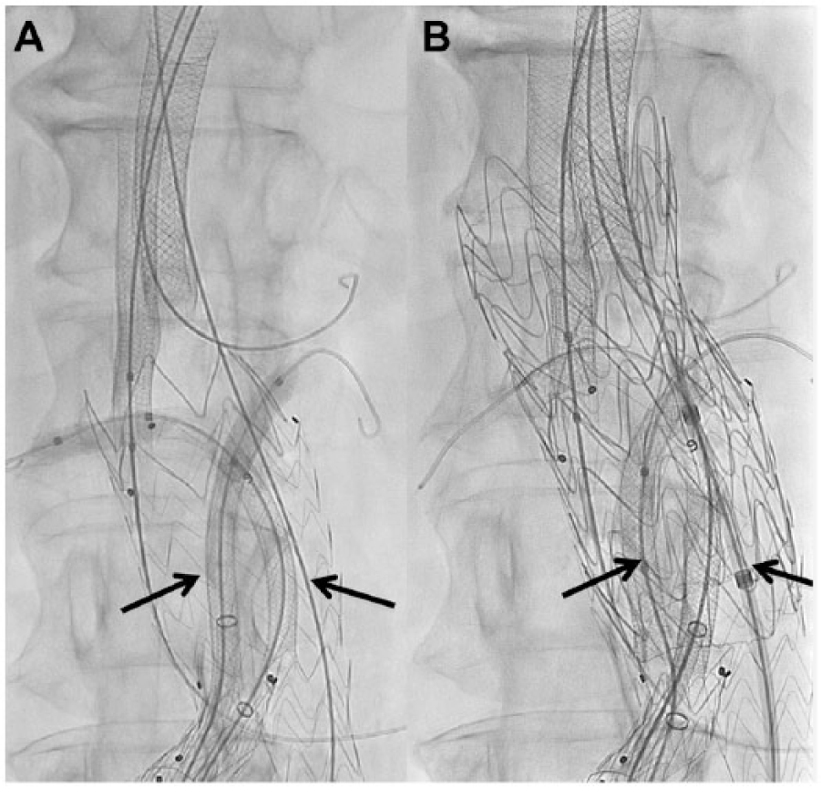
Four-vessel chimney periscope graft technique. A transfemoral periscope configuration was employed for renal arteries and a transaxillary chimney configuration for the celiac trunk and superior mesenteric artery. (A) Intraoperative angiogram showing final deployment of all renovisceral chimney and periscope grafts in the respective vessels. Balloons are inflated inside the periscope grafts (arrows) in the renal arteries to achieve anchoring. (B) Intraoperative angiogram showing cuff implantation and sandwich periscope for renal arteries (arrows).
The renovisceral branches were cannulated typically using a 45-cm Arrow sheath (Arrow International Inc, Reading, PA, USA) parked in the descending aorta close to the visceral orifice, a 5-F Chuang visceral reverse curve catheter (Cook Inc), and a Rosen wire (Cook Inc). After visceral branch cannulation, the CPG was positioned into the target artery, the aortic stent-graft was parked in the distal aorta, and the CPG was deployed with its distal end ~2 cm into the target vessel. In most cases, self-expanding covered stents (Viabahn; W.L. Gore & Associates, Flagstaff, AZ, USA) were used for CPG construction. Sporadically, bare metal stents (BMS) were employed [Wallstent (Boston Scientific Corporation, Natick, MA, USA) and Palmaz Blue or Corinthian SES (Cordis Corporation, Bridgewater, NJ, USA)]. The CPGs in the last 6 patients were lined with BMS to increase fluoroscopic visibility.11,12
After CPG deployment, a 2-cm-long balloon catheter (Admiral Xtreme; Medtronic Vascular) was used to fully expand the CPGs at their anchoring/landing zone. The deflated balloons remained in the target arteries, where they were reinflated and tensioned to hold the CPGs straight and parallel to the aortic wall during deployment of the new aortic stent-grafts [Excluder and TAG (W.L. Gore & Associates), Evita (Jotec, Hechingen, Germany), and Endurant (Medtronic Vascular)]. The design and anatomical details of the stent-grafts are reported in Table 2. Diameter sizing of the aortic stent-graft was based on the mean aortic diameter and the mean diameter of the CPGs as reported elsewhere. 13 A kissing balloon technique completed the procedure by achieving full and simultaneous expansion of the aortic graft and CPGs, especially in the overlapping areas. Completion angiography and selective pressure measurements (proximal and distal to the aortic stent-graft and in all CPGs) were performed to exclude endoleaks and/or significant pressure gradients.
Main Graft Details.

Abbreviations: AUI, aortouni-iliac; Bif, bifurcated; CG, chimney graft; PG, periscope graft; TEVAR, thoracic endovascular aortic repair.
Type Ib endoleak in patients with previous TEVAR and endovascular aneurysm repair.
Follow-up Protocol
A standardized follow-up protocol, including CTA, laboratory testing, and clinical examination was executed at 3, 6, and 12 months and annually thereafter. In case of endoleak or neck degeneration detected during follow-up, imaging frequency was tailored to the finding. In case of CTA contraindication, a native CT and duplex ultrasonography (eventually with contrast enhancement) were combined to assess stent-graft function. Medications consisted of aspirin (100 mg/d) and full heparinization during hospitalization; the heparin was switched to clopidogrel (75 mg/d) or an oral anticoagulant at discharge and continued for at least 3 months.
Outcomes
Evaluated outcomes included technical success, perioperative mortality, and morbidity. Technical success was defined as a procedure completed as intended with no secondary procedures within 30 days. Midterm outcomes were survival, CPG patency, endoleaks, and freedom from reintervention; Kaplan-Meier curves were employed to estimate survival and patency.
Mean and standard deviations are reported for parametric data; absolute values and percentages for nonparametric data. Differences were assessed using the t test. Statistical significance was assigned at p<0.05. Data analysis was performed using SPSS (version 16.0; IBM Corporation, Somers, NY, USA).
Results
Overall, 55 target vessels (2.3/patient) were addressed [44 renal arteries, 8 superior mesenteric arteries (SMAs), and 3 celiac trunks]. The chimney graft configuration was used in 31 and the periscope/sandwich graft configuration in 24. In 3 vessels, the chimney configuration was achieved with the lift technique from the femoral artery. No CPG was performed as a bailout procedure. Branches and CPG details are reported in Table 3. The intraoperative target vessel perfusion success rate with the CPGs was 100%. Technical success was achieved in 96% of the patients. One patient with a high-flow type Ia endoleak at postoperative CTA was managed successfully by proximal aortic cuff, CPG extension, and CPG gutter embolization with ethylene vinyl alcohol copolymer (Onyx) early after the first procedure (Figure 2).
Parallel Graft Details.

Abbreviations: CT, celiac trunk; LRA, left renal artery; RRA, right renal artery; SMA, superior mesenteric artery.

(A) Computed tomography (CT) showing abdominal aortic aneurysm rupture due to proximal type Ia endoleak after failed previous standard endovascular repair. (B) Intraoperative angiogram showing persistent type Ia endoleak after parallel graft implantation. (C) Persistent type Ia endoleak management with cuff implantation, superior mesenteric artery stenting, and Onyx embolization. (D) CT control showing complete sealing of the type Ia endoleak. (E) One-year CT showing significant aneurysm sac shrinkage. (F) Three-dimensional CT angiography volume rendering.
Mean procedure duration was 240±107 minutes (range 105–480). Mean intensive care unit stay was 1.9±4 days and mean in-hospital length of stay was 8.7±9 days. The mean postoperative eGFR was 58±17 mL/min/1.73 m2 and did not differ significantly from the preoperative eGFR (p=0.78).
Thirty-day mortality was 4.1% (1/24); this patient had double renal chimney grafts and died from multiple organ failure after SMA occlusion on the seventh postoperative day. Six (25%) patients experienced a complication. An iliac occlusion, a femorofemoral crossover bypass occlusion, and an axillary hematoma with neurological symptoms required reinterventions, while 2 renal hematomas and a case of postimplantation inflammatory syndrome were managed conservatively with prolonged hospitalization.
Mean follow-up after CPG placement was 23±28 months (range 0.2–122). During this time, 3 patients died of unrelated causes. Estimated survival at 12, 24, and 36 months was 83% (Figure 3A). Two CPGs occluded at 1 and 10 months postoperatively. Estimated CPG patency at 12, 24, and 36 months was 94% (Figure 3B). Four type I endoleaks (3 type Ia and a type Ib) were detected during follow-up; in 2 cases these were related to neck degeneration and in 2 cases to persisting gutter.

Kaplan-Meier estimates of (A) cumulative overall survival and (B) parallel graft patency. The standard errors did not exceed 10% at 36 months.
During follow-up, 6 (25%) reinterventions were performed at a mean of 15±12 months after CPG placement. All but one reintervention were performed endovascularly. Four reinterventions were related to type I endoleak and consisted of an infrarenal neck banding, 2 perileak embolizations with Onyx, and a proximal cuff extension and SMA stenting. The remaining 2 reinterventions were related to CPG occlusion and consisted of bypass surgery (mesenteric artery-to-renal artery bypass) and balloon dilation (Table 4).
Reinterventions During Follow-up.

Abbreviations: CG-EVAR, chimney graft endovascular aneurysm repair; RRA, right renal artery; SMA, superior mesenteric artery.
Mean maximal aneurysm sac diameter decreased from 88±26 to 85±33 mm (p=0.49) over the course of follow-up. Aneurysm sac diameter remained stable or decreased in 21 (87%) patients, while the other 3 had significant sac diameter increases (>5 mm). One of the latter cases was related to a recurrent type Ia endoleak.
Discussion
Despite significant improvements in stent-graft technology and surgeon experience, standard EVAR is still associated with a significant reintervention rate during follow-up. In 2 recent published experiences, Chang et al 14 and Mehta et al 15 reported reintervention rates of 14.5% and 19% at 3 and 2.8 years, respectively. Tadros et al 16 reported that the type I endoleak incidence, although reduced, was still present in the different device generations, with an incidence ranging from 3.5% to 15.5%. Thus, the association of type I endo-leaks with an increased aneurysm rupture risk is a strong recommendation for treatment.17,18
In such cases, an open aortic repair is generally performed in EVAR failure, despite a high mortality of up to 22%. 19 However, advanced age, suprarenal or supraceliac aortic clamping, and the technical challenge of endograft removal (stent incorporation into the aortic wall, presence of external stents or barbs, and periaortic inflammation) expose the patients to a high surgical risk, especially in the nonelective setting. 20 To reduce the operative risk, several options have been proposed. If an adequate proximal and/or distal landing zone is available, cuff implantation is a reasonable alternative to open surgery. Conversely, when a landing zone is missing, coil embolization, fenestrated EVAR, or CPGs have been used. Embolization of the sac with coils, n-butyl cyanoacrylate, 21 Onyx, 22 or fixation procedures with endoanchors 23 have been employed in isolated cases with good early results, but so far durability of such methods is not proven.
Fenestrated stent-grafts have been used in patients with type I endoleak. 24 However, the radiopaque stent skeleton and markers of the previous stent-graft make it challenging to recognize the fenestration markers, and as a consequence, positioning and accurate deployment of the fenestrated device are quite cumbersome. On the contrary, aortic devices used for the CPG technique are standard, nonfenestrated devices (tube or cuff) that do not have to be oriented or aligned to the renal and/or visceral orifices, which makes the procedure in this regard much easier. 25 When suprarenal fixation devices have been employed for the previous EVAR treatment, the struts are generally overlying the renal arteries. In such cases, renal catheterization can be hazardous after the deployment of a fenestrated stent-graft. Indeed, Katsargyris et al 26 reported technical difficulties in target vessel catheterization in 13% of cases.
By contrast, vessel catheterization in the CPG technique is achieved from above or below following the easier route to the visceral vessels, and it can be performed outside the existing failed stent-graft and before placement of a new aortic stent-graft. Moreover, in case of failure to catheterize the visceral arteries, a CPG procedure can be stopped before the placement of the new aortic stent-graft. In addition, the CPG technique employs standard nonfenestrated aortic and bridging stents-grafts, making this procedure feasible even in nonelective cases. 27
To date, there are only a few experimental and no consistent clinical data supporting the use of one device over the other for the parallel graft technique. 28 In our experience, we have employed different standard aortic stent-grafts (Excluder, Endurant, and Evita), but for chimney and/or periscope endografts, we essentially used self-expanding covered stents such as the Viabahn because of its low profile, flexibility, and heparin coating. We prefer combining the Viabahn with the Excluder aortic stent-graft, but overall, there were no significant differences in terms of CPG patency rate or endoleak incidence between the different aortic stent-grafts used. Balloon-expandable stent-grafts (BESG) may be used as chimney grafts, as the Münster group 9 showed in a study comparing self-expanding and BESGs. 9
Technical success after endovascular repair of type I endoleak using CPGs is good, with no aneurysm-related mortality, no secondary rupture, overall aneurysm sac reduction, and high target vessel patency. Reintervention for persisting or secondary endoleak was necessary in a significant percentage of the patients in this study, but this remains true for cases treated with fenestrated EVAR as well. Therefore, long-term imaging follow-up remains mandatory.
Conclusion
Use of the CPG technique avoids open aortic repair in patients presenting attachment site endoleak after EVAR. In comparison with open surgery, CPGs were safer and effective, even in nonelective patients, with high primary and midterm patency rates at 3 years. The CPG technique has a high initial success rate, with the occasional need for secondary intervention in patients presenting recurrent or de novo endoleaks. Close imaging follow-up is warranted.
Footnotes
Declaration of Conflicting Interests
The author(s) declared no potential conflicts of interest with respect to the research, authorship, and/or publication of this article.
Funding
The author(s) received no financial support for the research, authorship, and/or publication of this article.