






Abstract
Keywords
Introduction
Endovascular aneurysm repair (EVAR) has revolutionized the management of patients with aneurysmal disease of the abdominal aorta over the past 2 decades. The safety and efficacy of this less invasive modality has been well established among patients with favorable aortic anatomy 1 ; however, it is estimated that up to 40% of patients are anatomically unsuitable for conventional EVAR, most commonly due to limitations in proximal aortic neck anatomy.2,3 Several strategies have emerged to combat the issues of proximal neck fixation, ranging from simply deploying conventional infrarenal aortic stent-grafts outside the instructions for use (IFU) of the device4,5 to homemade and physician-modified endografts, 6 snorkel/chimney (s/c-EVAR) approaches with parallel covered stents,7–9 and fenestrated (f-EVAR) technology.9–11 The conceptual basis for these advanced endovascular approaches involves extension of the proximal seal zone with preservation of branch vessel patency, thereby expanding the applicability of EVAR to those with short-necked infrarenal, juxtarenal, and suprarenal abdominal aortic aneurysms (AAAs).
Initially described by Greenberg et al, 12 the snorkel/chimney approach utilizes one or more parallel renal stents combined with an aortic stent-graft in order to achieve a more proximal seal. Despite the immediate availability and excellent early results observed in s/c-EVAR,8,13 uncertainties remain regarding the durability of this approach, particularly with regard to branch vessel patency and the perceived increased potential for type Ia “gutter” endoleaks. By contrast, the applicability and utilization of f-EVAR is currently limited in the United States by its recent approval and lack of immediate availability to all practitioners. Both f-EVAR and s/c-EVAR have been used extensively at our institution, but the relative advantage of one approach over the other is unclear at the present time due to a paucity of reports directly comparing these two advanced EVAR techniques. 10 Available data to date, consisting of two single series and two meta-analyses, have been unable to demonstrate any reliable difference in these two approaches as it pertains to cannulation failure, target branch vessel patency, early mortality, type I endoleak, postoperative renal dysfunction, and need for secondary reintervention.9,14–16
As the debate continues regarding f-EVAR vs s/c-EVAR, one of the major differences between the two strategies is the cranial or caudal approach toward the target renal artery, which often makes cannulation a rate-limiting step in the efficiency and complexity of the repair. Moreover, the potential differences between the technical ease with which either procedure can be performed may have downstream effects on procedure time and radiation exposure that may ultimately favor one approach over another. As such, the purpose of this study is to evaluate the impact of renal artery geometry, specifically with regard to renal angulation, on renal cannulation times and procedure efficiency during f-EVAR and s/c-EVAR.
Methods
Study Design and Patient Population
For this retrospective study, which was approved by the local institutional review board, all patients with complex AAAs treated at a single institution between 2009 and 2013 with either f-EVAR or s/c-EVAR were retrieved from electronic medical databases and hospital records. Complex AAAs included short-necked (<15 mm) infrarenal, de novo juxtarenal, para-anastomotic juxtarenal, and expanding aneurysms, as well as those requiring revision of an existing stent-graft due to persistent type I endoleak.
Renal artery angulation was measured from the preoperative computed tomographic angiography (CTA) scans. Data on patient demographics (age, gender, and comorbidities), radiological imaging (AAA diameter, renal artery diameter, presence/absence of stenosis), procedures (use of a conduit for access, fluoroscopy and procedures durations, contrast volume, and blood loss), and clinical outcomes [length of stay, endoleak, mortality, adverse renal events (acute renal failure, renal artery dissection, renal stent thrombosis), and loss of renal artery patency] were retrieved, as were serum creatinine levels (baseline, postoperative maximum, discharge, and latest follow-up).
During the observation period, 77 consecutive patients (mean age 74.2 years; 63 men) with complex AAAs were treated with f-EVAR (n=24; first case 9/2012) or s/c-EVAR (n=53; first case 9/2009). All patients were considered anatomically unsuitable for conventional EVAR with a standard infrarenal stent-graft.
Preoperative Planning and Procedures
High-resolution computed tomographic angiography (CTA) of the abdomen and pelvis was obtained in all patients preoperatively. A dedicated 3-dimensional workstation (TeraRecon, San Mateo, CA, USA) with semiautomated centerline of flow reconstruction was used to characterize visceral segment geometry and aneurysm morphology. Anatomic parameters measured included maximum abdominal aortic diameter, neck length, neck diameter, neck angulation, neck shape, neck thrombus, ostial diameter of the renal arteries, maximum common iliac artery diameter, associated iliac aneurysm, and iliac tortuosity. For f-EVAR, clock positions for the visceral branches were calculated using the provided measurement tool and verified by the manufacturer. S/c-EVAR case planning was guided by the goal of placing the proximal edge of the main body device immediately caudal to the SMA and deploying 1 or 2 parallel adjacent renal snorkels from a transbrachial or transaxillary approach.
All procedures were performed in a hybrid endovascular suite with a fixed floor-mounted imaging Artis zee system (Siemens Medical Solutions, Malvern, PA, USA). The standardized techniques for both s/c-EVAR and f-EVAR have been previously described.8,10 Briefly, s/c-EVAR was performed by a 2-surgeon team working from the right femoral and left axillary positions. Antegrade renal/visceral access was obtained from open exposure of the left axillary or proximal brachial artery, and two 7-F, 90-cm-long sheaths (Terumo Medical Corp, Somerset, NJ, USA) were advanced into the visceral aorta. The renal vessels were catheterized using 260-cm-long hydrophilic guidewires and a 125-cm JB1 catheter (Cook Medical, Bloomington, IN, USA), and either covered iCAST (Atrium Medical Corp/Maquet Getinge Group, Hudson, NH, USA) or Viabahn (W.L. Gore & Associates, Flagstaff, AZ, USA) covered stents (dictated by the vessel tortuosity and intersecting angle with the aorta) were advanced into the target vessel. After the main body endograft was deployed, the snorkel stents were positioned for final deployment and balloon molding in a “triple kissing” fashion to optimize theoretical seal and minimize the potential for gutters.
The f-EVAR procedure was executed following the manufacturer’s IFU for the ZFEN stent-graft (Cook Medical) according to previously described techniques15,17 via bilateral 20-F femoral sheaths. The renal arteries were precannulated with a combination of 5-F, 70-cm-long van Schie 3 or 4 catheters (Cook Medical) and 0.018-inch, 260-cm-long hydrophilic guidewires. The proximal component of the main body ZFEN fenestrated device was introduced. Through an additional puncture of the contralateral 20-F sheath, 7-F, 55-cm-long Ansel sheaths (Cook Medical) were inserted through the distal end of the proximal ZFEN component and advanced to the level of the custom fenestrations. Renal artery cannulation was then performed through the fenestrations. The proximal ZFEN component was released and was balloon molded with the 7-F Ansel sheaths still within the fenestrations. Balloon-expandable covered iCAST stents were then deployed within each renal artery.
Angulation Measurement
Renal artery angulation was measured on all preoperative CTA studies using a semiautomated centerline in the abdominal aorta. The centerline was analyzed with multiplanar reconstruction views perpendicular to the centerline of flow. Renal artery angulation was defined as an angle above (positive angulation) or below (negative angulation) the orthogonal plane perpendicular to the aortic wall at the midpoint of the renal ostia (Figure 1). Quantitative angulation was calculated using the angular measurement tool featured in the AquariusNET software, a method similar to that used by several other studies examining visceral artery angulation.18–20 Renal artery angulation measurements were verified by consensus of all authors experienced with manipulation and preoperative planning CTA images (B.W.U., V.C., J.T.L.). Accessory renal arteries were not measured.

Measurement of renal artery angulation relative to the horizontal plane perpendicular to the aortic wall at the midpoint of the renal ostium. The figure demonstrates an example of a right and left renal artery with -45° and +30° angulations, respectively.
Efficiency Metrics
Renal artery cannulation times were taken from electronic time-stamped procedure logs and corroborated by review of available angiographic images. For s/c-EVAR, renal cannulation time was from the initial introduction of the sheath into the exposed high brachial or axillary artery until the 7-F sheath was delivered into the renal ostium. In f-EVAR cases, renal cannulation time began with the introduction of the 7-F sheath into the contralateral femoral sheath and ended when the 7-F Ansel sheath was placed into the renal ostium through the corresponding renal fenestration.
Statistical Analysis
The f-EVAR and s/c-EVAR cases were stratified around their median renal artery angulation values for comparison of renal cannulation times and other variables. Cases involving stenting of the celiac trunk or superior mesenteric artery (SMA), combined fenestrated and snorkel techniques, or failed renal artery cannulation were excluded from subsequent analysis in order to fairly evaluate total procedure time, fluoroscopy time, and contrast utilization in a relatively homogenous cohort.
Differences in branch vessel cannulation times and other metrics were assessed using the Student t test, Wilcoxon rank sum test, and Fisher exact test as appropriate. All p values were 2-sided, and p<0.05 was considered statistically significant.
Results
Group Comparison
There was no difference in the distribution of aneurysm type (eg, short-necked infrarenal, de novo juxtarenal) or distance between the SMA and start of the aneurysm between the two groups (18.8±8.4 mm for s/c-EVAR, 20.8±8.0 mm for f-EVAR; p=0.23). The mean number of fenestrations/scallops in the f-EVAR group was 2.6±0.6, with the most common device configuration featuring one SMA scallop and two small renal artery fenestrations (n=15). The s/c-EVAR group had a mean of 1.9±0.6 reconstructed vessels per patient and included 11 patients with celiac and/or SMA snorkel stents. A mean of 1.6±0.5 renal stents per patient were implanted in the s/c-EVAR group; 34 patients required bilateral renal artery stents.
Renal Artery Angulation
A total of 111 total renal artery cannulations were performed (f-EVAR, n=39; s/c-EVAR, n=72) among the 77 complex EVAR cases, with renal cannulation failure in one patient (99.1% technical success). Laterality included 57 right-sided (f-EVAR, n=19; s/c-EVAR, n=38) and 54 left-sided (f-EVAR, n=20; s/c-EVAR, n=34) renal artery cannulations. Descriptive statistics of the right and left renal arteries are shown in Figure 2. Median renal artery angulation was -33° (range +37° to -60°) for f-EVAR and -32° (range +22° to -65°) for s/c-EVAR patients (p>0.05). When both complex EVAR procedure types were individually stratified by their respective median values (approximately -30° in both groups), no difference was observed with regard to baseline demographics or aneurysm morphology (Table 1). Renal artery ostial diameter and presence of any degree of ostial stenosis was not significantly different between groups.
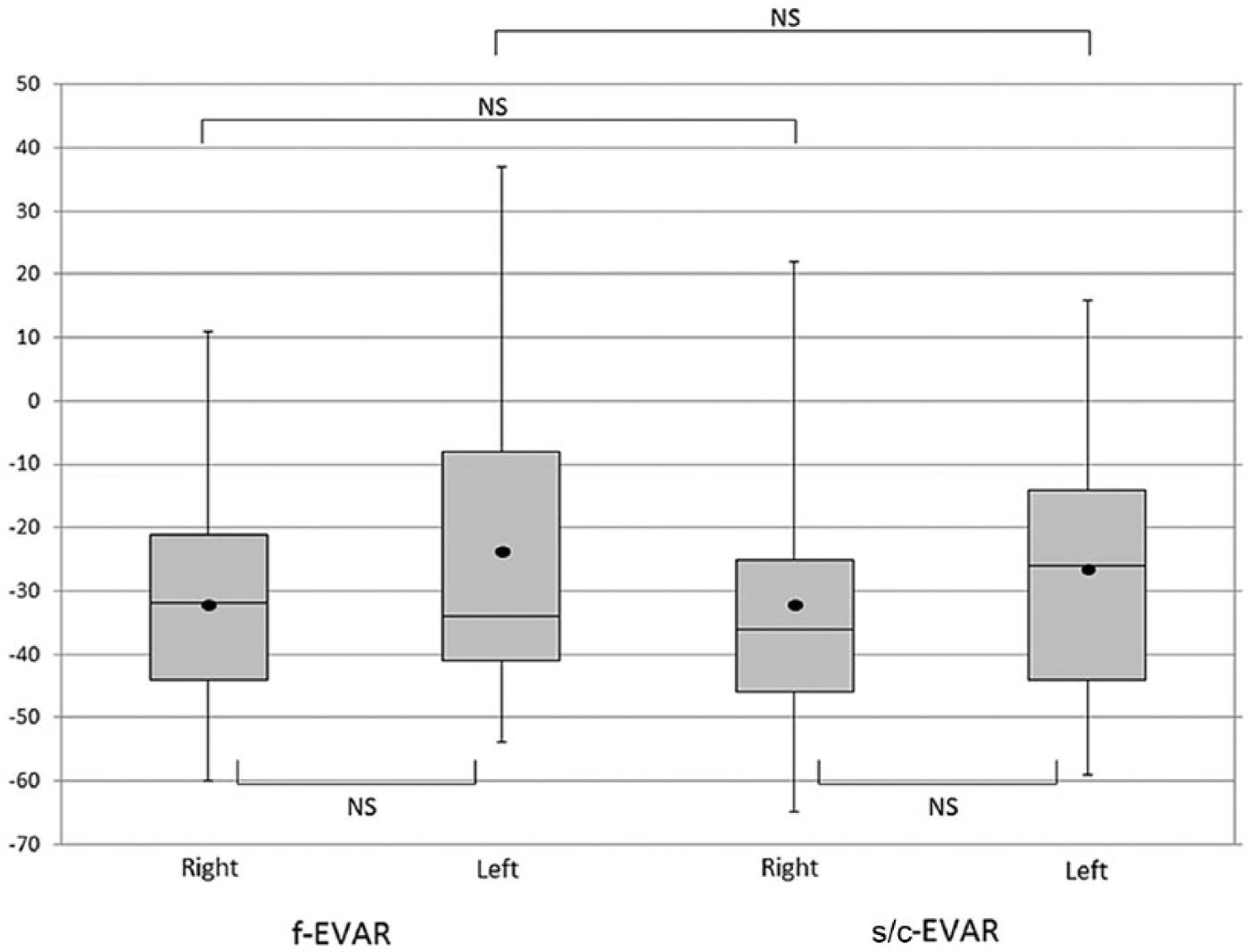
Distribution of right and left renal artery angulation in fenestrated (f-EVAR) and snorkel/chimney (s/c-EVAR) endovascular aneurysm repair. The upper and lower limits of the boxes represent the 75th and 25th percentiles, respectively. The horizontal line in the box corresponds to the median value, whereas the black circle indicates the mean. NS, not significant.
Baseline Demographics and Anatomic Data.

Abbreviations: AAA, abdominal aortic aneurysm; CAD, coronary artery disease; CHF, congestive heart failure; COPD, chronic obstructive pulmonary disease; CRI, chronic renal insufficiency; DM, diabetes mellitus; EVAR, endovascular aneurysm repair; f, fenestrated; HTN, hypertension; s/c, snorkel/chimney.
Continuous data are presented as the means; categorical data are given as the counts (percentage).
Defined as serum creatinine ≥1.5 mg/dL.
Impact of Angulation on Cannulation Time
After excluding 22 renal cannulations [concomitant celiac and/or SMA stenting (n=15), f-EVAR with renal snorkels (n=6), and renal artery cannulation failure (n=1)], 33 renal artery cannulations in 19 f-EVAR patients and 56 renal artery cannulations in 41 s/c-EVAR patients were available for the cannulation time analysis.
Renal artery cannulation in the f-EVAR group was performed significantly faster in cases with less downward (≥ −30°) renal artery angulation (16.0 vs 32.8 minutes, p=0.04; Table 2). In addition, f-EVAR cases involving ≥ −30° renal artery angulation were associated with shorter procedure time (187.7 vs 246.2 minutes, p=0.01) and decreased fluoroscopy time (70.3 vs 98.2 minutes, p=0.04). Contrast dose and estimated blood loss also tended to be lower among patients with renal arteries having less downward angulation. There was no significant difference between the f-EVAR subgroups in periprocedure adverse renal events or change in renal function over time relative to renal angulation (Table 3).
Procedure Metrics and Clinical Outcomes After Complex EVAR Stratified by Renal Artery Angulation.

Abbreviations: EVAR, endovascular aneurysm repair; f, fenestrated; s/c, snorkel/chimney.
Variables calculated according to each individual renal artery; as such, patients with bilateral renal artery cannulations are accounted for twice based on the angulation of each renal artery.
Continuous data are presented as the means; categorical data are given as the counts (percentage).
Defined as a 1.5-fold increase in the serum creatinine.
Mean Serum Creatinine Concentration (SCr) in Patients Undergoing Complex EVAR Stratified by Renal Artery Angulation.

In the s/c-EVAR group, renal artery cannulation was performed significantly faster in cases with greater downward (< −30°) renal artery angulation (10.9 vs 17.3 minutes, p=0.05). There was no significant difference in procedure or fluoroscopy times, contrast dose, estimated blood loss, adverse renal events, or change in renal function over time based on renal artery angulation.
Experience and Learning Curve Effect
When the renal angulation subgroups based on the -30° median value were bisected chronologically according to the median date of the procedure, there was no significant difference in renal artery cannulation times within each angulation group (Figure 3). In cases involving bilateral renal cannulations, the time required to successfully cannulate the second renal artery was not significantly different from the cannulation time of the first renal artery in the f-EVAR group (24.8 vs 21.9 minutes, p=0.72; Figure 4); however, cannulation time of the second renal artery tended to be shorter in the s/c-EVAR group (10.9 vs 16.9 minutes, p=0.07).

Renal artery cannulation time over the course of the study period. f-EVAR, fenestrated endovascular aneurysm repair; NS, not significant; s/c-EVAR, snorkel/chimney endovascular aneurysm repair.

Cannulation times for the first and second renal arteries during fenestrated (f-EVAR) and snorkel/chimney endovascular aneurysm repair (s/c-EVAR). NS, not significant.
Follow-up
Median follow-up for patients undergoing complex EVAR in this analysis was 18.2 months (range 0.5–45.0) for the s/c-EVAR group and 5.1 months (range 0–12.7) in the f-EVAR group. The f-EVAR group had a 30-day mortality rate of 5.3%. One patient in the f-EVAR group had a type Ia endoleak on completion angiography, but this resolved on interval postoperative imaging. One additional patient experienced early renal stent thrombosis, which prompted the only secondary intervention (thrombolysis and additional renal stent placement) in this group.
The s/c-EVAR group had a 30-day mortality of 2.4%; 3 patients had type Ia endoleaks on completion angiography (two resolved at follow-up). Three patients experienced loss of patency of 4 renal stents at a median of 5 months postoperatively (range 1–14). Two late reinterventions were performed in the s/c-EVAR group, including transcatheter embolization of a refractory type II endoleak and balloon angioplasty and additional stent placement for the treatment of bilateral renal artery stent stenosis. There was no statistical difference in either group in terms of early mortality, type Ia endoleak, loss of renal artery stent patency, or need for secondary reintervention based on renal angulation. The mean interval change in maximum AAA diameter from preoperative to latest postoperative imaging was -0.26 cm (p=0.018) in the f-EVAR group and -0.82 cm (p<0.0001) in the s/c-EVAR group.
Discussion
The current study is the first to investigate the impact of renal artery angulation on the technical ability and efficiency to successfully cannulate the renal arteries during fenestrated or snorkel/chimney EVAR. Comparative analysis of procedure metrics based on median renal artery angulation demonstrated faster renal cannulation in f-EVAR cases with less downward renal artery angulation and faster renal cannulation in s/c-EVAR cases with more renal artery downward angulation. Although these results are not necessarily surprising given the cranially directed approach of the branch vessels during f-EVAR and the antegrade approach of the branch vessels during s/c-EVAR, understanding the limitations and challenges for each strategy provides guidance to the optimal approach based on preoperative anatomy.
Meticulous preoperative planning and patient selection represent the most critical factors in optimizing procedure success and clinical outcome following complex EVAR. The selection of endovascular strategy (f-EVAR vs s/c-EVAR) for the treatment of complex AAA is determined after careful review of radiographic imaging, patient anatomy, comorbid burden, and functional status of the patient, as well as the surgeon’s comfort level and expertise. In our experience, renal artery cannulation represents the most common rate-limiting step in completing both f-EVAR and s/c-EVAR. 10 The current study suggests that renal artery geometry, specifically, angulation, has an impact on the choice of the more efficient endovascular strategy. The single renal artery that could not be cannulated in this series occurred in an f-EVAR case involving severe downward renal angulation (−53°); perhaps renal salvage could have been achieved using a s/c-EVAR approach.
In f-EVAR, the faster renal cannulation times observed in cases with less downward angulation to the renal arteries (ie, more horizontal or even upward going vessels) translated to other metrics of procedure efficiency, including shorter operative duration and fluoroscopy time, which all have positive influences on peri- and postprocedure outcomes. Similar findings would be expected in the more expeditious s/c-EVAR cases having more downward angulation of the renal arteries; however, this was not observed, perhaps due to other confounding factors during s/c-EVAR cases. Other potential metrics of procedure efficiency, such as volume of contrast agent administered and estimated blood loss, were not associated with faster renal cannulation, again implying other factors from complex EVAR that contribute to those metrics. Still, we believe that employing strategies that limit cannulation times could minimize multiple digital subtraction angiography runs and limit occult blood loss associated with multiple guidewire or catheter exchanges and manipulations, as well as peri-sheath bleeding in cases involving a longer sheath dwell time.
Renal dysfunction after complex EVAR21,22 theoretically could be improved with more efficient procedures. We were not able to associate challenging renal artery anatomy into any significant increase in the risk of early or late postoperative renal dysfunction. However, the potential for deleterious effects on renal function in cases involving challenging renal artery anatomy remains a valid and important issue given that the longer renal cannulation times observed in these cases frequently are associated with more wire or catheter exchanges and manipulations, increased contrast volume, and prolonged sheath placement in the origin of an initially cannulated renal artery (in cases with bilateral renal cannulation). The enhanced technical demand associated with these maneuvers is likely accompanied by an inherent increase in the risk of dissection, thromboembolic phenomena, and other related ischemic complications.
Interestingly, there was no learning curve effect observed over the study period with regard to improved efficiency of cannulating renal arteries with challenging angulation. Moreover, in cases involving bilateral renal artery cannulations, there was no advantage associated with a second renal cannulation following successful cannulation of the initial renal artery. These findings suggest that the barrier to efficient cannulation of renal arteries with challenging angulation is not entirely overcome with increased surgeon experience alone or even the perceived advantage of successfully cannulating the first renal artery when performing bilateral cannulations. As such, renal angulation remains a fixed variable that should be considered during the procedure planning process.
Limitations
This retrospective study has important limitations, and the results should be interpreted cautiously. Challenging renal artery anatomy and the impact it has on the technical demands of complex EVAR represents a multifactorial obstacle. While this analysis sought to address the influence of vertical (ie, cephalad or caudal) renal artery angulation on procedure efficiency in f-EVAR and s/c-EVAR, it did not specifically explore the impact of additional renal artery anatomic factors, such as concomitant renal artery angulation in the anterior-posterior orientation, severity of ostial stenosis, tortuosity, presence of accessory renal arteries, and length of the main renal artery proximal to the initial branching point. Each of these variables has some degree of influence on the procedure and should therefore be considered at all planning stages.
Defining the term “procedure efficiency” is also open to debate. While procedure efficiency likely involves the interplay of multiple variables and surgeon experience, as stated previously, successful renal artery cannulation has served as the most common rate-limiting step in our experience with both f-EVAR and s/c-EVAR. Therefore, we inherently associate faster renal artery cannulation times with more efficient complex EVAR procedures. In an effort to improve our own decision-making algorithm for f-EVAR vs s/c-EVAR, the results of this analysis provide some guidance as to which cases will be challenging to perform. For example, we can anticipate that a severe downward going renal artery of -65° will present a challenge if we were to employ a f-EVAR device; based on this study and prior reports that the two strategies produce similar midterm outcomes, we might now switch to a s/c-EVAR approach. Understanding the limitations of each endovascular strategy also provides the opportunity for future refinement of devices and techniques to make these procedures safer. Renal angulation will continue to have an impact on the procedure as we shift toward a primary fenestrated and branched EVAR paradigm.
Conclusion
Faster renal cannulation in f-EVAR cases is observed with less downward angulation to the renal arteries; in s/c-EVAR cases, cannulation is quicker in renal arteries with more downward angulation. Given that renal artery cannulation frequently serves as the rate-limiting step in complex EVAR, procedure efficiency may be optimized by considering renal artery angulation as one of several objective variables used in the selection of an endovascular strategy for treatment of complex AAAs. Future investigation is warranted to better elucidate the potential role of renal artery anatomy on other metrics of procedure efficiency, as well as to clarify the relationship between challenging renal artery anatomy and the development of postoperative renal dysfunction. Future device design and refinements of existing techniques should strongly consider the limitations of renal artery cannulation on the technical success and procedure efficiency associated with complex EVAR.
Footnotes
Authors’ Note
This study was presented at the 2014 Spring meeting of the Vascular and Endovascular Surgical Society (VESS) held in Boston, Massachusetts, June 4, 2014.
Declaration of Conflicting Interests
The author(s) declare no potential conflicts of interest with respect to the research, authorship, and/or publication of this article.
Funding
The author(s) received no financial support for the research, authorship, and/or publication of this article.