






Abstract
Keywords
Introduction
Stent-assisted angioplasty of the extracranial internal carotid artery (ICA) is already a well established option for treating atherosclerotic carotid stenoses.1,2 Although carotid endarterectomy (CEA) is still considered the gold standard in the therapy of carotid stenoses,3,4 stent implantation is less invasive, and many surgical complications can be avoided.5,6
Particular attention is focused on the stents used, whose design and materials are constantly being enhanced. Self-expanding stents are exclusively used today because most balloon-expandable stents had the disadvantage of low flexibility and a relatively high rate of irreversible stent collapse. 7 The mechanical behavior of self-expanding stents is determined by the varying design patterns and also by the material. 8 Analysis of the data available from clinical studies does not yield a clear advantage for the open-cell or closed-cell design, but there are trends that support recommendations of certain stents for specific lesion morphologies. 9
The perfect stent should safely cover the plaque and have good wall conformability. For this purpose, the stent must have the right balance between flexibility, collapse pressure, and radial force. 10 This study reports preliminary clinical data for a novel double-layer carotid stent after implantation in patients with symptomatic carotid artery stenosis and discusses stent mechanics. To better compare this double-layer stent with established carotid stents, the device was tested using well-established methods employed in various investigations of self-expanding stents11–13 with regard to bending stiffness, radial force, and collapse pressure, as well as wall adaptation in a step and curve model.
Methods
Clinical Experience
The double-layer CASPER RX stent (Microvention/Terumo, Saint-Germain-en-Lage, France) was deployed in 12 patients (median age 69 years; 8 men) with high-grade symptomatic ICA stenoses. Symptoms were transient ischemic attack in 10 and amaurosis fugax in 2 patients.
Patients were given aspirin (500 mg) and clopidogrel (300 mg) the day before the procedure. All interventions were done via a transfemoral approach using an 8-F Radiofocus Introducer II (Terumo Europe, Leuven, Belgium). Heparin (5,000 to 10,000 units) was given intra-arterially to achieve an activated clotting time between 250 and 300 seconds. A distal FilterWire EZ (Boston Scientific, Marlborough, MA, USA) was deployed in all patients after placing a long 8-F Vista Britetip support sheath (Cordis Corporation, Bridgewater, NJ, USA) in the common carotid artery. Twenty-millimeter-long stents in 8-mm (n=6), 9-mm (n=4), and 10-mm (n=2) diameters were placed without predilation. Postdilation was done with a 5-×30-mm Sterling Monorail balloon (Boston Scientific) at 10 atm after administration of 0.5 mg atropine (Braun, Germany). Intracranial digital subtraction angiography (DSA) was performed in 2 projections after stenting and after removing the distal filtration to rule out distal embolization. Hemostasis was achieved using the clip-based StarClose SE closure device (Abbott Vascular, Redwood City, CA, USA). After intervention, all patients were monitored neurologically in the stroke unit. At discharge, clopidogrel (75 mg/d) was prescribed for a minimum of 6 weeks; aspirin therapy (100 mg/d) was continued indefinitely. Patients were reevaluated with duplex sonography at 6 months.
Experimental Studies
The 8-×40-mm CASPER RX stent (Figure 1A) was used for experimental investigations; scanning electron microscopy (Figure 1B) shows the detail of the stent’s double-layer structure, with interconnections that produce a closed-cell design. The outer load bearing structure of the CASPER-RX stent is built of thick woven wires with an additional internal mesh beneath this structure (Figure 1B) consisting of woven but much thinner wires. Both the load bearing and the mesh structure are loosely connected by another fixation wire.

(A) The 8-×40-mm CASPER RX stent. (B) The distal tip of the stent delivery system of the 8-×40-mm CASPER RX stent; the mean profile in the stent region was 1.69 mm. (C) Scanning electron micrographs show the detailed structure of the expanded stent. The ~180-µm thick wires are woven and cover a woven mesh of thinner 42-µm wires. Additionally, there is an ~45-µm-thick wire added to connect the inner mesh with the outer structure.
The stent was evaluated for bending stiffness on the stent delivery system and in its expanded state, the latter with the stent freely deployed and expanded in a 7-mm vessel model. In addition, the radial force was measured as a function of expansion diameter. Collapse pressure and the change in length from mounted to an expanded state were also measured. The adaptation to changing vessel geometry, such as tapering or tortuosity, was also documented. Most of the test methods have been described for use in self-expanding stent systems and stents.11–14
Bending Stiffness/Flexibility
The flexibility defined as the inverse of the bending stiffness was measured with the stent mounted, freely expanded, and expanded within a flexible tube. The sample was fixed with a clamp at one side, leaving a free bending length of 12 mm. The force to deflect the stent was measured up to a maximum bending deformation of 0.5 mm. The recorded force distance curve is a valid approximation of the spring modulus for bending of the test object. The bending stiffness was calculated from the mean slope of the force and related deflection curve using the beam theory. The measurements were performed 3 times per direction to obtain an averaged value for bending stiffness. Taking into account possible asymmetric structures of the stent, bending stiffness was measured in 5 directions by rotating the sample by 45° for each direction around the circumference and then averaged.
Radial Force
The radial force during expansion and compression was measured using V-shaped clamps as described in Schmidt et al. 12 The distance of the prismatic clamping supports and the resulting force during stent expansion and compression were measured as a function of the expansion diameter using a universal testing machine (Zwick BZ2,5/TN1S; Zwick GmbH & Co. KG, Ulm, Germany). The resulting force distance curves were used to compare the radial force of the stent at the specific diameter of 7 mm, which is the typical target diameter for the given stent size. This test was performed in a 37°C atmosphere owing to the device’s temperature-dependent material properties.
Stent Length/Foreshortening
Stent length was measured with the device mounted on the delivery system, after stent release in vessel models of 5-, 6-, and 7-mm inner diameter, and in the completely expanded state to calculate the foreshortening during expansion.
Collapse Pressure
The stent was inserted into a thin and flexible polyurethane tube with an inner diameter of 7 mm. The wall thickness of the tube was about 100 µm and did not serve a support function but acted as a membrane separating the inner lumen of the stent from the surrounding pressure. A hydraulic radial outer load was applied to the stent by a pressure chamber. The pressure was increased in steps of 0.05 bar until the cross section of the stent was <50% of its initial state. This pressure was noted as the collapse pressure.
Vessel Wall Adaptation
The stent was implanted in 2 rigid tube models for assessment of vessel wall adaptation: (1) a double bent tortuous vessel model with a constant inner diameter of 7 mm and (2) a vessel model with a diameter gradient from 5 to 7 mm. Fluoroscopic images of each stent were taken in both configurations (SkyScan 1172; Bruker microCT, Kontich, Belgium). Imaging conditions were a maximum 80-×100-mm field of view, 80-kV acceleration voltage, and 100-µA current. A 0.5-mm aluminum filter was used for optimum image contrast of both the stents as well as the less radiopaque vessel models.
Results
Clinical Experience
The stents were implanted successfully in all patients without predilation, achieving <30% residual stenosis after postdilation (Figure 2). The distal filter was easily deployed, and no debris was observed after retrieval. Control angiography demonstrated no distal embolization or dissection. The activated clotting time during intervention was a median 264 seconds. There were no minor or major strokes, and all patients were neurologically asymptomatic at discharge and at 30 days. After 6 months, duplex ultrasound detected no restenosis in any stent. There were no deaths or major adverse events during follow-up to 6 months.
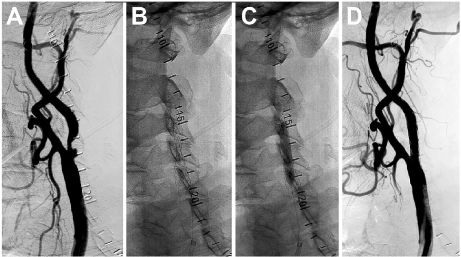
(A) A symptomatic high-grade stenosis of the right internal carotid artery in an 80-year-old man. (B) The stenosis was crossed with the distal protection device and then an 8-×20-mm CASPER Rx was implanted. (C) The stent was post-dilated with a 5-×30-mm balloon at 10 atm. (D) Final result showing no residual stenosis and good wall conformation.
Experimental Studies
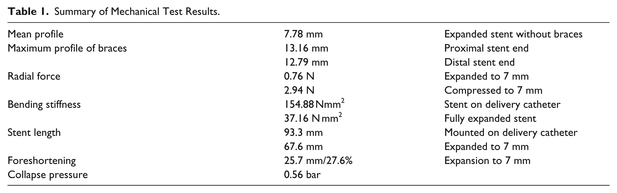
The bending stiffness (Table 1) differs remarkably between the mounted stent with delivery catheter (154.88 N mm2) and the more flexible free expanded stent (37.16 N mm2). The measurement of bending stiffness of the stent inserted in a flexible tube with negligible stiffness provided a more than 10 times higher stiffness of the stent than in its free state (Figure 3A). This is a typical result for woven stents if their length change is restricted by other means.
Summary of Mechanical Test Results.

(A) Bending stiffness of the 8-×40-mm CASPER RX stent. (B) Radial force as a function of stent diameter during the expansion process (normalized to the nominal stent length of 40 mm).
The radial force during stent expansion (Table 1) was measured as a function of stent expansion diameter (Figure 3B). The radial force decreased almost linearly from 5 to 8 mm. The residual force for diameters >8 mm resulted from the braces at the stent ends, which improve fixation but do not contribute to vessel support. The radial force on expansion of the stent to 7 mm was low (0.011 N/mm).
The stent exhibited a change in length (Table 1) from 93.3 mm on the delivery system to 67.6 mm when expanded to 7 mm, representing a significant 27.6% foreshortening. The collapse pressure was relatively high (0.56 bar) as a result of the stent’s particular structure.
The conformability to the wall in the step model was relatively smooth; in the curve model, straightening occurred with consecutive slight stenosis (Figure 4). The diameter of the device was reduced while following the zigzag shaped vessel; in both cases, a smooth transition is visible without abrupt changes in diameter or direction.

Adaptation of the 8-×40-mm CASPER RX device in (A) a curved vessel model (internal diameter 7 mm) and (B) a straight vessel model with an inner diameter taper from 7 to 5 mm.
Discussion
The purpose of carotid artery revascularization is to prevent a stroke. Carotid endarterectomy has been considered as standard treatment, but in the past 15 years, carotid artery stenting (CAS) has become a valuable minimally invasive alternative. 15 In contrast to surgical therapy, 50% to 66% of strokes after CAS occur within the first 4 weeks, which can be attributed to embolization of thrombus or plaque through the stent struts.16,17 However, only one study so far has shown a significant advantage of closed-cell over open-cell stents with respect to the late major adverse neurological event rate. 18 However, the influence of ischemia on cognitive functions after CEA or CAS did not differ significantly, 19 so the incidence of post-procedure ischemia has to be reduced.
In our small series, no peri- or postinterventional complications were observed with the new CASPER-RX double-mesh stent, which is in contrast to many other non-randomized trials that reported postprocedure ischemia. 20 Thus, these results suggest that further improvement has been achieved since the introduction of the relatively modern hybrid stents (eg, Cristallo Ideale; Medtronic/Invatec, Roncadelle, Italy) with a technical success rate of 100% and a 1.6% rate of neurological complications at 30 days, 21 much lower than those reported in other studies. In another small series with 7 patients, the Roadsaver double-layer stent (Terumo) has recently shown comparable clinical results. 22
In the experimental findings of the new double-layer stent, the mechanical behavior had some similarities to the Carotid Wallstent (Boston Scientific), with relatively low radial force (0.011 N/mm CASPER RX and 0.020 N/mm Carotid Wallstent), a rather high collapse pressure (0.56 and 0.48 bar, respectively), and nearly similar bending stiffness in a 7-mm tube (467.4 and 432.0 N mm2, respectively). 13 In contrast to the Carotid Wallstent, the adaptation to the wall in the curve and step model is much better with the CASPER RX and comparable to the modern hybrid stents. 13 A disadvantage of the CASPER RX is a foreshortening of 27.6% compared with the stent length on the delivery system, which must be taken into account during implantation and can sometimes cause problems with precise placement. The Cristallo Ideale is the only stent that shows no measurable foreshortening.10,13
Conclusion
The new double-layer CASPER RX shows promising results in this small clinical experience, with no ischemic events. The unique structure may provide better coverage of the plaque than other stents, as evidenced by the lack of distal embolization. The large foreshortening during implantation should be considered, as well as the higher bending stiffness, especially when used in elongated carotid arteries.
Footnotes
Declaration of Conflicting Interests
The author(s) declared no potential conflicts of interest with respect to the research, authorship, and/or publication of this article.
Funding
The author(s) received no financial support for the research, authorship, and/or publication of this article.