






Abstract
Keywords
Introduction
Percutaneous techniques to recanalize chronic total occlusions (CTOs) may be challenging, especially in long lesions below the inguinal ligament. The intraluminal approach may fail in up to a quarter of the cases in crossing the CTO lesion. 1 On the other hand, intentional subintimal dissection with the loop technique, which was first described by Bolia et al, 2 is the preferred method when the initial intraluminal approach fails. Subintimal recanalization is a reliable solution but still not a panacea, as it may dissect the distal healthy arteries and has reported failed true lumen re-entry rates between 10% and 20%. 3 Re-entry devices have been engineered to help cross CTOs in a reproducible subintimal manner when the initial spontaneous re-entry fails, offering not only a high success rate of recanalization but also accuracy and precision of the distal re-entry point, sparing the maximum possible healthy vessel.
The Outback re-entry catheter (Cordis Corporation, Bridgewater, NJ, USA) is one of the most popular devices to date. It is easy to use (0.014-inch platform) and may facilitate true lumen re-entry both in aortoiliac and infrainguinal CTOs, with reported success rates ranging between 65% and 100%.4–8 However, amassed experience to date is limited to relatively small series, and evidence about the actual accuracy of targeted re-entry into the preplanned vessel area is scarce. 4
Our aim was to review the immediate outcomes in a large consecutive series of cases in which the Outback re-entry device was used to assist targeted subintimal re-entry to the true lumen both in aortoiliac and infrainguinal CTOs. There was particular emphasis on accuracy and precision of the final successful re-entry target area. In addition, a systematic review was performed of published reports of the Outback device in the aortoiliac and infrainguinal arteries in order to synthesize available evidence on technical success and complication rates and provide an appropriate benchmark for comparison and clinical guidance.
Methods
Study Design
This was a retrospective single-center cohort study and as such the need for institutional ethics review was waived. Images and procedure reports from all eligible cases were retrieved from the radiology imaging and information databases; baseline demographics of all patients were retrieved from the electronic patient records system. Peripheral angioplasty cases involving the use of the Outback re-entry device following failure of other intraluminal or subintimal techniques from February 2011 to July 2013 were included for analysis. All patient records and radiology images were examined by 2 interventional radiology fellows to extract baseline procedure and demographic variables, including but not limited to age, gender, comorbidities, Rutherford category, lesion length, type of treatment (angioplasty or stent placement), and anatomy of the intervention. In addition, baseline lesion length, re-entry distance, and overall length of the treated CTO segment were estimated.
Outcome measures included minor and major complications according to well-accepted reporting standards.9,10 Minor complications were those that required no further treatment, while major events required further therapy or hospitalization or resulted in permanent adverse sequelae or death. For the purposes of this analysis, technical success was defined as successful targeted re-entry at the preplanned site in the distal true lumen. The re-entry distance (between the point of true vessel reconstitution at baseline and the eventual point of true lumen re-entry after successful lesion crossing) served as a metric of device re-entry accuracy.
Outback Procedure
Procedures were performed by different vascular interventional radiologists with a varying level of experience (3–10 years). The procedure for intraluminal and spontaneous subintimal approach, along with details about deployment and use of the Outback catheter, have been thoroughly reported elsewhere.1,4,6,11 In our institution, routine practice involves initial introduction of a 6-F sheath (preferably antegrade for femoropopliteal and retrograde for aortoiliac CTOs) and use of 4-F catheters and hydrophilic guidewires to recanalize the occlusion with the intraluminal approach if possible. 9 In case of unsuccessful intraluminal lesion crossing, the subintimal approach with a looped 0.035-inch J-shaped half-stiff wire (Terumo, Tokyo, Japan) is frequently employed, reserving the Outback catheter for cases of unsuccessful spontaneous true lumen re-entry. In general, selection of devices for intraluminal and subintimal attempts to cross a CTO is highly dependent on operator preference and individual device familiarity. In all cases, eventual use of the Outback device was decided either because of failure of the aforementioned methods or due to inadvertent extension of the subintimal lumen to a level that could compromise important collaterals.
The initial guidewire was exchanged with a 0.014-inch wire that was advanced to the preplanned level of desired re-entry, usually within 1 cm of the point of distal flow reconstitution in the healthy true lumen by visual estimate. The Outback device was advanced over the guidewire, and biplanar fluoroscopy images were acquired to properly orient the tip of the device per the “Instructions for Use.” Occasionally, single plane imaging was sufficient for the aortoiliac segment. Once re-entry was accomplished, the 0.014-inch guidewire was advanced into the true lumen and confirmed with selective angiography. Predilation was always performed at the point of re-entry followed by definitive lesion treatment. Treatment options included plain balloon angioplasty or deployment of either a bare metal or covered stent. Mode of treatment was decided by the operator, taking into consideration the site of treatment, baseline complexity and lesion length, and angiographic result following balloon predilation. In case of a suboptimal result or if a complication occurred after balloon angioplasty, a nitinol stent was inserted.
Patient Sample
In the 28-month period, 975 patients with symptomatic peripheral artery disease were treated. Of these, 91 patients (mean age 64±9 years; 57 men) experienced failed intraluminal and/or subintimal recanalization techniques in 100 vessels, and the Outback catheter was used to attempt true lumen re-entry (Figure 1). The majority of patients (63, 69%) had critical limb ischemia, while 28 patients suffered from lifestyle-limiting short distance claudication. Detailed patient demographics are outlined in Table 1. Overall, 52 re-entry attempts were made in the aortoiliac region (Figure 2), while the other 48 were infrainguinal (Figure 3) approaches. Cases were stratified between the anatomical regions: the aortoiliac group included the bifurcation, common iliac artery (CIA), and external iliac artery (EIA), and the infrainguinal group included the superficial femoral artery (SFA), popliteal artery, and tibial arteries. In many cases, however, lesions spanned more than one anatomical region. In the aortoiliac segment, most of the cases involved both the CIA and EIA, and in the infrainguinal segment, all popliteal artery occlusions extended also into the more proximal SFA (Table 1).

Flowchart shows selection and inclusion of 91 patients with 100 chronic total occlusions treated with Outback-assisted recanalization.
Baseline Characteristics of the 91 Study Patients Treated for 100 Chronic Total Occlusions (CTOs). a
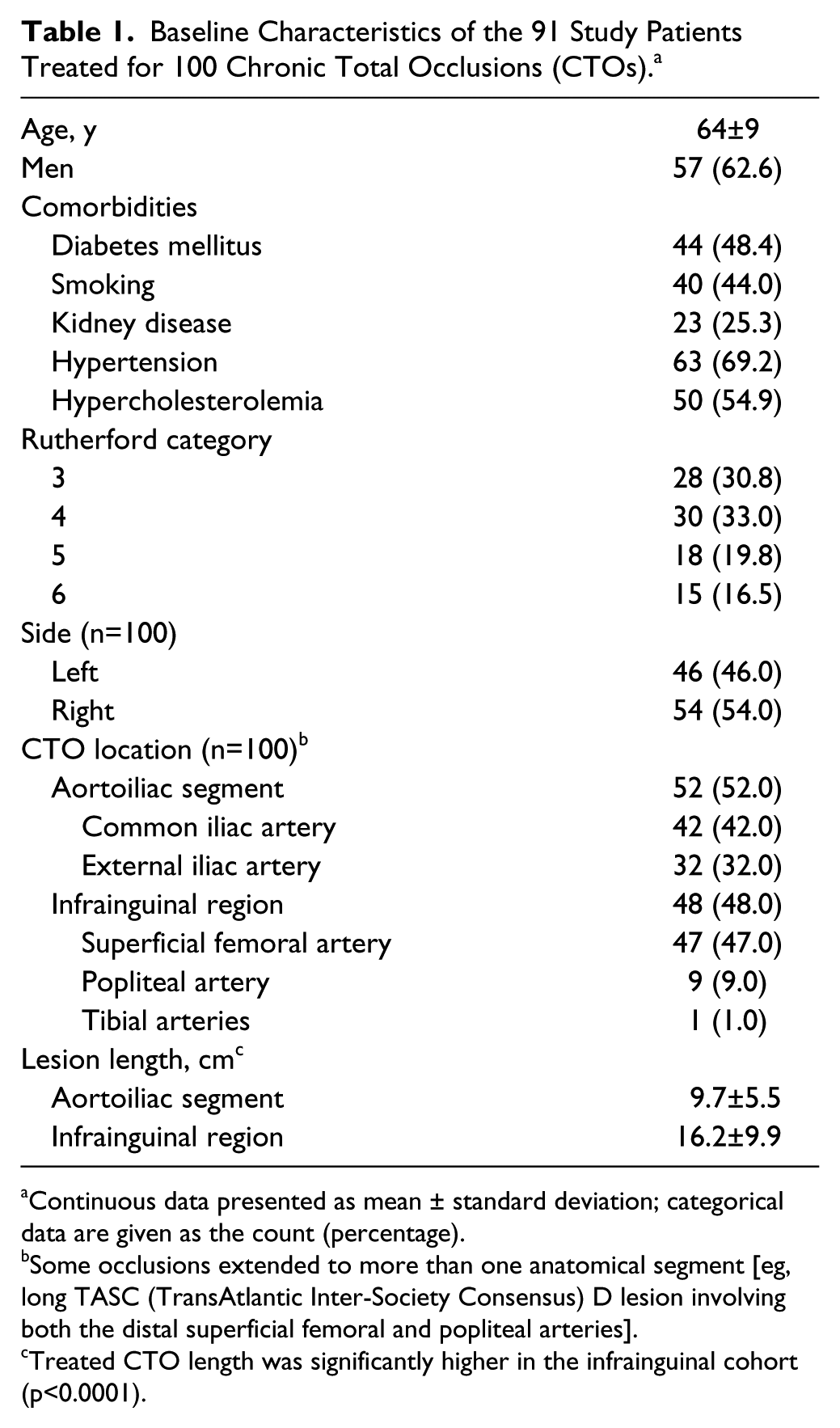
Continuous data presented as mean ± standard deviation; categorical data are given as the count (percentage).
Some occlusions extended to more than one anatomical segment [eg, long TASC (TransAtlantic Inter-Society Consensus) D lesion involving both the distal superficial femoral and popliteal arteries].
Treated CTO length was significantly higher in the infrainguinal cohort (p<0.0001).

(A) Targeted Outback-assisted re-entry in a long-segment left iliac occlusion. (B) A pigtail catheter was advanced from the right side to allow regular angiographic control and serve as a target for the Outback needle. (C) A guidewire has been advanced into the aorta after successful Outback true lumen re-entry, and (D) completion angiography following successful angioplasty and stent placement (the aortoiliac bifurcation was treated with kissing covered stents).

(A) Targeted Outback-assisted re-entry in a distal left femoropopliteal occlusion. (B) Flow reconstituted at the P1 segment of the popliteal artery. (C) After failure to re-enter spontaneously with the subintimal loop technique, the Outback catheter was advanced for targeted re-entry in order to avoid inadvertent distal extension of the subintimal dissection plane. (D) Successful Outback-assisted targeted re-entry with the 0.014-inch wire into the distal popliteal artery. (E) Completion angiography following successful angioplasty and nitinol stent placement.
Systematic Review and Evidence Synthesis
PubMed (MEDLINE), EMBASE, AMED, Scopus, and online content were searched in December 2014 for eligible clinical studies following the PRISMA (Preferred Reporting Items for Systematic reviews and Meta-Analyses) selection process. 12 Cohort and randomized clinical trials (RCTs) were eligible for inclusion as long as they included at least 10 cases that reported the use of the Outback device for true lumen re-entry in the aortoiliac and/or infrainguinal arteries. The title and abstract of 345 scientific records were screened for potential inclusion by 2 interventional radiology fellows; disagreements were resolved by consensus. Most of the abstracts were irrelevant, case reports, or described the use of the Outback in other anatomies. Finally, 11 observational studies were included for further analysis.4–7,13–18 Outcome measures included technical success of true lumen re-entry, any metrics of device re-entry accuracy (if reported), and periprocedure complications according to international reporting standards. 10 Pooled proportional outcomes were calculated with a random effects model to account for clinical and conceptual heterogeneity as described elsewhere in detail.19–21
Statistical Analysis
Discrete variables were expressed as counts (percentages); continuous variables were given as medians (interquartile ranges between the 25th and 75th percentiles) or as means ± standard deviations if normally distributed (Kolmogorov-Smirnov goodness-of-fit normality test). The unpaired Student t test was used to compare normally distributed continuous variables, while the Mann-Whitney test was used for qualitative variables and for non-parametric continuous variables. Comparison of proportions involving small counts (n<5) used the chi-square or Fisher exact test. A multiple comparison chi-square test was applied for >2 groups. Complications by treatment (sole angioplasty vs stent) and vessel segment were compared; the results are presented as the odds ratio (OR) and 95% confidence interval (CI). Outcome measures were stratified according to CTO anatomy (aortoiliac vs infrainguinal) and mode of treatment (balloon angioplasty vs nitinol stent vs covered stent). Statistical analysis was performed with the GraphPad Prism statistical software package (version 5; GraphPad Software, La Jolla, CA, USA).
For the pooled data analysis, quantitative data synthesis was performed with a DerSimonian and Laird random effects model using the Statsdirect statistical package (version 2.7.9; Statsdirect Ltd, Altrincham, UK). The threshold of statistical significance was set at p<0.05 for all comparisons.
Results
Case Series Outcomes
In total, 104 Outback devices were used with the intention to recanalize 100 CTO (52 aortoiliac and 48 infrainguinal) in 91 patients. In 9 aortoiliac cases, 2 iliac vessels were treated per patient. In 1 case, the Outback device was applied successfully in the infrapopliteal arteries (Figure 4). Overall technical success was 93% on a per lesion basis. Success was 90.4% (47/52) in the aortoiliac group and 95.8% (46/48) in the infrainguinal cases (p=0.44). The 7 Outback failures were due to heavy circumferential calcification at the vessel re-entry site; in these cases, 11 Outback devices were employed due to inability to advance and/or destruction of the tip of the first device. Despite repeated balloon predilation of the false lumen and the deployment of new devices, all 11 attempts were unsuccessful. The procedures were eventually aborted and referred for consideration of a vein bypass.

(A) Targeted Outback-assisted re-entry in a heavily calcified tibioperoneal trunk occlusion in a patient with critical limb ischemia. (B) Targeted re-entry was attempted with the Outback device aiming to re-enter the true lumen just proximal to the tibioperoneal bifurcation and maintain flow to >1 vessel to the foot (arrow in A–D). (C, D) Completion angiography following placement of a drug-eluting stent shows restoration of antegrade flow into both the peroneal and posterior tibial arteries as the final re-entry point was located above the tibioperoneal bifurcation.
The length of the treated CTO was significantly longer in the infrainguinal group (16.2±9.9 cm) compared with the aortoiliac segment (9.7±5.5 cm, p<0.001). Re-entry distance was similar between the groups (1.2±0.1 cm for aortoiliac vs 1.3±0.1 cm for infrainguinal, p=0.40). Sites of vessel re-entry were treated with placement of uncovered or covered stents or sole balloon dilation. In the aortoiliac group, primary treatment was balloon angioplasty in 2 (3.8%) cases, nitinol stents in 24 (46.1%) cases, and covered stent in 26 (50%). In the infrainguinal group, 16 (33.3%) vessels were treated with balloon angioplasty, 28 (58.3%) received a nitinol stent, and 4 (8.3%) had a covered stent (Table 2). There were no major complications, but 17 minor complications were identified. Nine of the minor complications were in the aortoiliac segment and included vessel perforation (3/52, 5.7%), dissection/flap (2/52, 3.8%), and distal embolic event (3/52, 5.7%). The remaining 8 minor complications occurred in the infrainguinal group and consisted of vessel perforation (2/48, 4.2%), dissection/flap (3/48, 6.2%), and distal embolic event (2/48, 4.2%; Table 2). The incidence of minor complications was similar between the aortoiliac and infrainguinal groups (p=1.0). However, there were significantly more minor complications in the infrainguinal group when sole balloon angioplasty was used as a definitive treatment of the re-entry zone (OR 4.1, 95% CI 1.3 to 14.2, p=0.01).
Mode of Treatment and Complications. a

Categorical data are given as the count (percentage).
Significantly more minor complications occurred in the infrainguinal group when balloon angioplasty was used as a definitive treatment of the re-entry zone (p=0.01).
There were no statistically significant differences between groups.
Systematic Literature Review
Including the present report, Outback-assisted CTO recanalization was reported in 119 aortoiliac segments (including 52 from the present report) and 464 infrainguinal vessels (including 48 from the present report); there were 249 claudicants and 273 CLI cases (Table 3). Pooled technical success was 90% (95% CI 85% to 94%) and the pooled periprocedure complication rate was 4.3% (95% CI 1.6% to 8.3%), including 1 death due to late femoral bleeding and ensuing myocardial infarction. Statistical heterogeneity (I2) was 71% to 73% (p<0.001); there was no evidence of publication bias.
Synthesis of Outback Outcomes in the Peripheral Arteries.

Abbreviations: AI, aortoiliac; CI, confidence interval; CLD, claudicant; CLI, critical limb ischemia; FP, femoropopliteal; MI, myocardial infarction; NA, not available.
Random effects model.
Discussion
Most interventionists prefer to pursue the intraluminal approach for recanalization of CTOs; however, it is not always a successful one. 9 Hence, subintimal recanalization, in which intentional dissection using the loop technique attempts to bypass the CTO, has emerged as an alternative approach, albeit with a steep learning curve to achieve good results. Admittedly, up to 25% of subintimal recanalizations fail primarily because of inability to re-enter the true lumen distal to the initial CTO. 8 Apart from occlusion chronicity, lesion length is probably also a major contributing factor increasing the technical complexity of the recanalization, as devices have a longer distance of solid degenerate atherosclerosis to travel before reaching the distal healthy vascular bed.
In addition, vessel calcification is a long recognized predictor of subintimal re-entry failure. 22 Circumferential calcium deposits may extend to the distal media vessel layer downstream of the baseline CTO, prohibiting spontaneous breakthrough into the true lumen as originally theorized by Bolia et al. 2 Hence, advancement of the looped wire may inadvertently dissect the distal landing zone and promote distal false lumen development that may compromise the true lumen, thereby wasting further healthy vessel until successful re-entry is eventually achieved. This is particularly important in cases of subintimal recanalization in the aortoiliac region, where the tract of false lumen dissection may extend to the inferior mesenteric or renal arteries or potentially compromise the origin of the lumbar and/or spinal feeding vessels. On the other hand, in the infrainguinal region, the confluence of important genicular collaterals routinely found at the adductor hiatus level may be compromised should the proximal popliteal artery develop a spiral dissection.
To date, dedicated re-entry catheters (with or without inert image guidance) have been developed to effectively assist operators in cases of unsuccessful distal spontaneous re-entry. The Outback re-entry catheter has been tested in several studies.4–7,13–18 To our knowledge, this is one of the largest retrospective analyses of Outback-assisted subintimal recanalization including CTOs in both the aortoiliac and infrainguinal regions. In the present report, the Outback re-entry catheter performed very well, with an overall 93% success rate and an associated 17% rate of minor complications without any major events. Failures were attributed to heavy vessel wall calcifications. This is well in line with previous reports having identified heavy calcifications as the sole determinant of Outback failure.4,8,17 Occasionally, calcification may be so heavy that it prohibits not only needle penetration but also needle deployment. 18
Our success rate exceeded the 90% threshold in both the aortoiliac and infrainguinal segments and, in fact, approximates the upper confidence interval of the pooled success rate calculated from the evidence synthesis. Of note, we had no major complications, but the rate of minor sequelae was the highest among the collated reports (17% vs the pooled 4.3%). This is due to the fact that the authors decided to report all the broad spectrum of minor complications regardless of the degree of severity. For example, severe intimal dissections were classified and reported as minor complications, even though they inherently result from the process of subintimal angioplasty.
Of interest, significantly more minor complications occurred in the infrainguinal region when balloon angioplasty was used as the primary mode of treatment of the subintimal lumen and re-entry site. We speculate that the presence of metallic scaffolding was critical in avoiding the dislodgment of plaque and inhibiting acute development of elastic recoil and/or flow-limiting intimal flaps. Currently, it is our routine practice to apply primary stent placement in the treatment of complex aortoiliac CTOs, as well as in long complex femoropopliteal occlusions.
Overall, the pooled analysis has shown that the Outback catheter is safe and very effective in accomplishing true lumen re-entry once the standard techniques have failed. Of note, most of the published experience related to femoropopliteal CTOs (464/ 583 lesions) and the present report includes the largest aortoiliac series to date.
The ability of the Outback catheter to achieve targeted re-entry at the preplanned vessel site is the primary advantage of the device. Extension of vessel dissection and sacrifice of distal healthy vessel is a major limitation of conventional subintimal angioplasty techniques, especially when distal re-entry relies solely on spontaneous intimal breakthrough by the guidewire. In our study, the re-entry distance was around 1 cm in both the aortoiliac and infrainguinal vessels, which represents no more than 10% of extra lumen wasted compared with the baseline CTO lesion length. Thus, Outback-assisted recanalization was very accurate, with minimal true vessel sacrifice both above and below the inguinal ligament.
In the only RCT available to date, Gandini et al 4 compared the Outback device with a manual re-entry technique in 52 CTOs in the femoropopliteal artery. Technical success, defined as successful re-entry within 5 cm of the preplanned landing site, was 100% in the Outback arm vs <50% in the control arm. In addition, procedure and fluoroscopy times were significantly reduced in the Outback study arm. Consequently, the utility of the Outback re-entry catheter in achieving accurate targeted re-entry when crossing CTOs cannot be overstated.
Limitations
The fact that this was a retrospective data analysis of cases performed in a single center constitutes the major limitation of the present study. Of note, cases were performed by several different operators and included both the aortoiliac and the infrainguinal vessels. Thus the present experience may be considered representative of expected success and minor complication rates in everyday practice. The absence of a comparator group, the lack of independent core lab analysis, and no objective standardized analysis of the extent of vessel wall calcification are further study limitations. In addition, the systematic review of amassed evidence on the Outback device provides only a low level of evidence because it relied on mostly observational studies and only a single RCT. Finally, cost utility concerns were not addressed.
Conclusion
The Outback catheter is safe and has a very high technical success rate in achieving targeted true lumen re-entry after failed spontaneous subintimal recanalization. It is highly accurate and avoids inadvertent extension of distal true lumen dissection, thereby minimizing the sacrifice of healthy vessel and maintaining options for future repeat endovascular procedures or surgical bypass.
Footnotes
Authors’ Note
In memory of Dr Richard Salter, who was a dear colleague, a great teacher, and an amazing endovascular specialist.
Declaration of Conflicting Interests
The author(s) declare no potential conflicts of interest with respect to the research, authorship, and/or publication of this article.
Funding
The author(s) received no financial support for the research, authorship, and/or publication of this article.