
Abstract
Keywords
Atherosclerotic carotid artery stenosis is a main cause of transient ischemic attack and stroke.1,2 Evidence is accumulating that plaque composition determines the vulnerability of the lesion and relationship to major stroke.3,4 Features of the vulnerable carotid plaque, including plaque thrombus, low fibrous content, macrophage infiltration, and microvessel density, correlate with predicted stroke risk. 4
Carotid artery stenting (CAS) has emerged as a therapeutic alternative to carotid endarterectomy for the treatment of extracranial carotid artery stenosis5,6; however, stent malapposition, plaque prolapse, and fibrous cap rupture may occur after CAS. 7 In the coronary artery, stent strut apposition has been linked to the risk of stent thrombosis, 8 and acute stent thrombosis is associated with residual exposed lipid from the culprit plaque. 9 However, the impact of plaque morphology on stent apposition is still unquantified because available imaging systems [angiography, intravascular ultrasound (IVUS), and duplex ultrasound] may not be able to detect such microdefects.
Optical coherence tomography (OCT) is an optical analogue of intravascular ultrasound that allows high-resolution (10 µm) tomographic intra-arterial imaging.10,11 OCT has been shown to accurately identify plaque components, such as lipid, calcium, fibrous tissue, thin-cap fibroatheroma (TCFA), intraluminal thrombus, calcified nodules, and vascular inflammation.12–14 The superiority of OCT in the setting of coronary stent apposition has been because of its ability to resolve small gaps between the stent strut and the vessel wall, which is often missed by IVUS.13,15 Recent reports have suggested that the evaluation of carotid plaque characteristics by OCT has the potential to alter the understanding and treatment of carotid artery disease.16,17
This study examined stent strut apposition after CAS based on the presence of lipid-rich plaque with the aim of shedding light on whether stent malapposition, plaque prolapse, and fibrous cap rupture could be linked to in-stent restenosis (ISR).
Methods
Study Population
Under a study protocol approved by our hospital’s ethics committee, data on 26 consecutive patients who underwent CAS with OCT imaging between April 2014 and May 2015 at a single center were retrieved from the prospectively maintained Nanjing Stroke Registry Program (NSRP) database. 18 Patients were eligible for CAS if there was a >70% asymptomatic internal carotid artery (ICA) stenosis or a >50% symptomatic ICA stenosis according NASCET (North American Symptomatic Carotid Endarterectomy Trial) criteria.19,20 Patients were ineligible if there was renal insufficiency, severe arrhythmia, arteriovenous malformation, carotid tortuosity, severely disabling stroke or dementia, critical intracerebral stenosis, cerebral tumor, or significant thrombus at the lesion site. Duplex ultrasound imaging and independent neurological examinations of all patients were performed before the intervention.
CAS Procedure and OCT Imaging Protocol
Carotid artery stenting was performed according to standard protocol by an interventional cardiologist with OCT experience (Y.J.) and an interventional neuroradiologist (M.L.). The degree of carotid artery stenosis was confirmed by digital subtraction angiography (DSA) prior to the procedure using a dynaCT angiography scanner (Siemens Axiom Artis dTA; Siemens Healthcare, Erlangen, Germany). A cerebral protection device (NAV-6 Filterwire; Abbott Vascular, Redwood City, CA, USA) was deployed distal to the culprit lesion in the extracranial ICA. Carotid OCT images were acquired before stent deployment using a 2.7-F Dragonfly OCT imaging catheter (St. Jude Medical, St. Paul, MN, USA) inserted through an 8-F sheath over the 0.014-inch guidewire of the filter and navigated past the ICA lesion. Images were calibrated by adjustment of the Z-offset. Before image acquisition, blood was displaced by automatic injection (Medrad Mark V ProVis; Bayer Healthcare, Whippany, NJ, USA) of 15 mL of undiluted contrast (iodixanol 320) with a velocity of 8 mL/s and automatic pullbacks covering 54 mm of the vessel at a velocity of 20 mm/s. 16 After deployment of the self-expanding Precise stents (Cordis Corporation, Bridgewater, NJ, USA), the same OCT maneuvers were performed after stent dilation. Technical success of the CAS procedure was determined by ≤30% residual stenosis on the final angiogram.
Patients were prescribed aspirin (100 mg/d) and clopidogrel (75 mg/d). The degree of ISR was measured on DSA images or estimated from the computed tomography (CT) scans at 6 months in all patients.
Imaging Analysis
Extracranial vessel analysis on the DSAs was performed on a segmental basis according to the NASCET criteria by a single operator (Q.Y.) and reviewed by a second operator (M.L.) before OCT analysis. Angiographic and OCT analyses used co-registration based on the carotid artery bifurcation to ensure that measurements were made at identical sites.
Optical coherence tomography data were stored using available OCT systems (C7-XR or Ilumien Imaging System; St. Jude Medical) and analyzed by 2 experienced OCT readers (R.L. and Y.J.) using dedicated software with an automated contour-detection algorithm (Off-line Review Software, version C.0.2; St. Jude Medical). The OCT images were evaluated according to a previously reported protocol based on the accuracy of vessel wall identification. 16 Images were considered nonanalyzable if any portion of the cross-sectional image was out of the screen, if there was a fold-over artifact, or if intraluminal blood impaired the assessment of a continuous 270° arc. 16
The lesion location was identified, and the distal reference was defined as the most normal-appearing segment distal to the lesion shoulders by OCT. On the basis of previously published pathological and noninvasive imaging data,10,16 a lipid-rich plaque was defined by lipid present in ≥2 quadrants; a non–lipid-rich plaque referred to lipid present in <2 quadrants in any of the images. For each patient, the cross-sectional image with the highest number of lipid quadrants was used for analysis.
The fibrous cap thickness was measured at its thinnest part for all images with an OCT-determined lipid pool. 21 Cap thickness of each image was measured 3 different times and averaged. If a patient had >1 image within a plaque, the thinnest cap measurement was used for measurement. TCFA was defined as a plaque with lipid content in ≥2 quadrants and the thinnest part of a fibrous cap measuring ≤65 µm. The presence of plaque disruption, calcium, or thrombus was also noted (Figure 1). Thrombus was any mass protruding into the lumen, with an irregular surface and a sharp intensity gap between the mass and surrounding tissue. 10
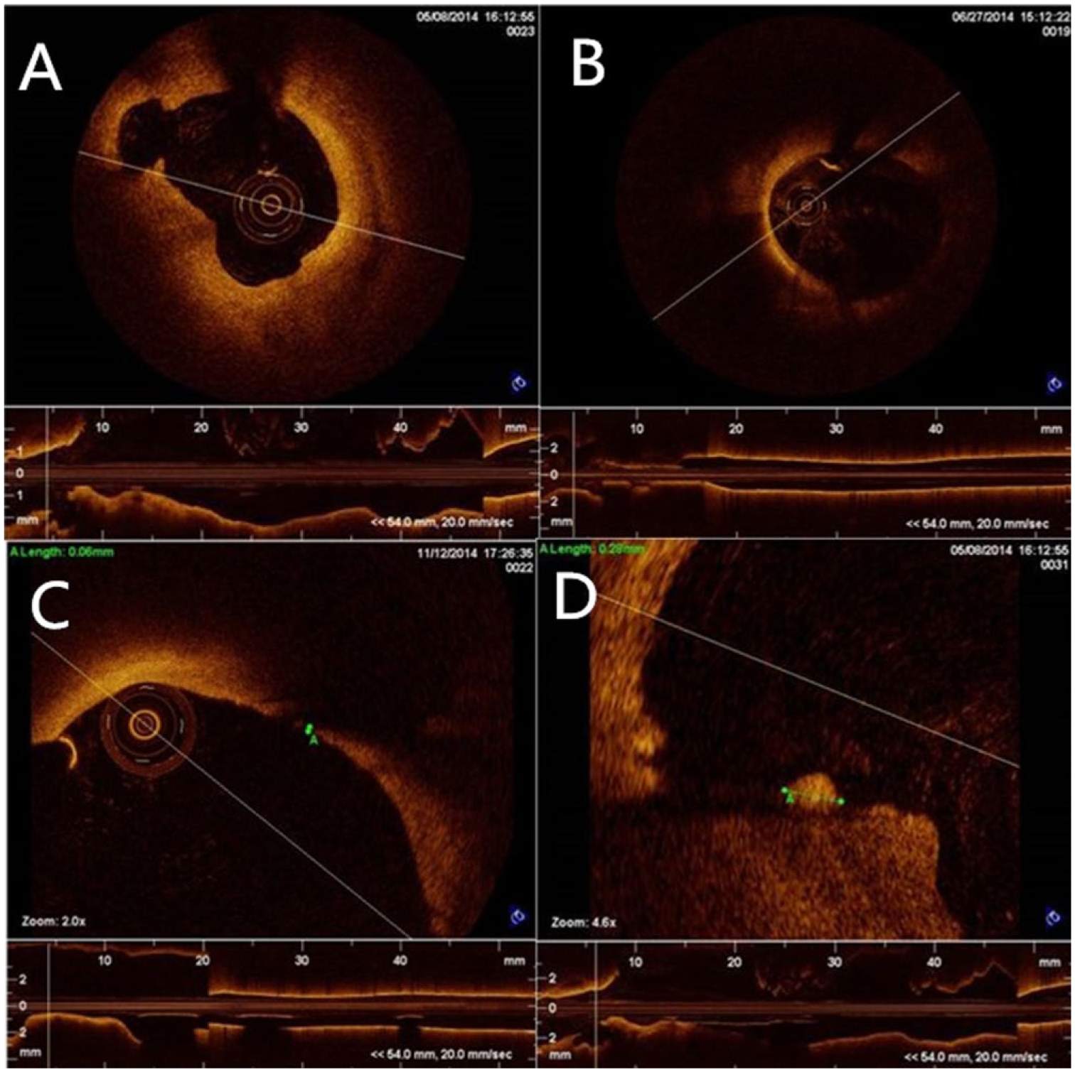
Optical coherence tomography images of (A) plaque disruption, (B) lipid-rich plaque, (C) thin-cap fibroatheroma, and (D) thrombus.
Cross-sectional OCT images within the stented ICA segment were evaluated at 1-mm intervals for the presence of malapposition, plaque prolapse, and fibrous cap rupture (Figure 2). Malapposition was defined when the distance from the surface of the blooming (the inner and outer contours of each strut reflection) to the lumen contour was greater than the total thickness of the stent strut plus one half of the blooming. According to the carotid stent strut thickness, a well apposed strut referred to a distance ranging from 10 to 200 µm, while a malapposed strut was indicated by a distance >200 µm. Stent struts were defined as embedded when buried into the intima for almost their entire thickness (distance measured <10 µm). Any appreciable tissue prolapse between the stent struts was considered as plaque prolapse after stenting, while a rupture of the fibrous cap was defined as any discontinuity of the inner layer of the plaque profile. 7 Data on stent malapposition were expressed as the percentage of struts analyzed, while the results of stent malapposition, plaque prolapse, and rupture of fibrous cap were based on the number of slices evaluated.
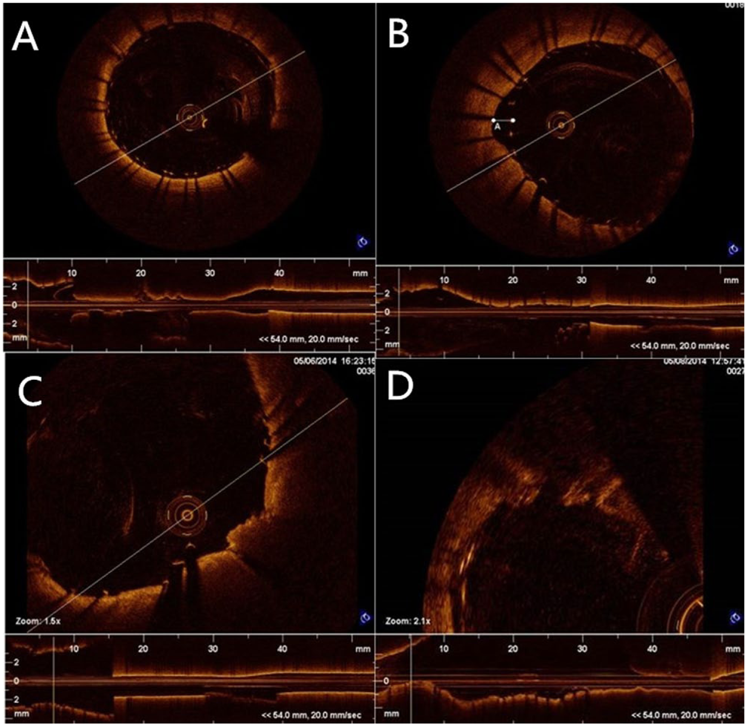
Optical coherence tomography classification of stent strut apposition: (A) well apposed, (B) malapposed, (C) embedded and plaque prolapse, and (D) rupture of fibrous cap.
Statistical Analysis
Data were expressed as mean ± standard deviation or median with interquartile range based on distribution. Differences in baseline characteristics between the lipid-rich plaque group and the non–lipid-rich plaque group were analyzed using a chi-square test, Fisher exact test, or one-way analysis of variance. The frequencies of various types of plaques or of TCFA were compared between groups using a chi-square or Fisher exact test. Significant differences of cap thickness were tested using a Kruskal-Wallis test.
Inter- and intraobserver variabilities were measured for evaluation of all OCT images by 2 independent readers and by the same reader at 2 separate time points using the Cohen kappa (κ) test of concordance. All statistical analyses were performed using SPSS software (version 20.0; IBM Corporation, Somers, NY, USA).
Results
OCT and CAS Procedures
Of the 26 patients in whom OCT imaging was attempted, adequate imaging quality could not be obtained in 6 patients due to technical difficulties (vascular tortuosity, inadequate blood displacement). Of the interpretable images acquired in 20 patients (Table 1), the majority (91.8%) of the OCT frames were analyzable. The reasons for nonanalyzable frames were out-of-screen images (7.0%) and the presence of residual blood impairing proper assessment (1.2%).
Baseline Characteristics of the Study Population.
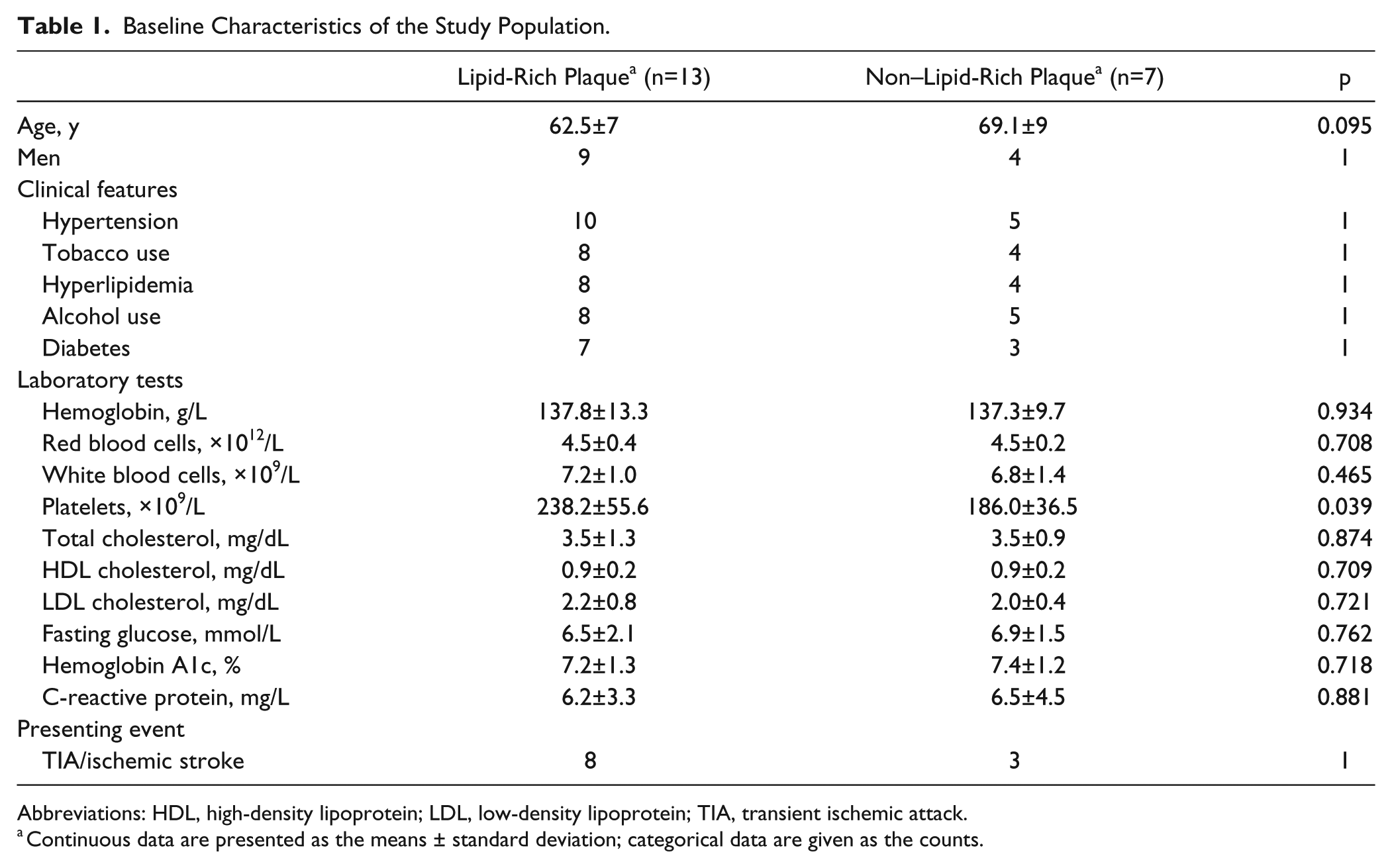
Abbreviations: HDL, high-density lipoprotein; LDL, low-density lipoprotein; TIA, transient ischemic attack.
Continuous data are presented as the means ± standard deviation; categorical data are given as the counts.
Successful revascularization with <30% residual stenosis in each case was confirmed by completion angiography. No technical or neurological complications occurred during OCT pullbacks. The mean time for angiography, CAS, and OCT imaging was 66±12 minutes, during which 97±16 mL of contract (range 74–143) was used per patient. No stroke or death was recorded at 30 days.
OCT Results
There were no significant differences between the groups with (n=13) and without (n=7) lipid-rich plaques in terms of baseline data (Table 1). OCT analysis within the ICA lesion segment and stent segment at 1-mm intervals produced a mean of 50±5 slices in each patient. A total of 500 cross-sectional OCT images were analyzed to assess plaque characteristics, and 500 images were chosen to assess the rate of stent apposition, plaque prolapse, and fibrous cap rupture (Table 2). A mean of 28±2 stent struts was detected in each slice for a total of 13,910 struts evaluated.
Plaque Morphology and Stenosis Grade in Patients With vs Without Lipid-Rich Plaques.
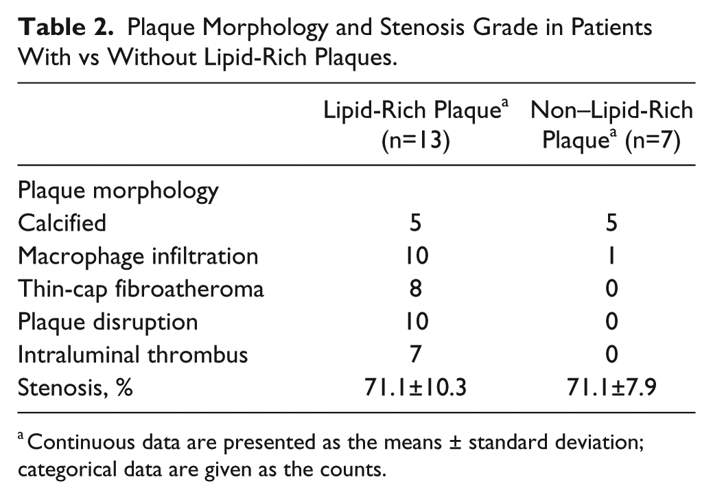
Continuous data are presented as the means ± standard deviation; categorical data are given as the counts.
Intra- and interrater agreement for OCT qualitative assessments of plaque components was high (κ=0.92 and κ=0.91, respectively; both p<0.001). Interobserver variability was very good for identification of stent apposition (κ=0.84), plaque prolapse (κ=0.85), and rupture of the fibrous cap (κ=0.81). Intraobserver agreement, evaluated for one observer (R.L.) assessing images twice within a 7-day interval, was good (stent apposition κ=0.95, plaque prolapse κ=0.85, and fibrous cap κ=0.81).
In the strut-based analysis of stent apposition (Table 3), an average of 16.0% of the struts were malapposed in the lipid-rich plaque group compared with 16.7% in the non–lipid-rich plaque group (p=0.953). Stent struts were found to be embedded into the intima in 29.4% of the lipid-rich plaque group compared with 23.7% of the non–lipid-rich plaque group (p<0.001), but well-apposed struts were less frequent in the lipid-rich group vs the non–lipid-rich plaque group (54.6% vs 59.6%, respectively; p<0.001).
Strut Apposition to the Arterial Wall Based on Lipid-Rich Plaque.
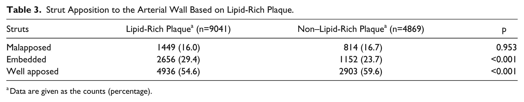
Data are given as the counts (percentage).
In the slice-based analysis, plaque prolapse was significantly less frequent in patients with non–lipid-rich plaques (49.1%) compared to lipid-rich plaque patients (65.5%; p<0.001). Significant differences were also noted in the rates of fibrous cap rupture between the lipid-rich and non–lipid-rich groups (49.8% vs 33.7%; p<0.001). The rate of malapposition between the lipid-rich (12.3%) and non–lipid-rich (14.9%) groups was not significant (p=0.489).
Table 4 gives the OCT outcomes and 6-month follow-up evaluation of ISR by CTA (n=11) or DSA (n=9). No patient had >50% ISR, but 12 patients had some degree of restenosis. In one patient who had 21% residual stenosis after the procedure, follow-up angiography demonstrated 42% ISR at 6 months. In 3 patients with no residual stenosis, angiography documented 8%, 17%, and 20% ISR. In the patients having CTA, mild ISR was found in 6 patients; 5 of them had >20% residual stenosis after the CAS procedure. In the 8 patients with no obvious restenosis by either imaging technique, malapposed struts, embedded struts, plaque prolapse, and fibrous cap rupture were still found.
Optical Coherence Tomography Outcomes and 6-Month Follow-up in 20 CAS Patients.

Abbreviations: CAS, carotid artery stenting; CTA, computed tomography angiography; DSA, digital subtraction angiography; ILT, intraluminal thrombus; ISR, in-stent restenosis; TCFA, thin-cap fibroatheroma; TIA, transient ischemic attack.
N per total evaluated slices in parentheses.
Discussion
To the best of our knowledge, OCT has not heretofore been used to assess the rate of stent malapposition, plaque prolapse, and fibrous cap rupture relative to characteristics of carotid artery plaques. While malapposed stent struts were not significantly different in patients with vs without lipid-rich plaques, the rate of embedded stent struts was more common in the lipid-rich plaque group. Patients with non–lipid-rich plaque had less frequent prolapse and fibrous cap rupture. There were no higher rates of malapposed stent struts or fibrous cap ruptures in patents with measurable restenosis in this study.
A number of potential variables in addition to stent design may influence the result detected by OCT. For example, calcified plaque results in more malapposed struts, while soft plaques may generate more plaque prolapse. 7 Complicated plaques, defined by intraplaque hemorrhage, surface disruption, or intraluminal thrombus, are a type of extensive histological classification of atherosclerosis. 22 In the present study, uncomplicated plaques were more common in asymptomatic than symptomatic patients, as was seen by Jones et al. 16 Howard et al 4 discovered complicated plaques in nearly 75% of patients with symptomatic disease.
Current consensus guidelines cite only the degree of stenosis as an indication for carotid revascularization. In fact, the identification of patients with asymptomatic disease who are at risk of stroke remains challenging as there is an increasing awareness that the degree of carotid stenosis is a poor predictor of stroke risk. 23 But this classification scheme of plaque is limited by its reliance on pathological specimens or in vivo plaque assessment. The 10-µm resolution of OCT is recognized to be accurate in the identification of all the important features of plaque vulnerability, including vascular inflammation, intraluminal thrombus, and plaque rupture.12,14
de Donato et al 7 reported carotid stent apposition, plaque prolapse, and fibrous cap rupture systematically following CAS according to the stent design using OCT. The study suggested that OCT, with its high-resolution capability, can detect complex interaction between carotid plaques and stents. Some coronary stent studies demonstrated the beneficial effects of strut coverage in embedded struts vs apposed or malapposed struts.24,25 The percentage of uncovered struts in the presence of tissue prolapse within the stent was found significantly more often in the subgroup of embedded struts. Both the abluminal and luminal thrombi were generally resolved by the time of follow-up in the embedded struts. 24 Incomplete coronary stent strut apposition has recently received attention, with studies suggesting an association with an increased rate of long-term stent thrombosis. 25
In-stent restenosis is an important factor endangering the long-term safety and efficacy of CAS. It is plausible that soft vulnerable plaques are more likely to be injured during the CAS procedure and are therefore more likely to initiate the cascade that leads to ISR. Increased calcification in the plaque before treatment is associated with a decrease of stent expansion, which is known as a risk factor for the development of ISR.26,27
In previous studies, stent malapposition was more frequent with closed-cell stents, while plaque prolapse was more common with open-cell stents.7,26 In our study, open-cell stents were used in all the patients; only ~50% of stent struts analyzed by OCT were considered well apposed to the arterial wall. Moreover, plaque prolapse was common (>60%) too. It is known that the constant radial force of self-expanding stents and the re-endothelialization process might both play roles in this remodeling process after CAS, so OCT analysis might show better stent strut apposition and less prolapse of plaque, the relationship of plaque characteristics and stent apposition is still undefined. However, it is obvious that soft plaque may be responsible for more embedded stent struts, plaque prolapse, or fibrous cap rupture, as our research has confirmed.
Calcified plaque may cause higher rates of strut malapposition. Platelet micro-aggregation can occur behind malapposed stent struts, which may cause thrombus formation and ISR. Finally, rupture of the fibrous cap, even in a nonprolapsing plaque, may also be the reason for restenosis, but this study did not find higher rates of malapposed stent struts or fibrous cap ruptures in patents with obvious restenosis, which may be due to the short follow-up and the limited number of patients.
Limitations
This was a single-center study with a limited number of patients because OCT is a relatively new technology in China. Surrogate parameters were alternatives to clinical events. Any baseline patient factors could have influenced the reported findings. The events of stent malapposition, plaque prolapse, or fibrous cap rupture after CAS can be affected by several factors, but the small number of cases and the large number of potential determinants could not allow powerful multivariate analysis.
Microdefects after stent deployment are frequent and are related to the design of implanted stents. In this study, according to a tailored strategy that takes into account plaque type, anatomy at the level of the culprit lesion, and patient symptoms, open-cell stents were used in all the 20 patients, which may have introduced a selection bias. In this study, only a 5-cm-long section of the stent was involved with the most diseased plaque zone. The key information in terms of plaque injury and the strut-plaque relationship may be at the zone of maximum stenosis and/or plaque mass.
Conclusion
Embedded stent struts were more common in lipid-rich plaque patients, while plaque prolapse and fibrous cap rupture were less common in non–lipid-rich plaque patients. In this study, we did not find higher rates of malapposed stent struts or fibrous cap rupture in patents with obvious restenosis. Longer follow-up is needed to evaluate the long-term effect of stent apposition on ISR.
Footnotes
Declaration of Conflicting Interests
The author(s) declared no potential conflicts of interest with respect to the research, authorship, and/or publication of this article.
Funding
The author(s) disclosed receipt of the following financial support for the research, authorship, and/or publication of this article: The project is supported by the National Natural Science Foundation of China–the Major International (Regional) Joint Research Program of China (Grant Number: 81220108008), the National Natural Science Foundation of China (Grant Number: 31171016 and 81400898), and the Jiangsu Provincial Special Program of Medical Science (Grant Number: BL2013025).