





Abstract
Keywords
Introduction
The femoropopliteal arterial segment is a particularly challenging location for implantation of a permanent endoprosthesis due to extensive plaque burden and the extreme mechanical forces sustained during the activities of daily living. Newer nitinol stents with enhanced flexibility have been developed for femoropopliteal use and offer the promise of a reduced rate of stent fracture and improved long-term patency.1,2 Recent trials with these newer generation stents have demonstrated superior outcomes compared to percutaneous transluminal angioplasty in femoropopliteal lesions, with excellent freedom from target lesion revascularization (TLR) up to 3 years.3–6 Sustained benefit of a paclitaxel-eluting nitinol stent has been demonstrated to 5 years. 7 Most of these clinical trials have excluded lesions >15 cm in length. Analyses have suggested a linear relationship between lesion length and risk of restenosis, and the safety and effectiveness of stenting for long femoropopliteal lesions is uncertain.8,9
The TIGRIS Vascular Stent (W.L. Gore & Associates, Flagstaff, AZ, USA) is a new-generation nitinol stent with enhanced flexibility and thromboresistance, which may make this stent design attractive for femoropopliteal use in terms of fewer stent fractures and improved long-term outcomes. To test this potential, an international, multicenter, randomized controlled trial (RCT) was initiated to compare the TIGRIS Vascular Stent to the LifeStent (Bard Peripheral Vascular, Tempe, AZ, USA), a second-generation nitinol stent, for the treatment of atherosclerotic lesions in the superficial femoral artery (SFA) and proximal popliteal artery (PPA) in patients with symptomatic peripheral artery disease (PAD).
Methods
Study Design
This safety and efficacy trial was conducted under a Food and Drug Administration (FDA)–approved clinical protocol in support of an investigational device exemption (IDE). The objective was to test the hypothesis that the 30-day safety and 12-month primary patency of the TIGRIS Vascular Stent were statistically noninferior to the control device (LifeStent) when used to treat de novo or restenotic PAD lesions ≤24 cm long in the SFA/PPA. The multicenter RCT was designed to randomly assign eligible patients in a 3:1 ratio to treatment with either the TIGRIS or LifeStent at 33 sites in the United States and 3 sites in Europe. Randomization, which was not complete until the intraprocedural angiographic criteria had been met, was stratified by site and lesion length (≤16 or >16 cm) in blocks to assure that the proper ratio of subjects was assigned to each treatment group. The institutional review board or ethics committee at each study site approved the protocol.
The study was conducted in accordance with International Conference on Harmonization Guidelines on Good Clinical Practice and applicable regulatory requirements in the United States and Europe. An independent data and safety monitoring board (DSMB) was established to review data, including all adverse events and protocol deviations, on an approximately quarterly basis.
All imaging data collected during the study were analyzed by independent angiographic (Yale University Angiographic Laboratory, New Haven, CT, USA) and ultrasound/radiography (VasCore, Boston, MA, USA) core laboratories.
Study Devices
The TIGRIS Vascular Stent is a flexible, self-expanding, wire-wound nitinol frame covered with a lattice of fluorinated ethylene propylene/expanded polytetrafluoroethylene extending along its entire length (Figure 1). Gore’s thromboresistant CBAS Heparin Surface, a stable, reduced molecular weight heparin of porcine origin, is covalently bonded to the stent surface. A triaxial delivery catheter allows accurate stent deployment. The TIGRIS Vascular Stents used in this study were 5, 6, and 7 mm in diameter and 3, 4, 6, 8, and 10 cm in length.

The TIGRIS stent platform consists of (A) a wire-wound nitinol frame with (B) a fluoropolymer lattice extending along its entire length.
The control device (LifeStent) is an FDA-approved self-expanding bare nitinol stent intended to improve luminal diameter in the treatment of symptomatic de novo or restenotic lesions up to 24 cm in length in the native SFA/PPA, with reference vessel diameters ranging from 4.0 to 6.5 mm. Sizes of control devices implanted in the study were 6 and 7 mm in diameter and 2, 3, 4, 6, 8, 10, 12, 15, and 17 cm in length.
Inclusion/Exclusion Criteria
Patients were eligible for inclusion if they had symptomatic PAD classified as Rutherford category 2 to 4 ischemia, an abnormal ankle-brachial index (ABI) ≤0.9 or toe-brachial index (TBI) ≤0.7, and intraprocedural angiographic evidence of a single de novo or restenotic lesion in the SFA/PPA with a cumulative length visually estimated to be ≤24 cm, with ≥1 region of focal luminal narrowing ≥50% and at least 1 patent tibial artery. The angiographic inclusion criteria had to be met before randomization was completed.
Patients were ineligible for the study if they had vascular access/catheterization in the target leg within 30 days of study enrollment, prior treatment (stenting or bypass) of the SFA/PPA in the target leg, flow-limiting aortoiliac disease (uncorrected), or additional ipsilateral femoropopliteal disease outside of the lesion to be stented.
Procedure and Follow-up Evaluation
After all angiographic inclusion criteria had been met and randomization was complete, the investigator proceeded with stent implantation in accordance with the assigned treatment arm. The study protocol dictated that lesions should be fully stented (no spot stenting allowed), with stent coverage extending 1 cm into healthy tissue on either end of the lesion. Images of the stent and stent delivery system were obtained before and after stent deployment and used to quantify any stent elongation that occurred during the deployment process.
Follow-up visits were scheduled for 1, 6, 12, 24, and 36 months postprocedure. Each visit included a physical examination, assessment of the Rutherford category, ABI or TBI measurement (whichever was collected at baseline), duplex ultrasound, and quality of life (QoL) assessment with the EQ-5D (EuroQol health status measurement tool in 5 dimensions; EuroQol, Rotterdam, the Netherlands; available at http://www.euroqol.org/eq-5d-products/eq-5d-3l.html) for overall QoL and the Peripheral Artery Questionnaire (PAQ) for PAD-specific QoL. 10 Radiography of the treated region was also obtained at 12, 24, and 36 months. Ultrasound images were examined by the core laboratory to determine patency within the stented region; radiographs (straight and bent knee) were analyzed to identify stent fractures, which were classified according the schema of Jaff et al. 11 Angiographic images were analyzed by the angiographic core laboratory for relevant pre- and postprocedure characteristics of the target lesion and vessel.
Patient Enrollment
Between May 2012 and June 2014, 274 patients were randomized to treatment with the TIGRIS stent (n=197; mean age 66.7±9.28 years, 141 men) or LifeStent (n=70; mean age 67.9±8.87 years, 49 men), forming the intent-to-treat (ITT) population. The baseline demographics and risk factors are presented in Table 1. Both groups had >85% current or former smokers and other comorbidities typical of a PAD population. There were more non-whites in the study group.
Patient Characteristics. a

Continuous data are presented as the means ± standard deviation; categorical data are given as the counts/sample (percentage).
The baseline lesion characteristics are summarized in Table 2. The mean lesion lengths were 107.6±68.6 mm for the TIGRIS group and 117.9±75.4 mm for the controls. For both groups, 44% of lesions were located in the distal SFA or proximal PPA. Both groups also had ~40% occlusions and 50% moderately or severely calcified lesions.
Lesion Characteristics. a

Abbreviation: SFA, superficial femoral artery.
Continuous data are presented as the means ± standard deviation; categorical data are given as the counts/sample (percentage).
Endpoints and Outcomes
The primary safety endpoint was the 30-day major adverse events (MAE), which included death, target vessel revascularization (TVR), and amputation above the metatarsals in the treated limb. The primary efficacy outcome was primary patency at 12 months after implantation, defined as uninterrupted patency without TLR or restenosis as documented by a peak systolic velocity ratio ≤2.5 on duplex imaging.
Secondary outcomes included device success (successful delivery of the stent to the intended site and successful deployment), procedure success (device implantation with residual stenosis <30% and no serious adverse events), freedom from TVR, freedom from TLR (treatment for restenosis or occlusion by any means), Rutherford category improvement over the baseline value, ABI/TBI improvement, QoL changes reflected in the EQ-5D and PAQ, primary patency at 1 and 6 months after implantation, assisted primary patency (uninterrupted patency regardless of reinterventions performed), secondary patency (patency restored by endovascular means after occlusion), and freedom from stent fracture as adjudicated by an independent core laboratory.
Statistical Analysis
Continuous data were compared using the Student t test and are presented as the means ± standard deviation. Categorical data were compared using the Fisher exact test and are given as the counts (percentage). Time-to-event analysis of patency and freedom from TLR was performed using the Kaplan-Meier method; the groups were compared using the log-rank text. Cox regression analyses were used to investigate the impact of lesion length or number of stents on primary patency. The results are presented as the odds ratio (OR) with 95% confidence interval (CI) and the Wald chi-square p-value. The threshold of statistical significance was p<0.05. Statistical analyses were performed using SAS software (version 9.3; SAS Institute, Inc, Cary, NC, USA).
Results
While the DSMB made no recommendation to modify or discontinue the study, it did identify 7 subjects as protocol deviations that could affect the evaluation of primary endpoints, leaving 267 patients in the per-protocol group. Results presented below reflect analysis of the per-protocol population; however, the data analyses for the per-protocol and as-treated patient cohorts demonstrated similar results.
The procedure was successful in 196 (99.5%) of the 197 TIGRIS subjects and 68 (97.1%) of the 70 control subjects. One TIGRIS device could not be deployed within the target lesion; the subject was successfully treated using balloon angioplasty alone. In the control group, there was >30% residual stenosis after the index procedure in 1 subject and the other experienced a myocardial infarction immediately following the procedure. Neither the TIGRIS nor LifeStent demonstrated significant elongation during deployment (0.03%±0.52% vs 0.12%±0.36%, p=0.36). The only statistically significant difference between the test and control devices was the number of stents needed to cover the lesion (Table 2). Of the 196 lesions stented in the study group, 40 (20.4%) required 3 or 4 stents compared with only 4 (5.7%) of 70 control lesions requiring 3 stents (p=0.033). This difference was due to the availability of longer control devices (up to 17 cm) compared with study devices (up to 10 cm).
The safety of the TIGRIS Vascular Stent was found to be statistically noninferior (p>0.99) to that of the control device at 30 days after implant. There was a single MAE in the study group that required a TVR on the day of the index procedure to treat a procedural complication outside the margins of the deployed TIGRIS stent.
By the 12-month follow-up window 25 patients were no longer in the study: 11 (6 TIGRIS) withdrew consent, 1 (TIGRIS) was removed by the investigator, 6 (all TIGRIS) were lost to follow-up, 3 (2 TIGRIS, 1 LifeStent) had surgical bypass procedures, and 4 (4 TIGRIS) died. No deaths were determined to have a relationship to the study device or procedure.
Kaplan-Meier survival curves identified no difference in primary patency (Figure 2A) between the TIGRIS and LifeStent groups at 12 months (60.6% vs 63.2%, p=0.73) or 24 months (56.3% vs 50.2%, p=0.60), nor was there any difference in freedom from TLR (Figure 2B), assisted primary patency, or secondary patency at 1 or 2 years (Table 3).

Kaplan-Meier curves of (A) primary patency and (B) freedom from target lesion revascularization (TLR) for the study and control devices to 24 months. The standard error did not exceed 10% at 24 months.
Safety and Efficacy Outcomes.
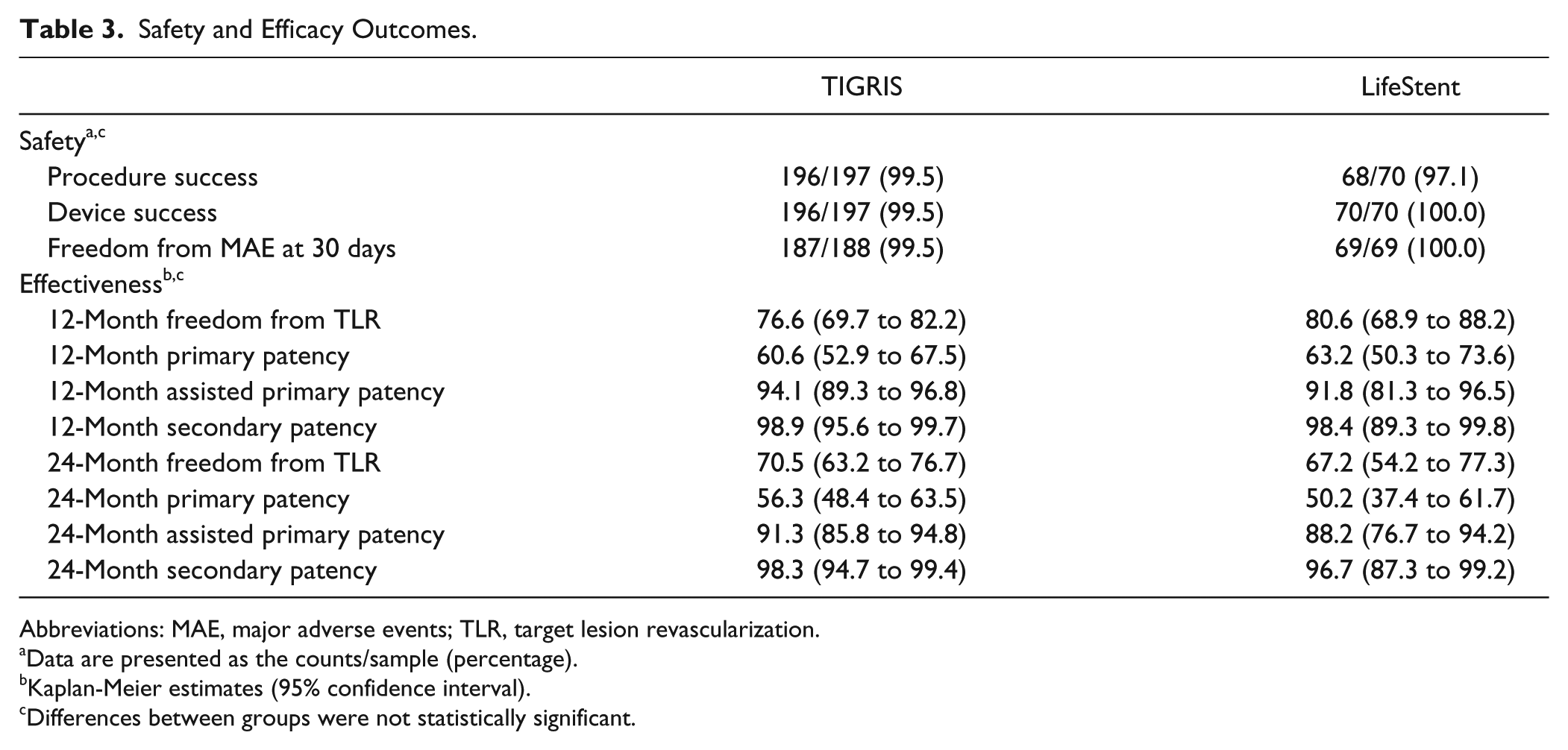
Abbreviations: MAE, major adverse events; TLR, target lesion revascularization.
Data are presented as the counts/sample (percentage).
Kaplan-Meier estimates (95% confidence interval).
Differences between groups were not statistically significant.
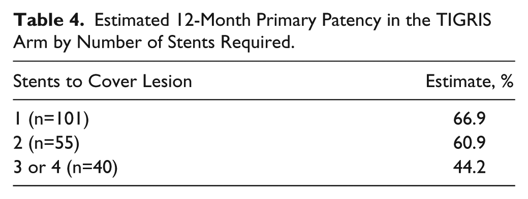
There was a downward trend in TIGRIS primary patency associated with an increasing number of stents implanted (Table 4). Separate Cox regression analyses of the TIGRIS arm showed a statistically significant impact of lesion length (OR 1.004, 95% CI 1.001 to 1.008, p=0.012) and number of stents (p=0.008) on primary patency. Similarly, the control device had lower primary patency when the lesion required 2 stents compared with when 1 stent was required (all but 4 lesions were treated with 1 or 2 devices). Cox regression analyses of the LifeStent arm of the study showed statistically significant impact of lesion length only (OR 1.005, 95% CI 1.000 to 1.010, p=0.045).
Estimated 12-Month Primary Patency in the TIGRIS Arm by Number of Stents Required.
QoL measures including EQ-5D, PAQ, and changes in the Rutherford category and ABI/TBI all demonstrated improvement over baseline for subjects in both the study and control arms of the trial with no statistically significant differences for any indicators. The mean EQ-5D improvement at 6, 12, and 24 months was 0.094, 0.104, and 0.110 for the study device and 0.122, 0.133, and 0.126 for the control device. The PAD-specific PAQ score improvement at 6, 12, and 24 months was 28.23, 29.84, and 31.55 for the study device and 32.81, 32.03, and 34.63 for the control device. As outlined in Table 5, >85% of patients participating in the study demonstrated sustained Rutherford category improvement at 12 and 24 months postprocedure. Similarly, the mean ABI was improved over baseline (mean 0.66 TIGRIS and 0.65 controls) by at least 0.23 at all time points analyzed.
Clinical Outcomes and Quality of Life Improvements at 24 Months. a
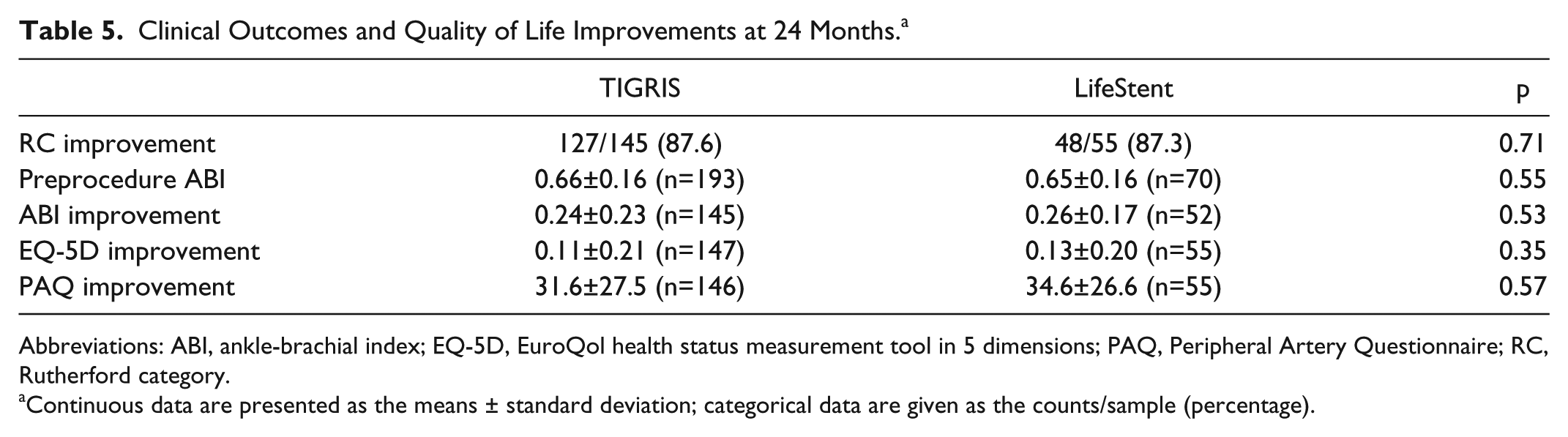
Abbreviations: ABI, ankle-brachial index; EQ-5D, EuroQol health status measurement tool in 5 dimensions; PAQ, Peripheral Artery Questionnaire; RC, Rutherford category.
Continuous data are presented as the means ± standard deviation; categorical data are given as the counts/sample (percentage).
There were 0 fractures in the study device at 12 and 24 months (Table 6). It should be noted that 1 study device was determined to have a grade 1 fracture immediately after implantation; however, no fracture could be seen in subsequent follow-up images of this subject’s stent. The control device had a significantly higher fracture rate of 32.7% at 24 months (p<0.001), with the majority of fractures being high grade (class 4 and 5; Figure 3).
Stent Fracture Rates at 12 and 24 Months. a
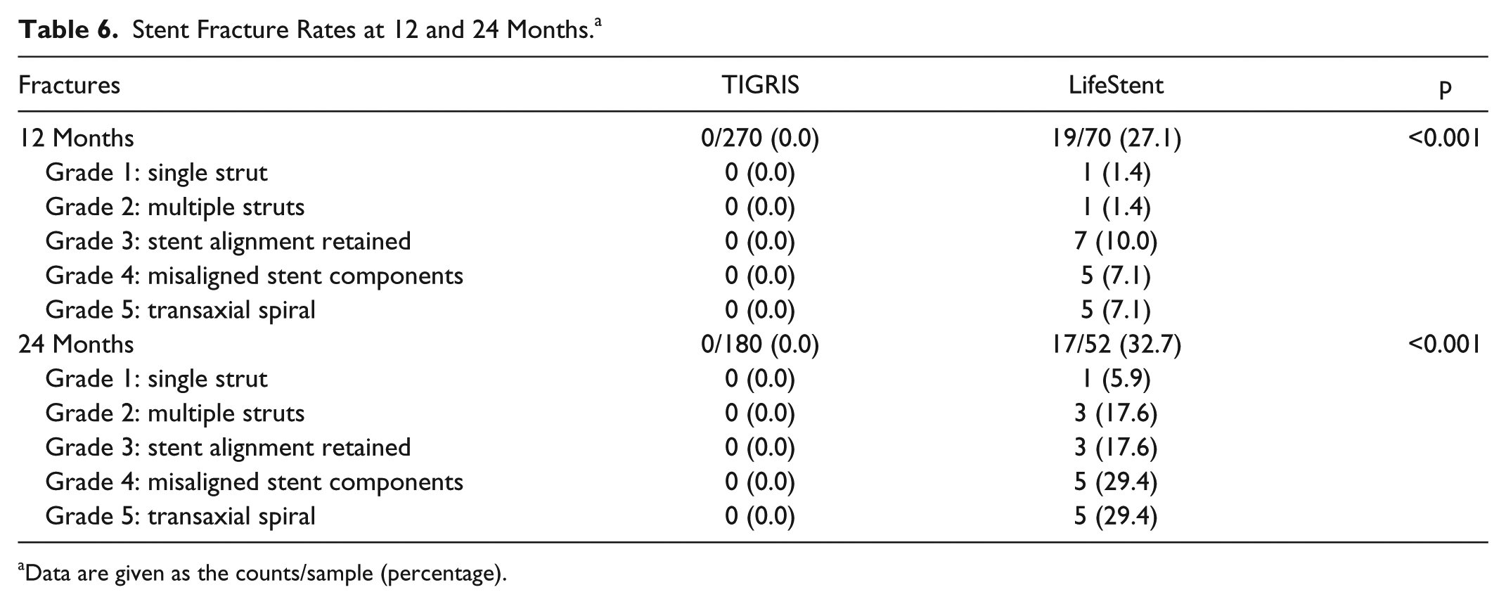
Data are given as the counts/sample (percentage).

Fractures in the LifeStent. The images below show multiple high grade LifeStent fractures (arrows) in the mid SFA (A) as well as multiple high grade LifeStent fractures in the distal SFA and proximal popliteal (B).
Discussion
In the current study, the TIGRIS stent and LifeStent were similarly effective for the treatment of long lesions in the SFA and PPA. The TIGRIS stent was found to be noninferior to the LifeStent for both the primary safety and efficacy outcome measures. Despite the complex nature of the lesions treated in this study, both stents demonstrated satisfactory primary patency at 12 and 24 months. There was sustained clinical benefit for both treatment groups as demonstrated by a decrease in Rutherford category, improvement in QoL measures, and increase in the ABI/TBI. Both devices were highly resistant to elongation or compression during deployment; however, the TIGRIS stent was found to be fracture resistant, with no stent fractures identified by the core laboratory on straight and bent knee radiographs out to 24 months. Surprisingly, there was a high rate of stent fracture in the LifeStent group, with a significant percentage of high-grade fractures.
Piorkowski and colleagues 12 published a single-center experience with the TIGRIS stent in 32 consecutive patients with 40 femoropopliteal lesions. The mean lesion length was 62.3 mm and 5% were chronic total occlusions. They reported a favorable 85.5% 1-year primary patency following treatment of these relatively straightforward lesions. Parthipun and colleagues 13 subsequently reported outcomes following use of the TIGRIS stent for a more complex lesion and patient cohort. They treated 48 patients of which two-thirds had critical limb ischemia and 74% presented with chronic total occlusion (CTO). The mean lesion length was 114.2 mm. Following successful treatment with the TIGRIS stent, primary patency was 70% at 1 year and freedom from TLR was 86%.
The current study is unique among contemporary femoropopliteal stent IDE trials with regards to its randomized trial design. The comparator stent was FDA approved and considered a gold standard at the time the study was designed.4,5 The study design is also unique with regards to the length of the lesions included in the trial. Most of the previous FDA-approved femoropopliteal stent protocols in support of an IDE were restricted to lesions up to 150 mm in length.4,7,14–17 The TIGRIS inclusion criteria reflected a more “real world” patient population with lesion lengths up to 240 mm. The mean lesion lengths for the TIGRIS stent (107.6 mm) and LifeStent (117.9 mm) in the current trial are considerably longer than those treated in recent femoropopliteal stent trials: ZILVER PTX (54.6 mm) 7 , DURABILITY II (89.1 mm), 14 RESILIENT (70.5 mm), 4 COMPLETE SE (60.7 mm), 17 STROLL (77.3 mm), 18 and SUPERB (78.1 mm). 15 In addition, the TIGRIS study population had a high incidence (42%) of CTO, which is independently associated with a higher rate of in-stent restenosis. 19
Little data are available regarding the outcomes of stenting of longer femoropopliteal lesions. The DURABILITY-200 study included 100 patients with long lesions (average lesion length of 242 mm) who were treated with at least one 200-mm-long Everflex stent. 20 The primary patency rate at 12 months was 64.8%, with a TLR rate of 32%. The STELLA study of TransAtlantic Inter-Society Consensus (TASC) C and D lesions also reported a primary patency rate of 66% among patients with a median lesion length of 220 mm. 9 In a single center retrospective study, 115 patients underwent stenting of long (>150 mm) lesions (mean lesion length 254±58 mm). One-year primary patency was 49% following implantation of a variety of commercially available nitinol stents. 21
Stent fracture following endovascular treatment of SFA lesions is a concern, in view of the association between fractures and restenosis in some trials.22,23 High rates of stent fracture were observed in early SFA stent trials, including 20% in SIROCCO I trial 24 and 12% in FAST. 25 It has been speculated that longer lesions are more prone to stent fracture, possibly because of the use of multiple overlapping stents. Scheinert et al 23 reported a 37% stent fracture rate in a registry of patients with longer SFA lesions treated with a variety of nitinol stents; the fracture rate increased with the length of the stented segment and number of stents implanted. Contemporary stent trials, including the FDA-approved IDE trials that included lesions <150 mm in length, have demonstrated low rates of stent fracture at 1 year: RESILIENT (3.1%), 4 ZILVER PTX (1%), 7 DURABILITY II (0.4%), 15 COMPLETE SE (0%), 17 SUPERB (0%), 15 and STROLL (2.0%). 18 The 0% fracture rate noted with the TIGRIS stent in the current study, despite the complexity of the lesions being treated, is a testimony to the flexibility and fracture resistance of this device.
The high 2-year fracture rate (32.7%) associated with the LifeStent is in sharp contrast to the results of the RESILIENT trial. 5 This high fracture rate occurred despite the fact that longer stents (up 17 cm) were available for use in this trial, minimizing the need for multiple stents and reducing the number of overlap zones. Of particular concern, a majority of LifeStent fractures were complex types 4 and 5. While these fractures were not clearly associated with restenosis in the current study, it is important to note that the total number of fractured devices was relatively small, and this study was not designed to evaluate the relationship between stent fracture and patency. Longer term follow-up with both the study and control arms may provide important insights regarding the late impact of types 4 and 5 fractures.
Uncertainties remain about the optimal endovascular treatment strategy for femoropopliteal occlusive disease because there is no “evidence-based” standard of care in this vascular segment. Numerous options are available to the practicing interventionist, including balloon angioplasty, cutting/scoring balloon angioplasty, atherectomy, bare nitinol stents, covered stents, drug-coated balloons (DCB), and drug-eluting stents (DES). Recent randomized trials have demonstrated that both paclitaxel-coated balloons and a paclitaxel-eluting stent are superior to balloon angioplasty for moderate length lesions in the SFA and PPA.7,26–28 The paclitaxel-eluting ZILVER PTX stent performed better than a bare Zilver stent after failed/suboptimal dilation alone. 7 There is also limited data regarding atherectomy and DES for femoropopliteal lesions longer than 150 mm. DCBs and DES have been compared in retrospective studies, with equivalent outcomes in long femoropopliteal lesions, 29 but the ongoing FOREST trial is the first to randomize DCB vs DES. 30
Recent publications of DCB use for long femoropopliteal lesions have shown promising early results31,32; however, concerns exist about the longer-term results of this approach. Experience from the global registries suggests that when long lesions or CTOs are treated with DCB, the provisional stent rate may be as high as 25%.32,33 When provisional stenting is required following DCB for long lesions, use of a flexible, fracture-resistant stent such as TIGRIS would be preferred.
Limitations
One limitation was the lack of availability of longer TIGRIS devices compared to the 17-cm devices that were available in the LifeStent arm of the study, resulting in imbalances with regards to the number of stents implanted. The availability of 150- and 170-mm devices may also have influenced the (statistically insignificant) differences between lesion length and stented length. An additional limitation of the current study was the 3:1 randomization scheme with a relatively small number of patients in the control group, which limited subsequent subgroup analysis. Despite this limitation, there were still a sufficient number of patients in both arms to draw conclusions about the safety and efficacy of the 2 devices.
Conclusion
The TIGRIS RCT has demonstrated that the TIGRIS stent and LifeStent were similarly effective for the treatment of lesions up to 24 cm in length in the SFA and PPA. The high flexibility and zero fracture rate associated with the TIGRIS stent make this stent favorable for use in high-flexion arterial segments.
Footnotes
Appendix
The following institutions (principal investigators) presented in alphabetical order participated in the TIGRIS study: Advanced Vascular Associates Vascular Research Institute, Morristown, NJ (Amit Patel, MD); Advocate Christ Medical Center, Oak Lawn, IL (Jaafer Golzar, MD); Arkansas Heart Hospital, Little Rock, AR (Ian Cawich, MD); Baylor University Medical Center, Dallas, TX (Dennis Gable, MD); CAMC Health Education and Research Institute, Charleston, WV (Aravinda Nanjundappa, MD); Cardiology Associates of Mobile, Fairhope, AL (Frank Bunch, MD); Cardiovascular Associates, Elk Grove Village, IL (Jack Chamberlin, MD); Coastal Vascular and Interventional, Pensacola, FL (Christopher LeCroy, MD); First Coast Cardiovascular Institute, Jacksonville, FL (Yazan Khatib, MD); Genesis Medical Center, Davenport, IA (Eric Dippel, MD); Heart Care Midwest, Peoria, IL (Marco Barzallo, MD); Heart Care Research Foundation, Berwyn, IL (Ronald Stella, MD); Heritage Valley Health System, Beaver, PA (Richard Begg, MD); Herz-Zentrum Bad Krozingen, Germany (Thomas Zeller, MD); Indiana University Vascular Surgery, Indianapolis, IN (Raghu Motaganahalli, MD); Kaiser Permanente Honolulu, HI (Peter Schneider, MD); Kaiser Permanente San Francisco Medical Center, San Francisco, CA (Hong T. Hua, MD); Medical Center of Central Georgia, Macon, GA (John William Mix, MD); Medical University of Vienna, Austria (Christian Loewe, MD); Minneapolis Radiology and Vascular Research Foundation, Plymouth, MN (Scott Schultz, MD); The Miriam Hospital, Providence, RI (Peter Soukas, MD); Munroe Regional Medical Center, Ocala, FL (Robert Feldman, MD); North Central Heart Institute, Sioux Falls, SD (Michael J. Bacharach, MD); North County Radiology, Oceanside, CA (Richard Saxon, MD); Northwestern/Feinberg School of Medicine, Chicago, IL (Mark Eskandari, MD); Ohio Health Research Institute, Columbus, OH (Charles Botti, MD); Orlando Regional Healthcare System, Orlando, FL (Vijaykumar Kasi, MD); Providence Heart & Vascular Institute, Portland, OR (Ethan Korngold, MD); SSM Health Care, St. Charles, MO (Thomas Schneider, MD); Universität Leipzig, Germany (Dierk Scheinert, MD); University of California Davis Health System, Sacramento, CA (John Laird, MD); University of Iowa Healthcare, Iowa City, IA (Mel Sharafuddin, MD); University of Pennsylvania, Philadelphia, PA (Arthur Topoulos, MD); University of Pittsburgh Medical Center, Pittsburgh, PA (Efthymios Avgerinos, MD); Vascular Surgical Associates, Austell, GA (Arun Chervu, MD); Yale University School of Medicine, New Haven, CT (Carlos Mena-Hurtado, MD).
Declaration of Conflicting Interests
The author(s) declared the following potential conflicts of interest with respect to the research, authorship, and/or publication of this article: John Laird has received research grants from W.L. Gore & Associates and Medtronic; is a compensated consultant for Abbott Vascular, Bard Peripheral Vascular, Boston Scientific, and Medtronic; and is a board member of VIVA Physicians, a 501c3 not-for-profit education and research organization. Thomas Zeller received honoraria from Abbott Vascular, Bard Peripheral Vascular, Veryan, Biotronik, Boston Scientific, Cook Medical, Cordis, W.L. Gore & Associates, Medtronic, Spectranetics, Straub Medical, TriReme, VIVA Physicians, GLG, and Philips. He has consulted for Abbott Vascular, Bard Peripheral Vascular, Boston Scientific, Cook Medical, W.L. Gore & Associates, Medtronic, and Spectranetics. He has received research, clinical trial, or drug study funds from 480 Biomedical, Bard Peripheral Vascular, Veryan, Biotronik, Cook Medical, Cordis, W.L. Gore & Associates, Abbott Vascular, Medtronic, Spectranetics, Terumo, TriReme, and Philips. Christian Loewe is on the speaker bureaus for Siemens, GE Healthcare, Guerbet, Medtronic, and Covidien-Mallinkrodt and receives research grants from Biotronik and Boston Scientific. Jack Chamberlin reports associations with Jansen, Boston Scientific, W.L. Gore & Associates, Endologix, Amgen, and Covidien. Peter Schneider has received royalties from Cook; is a noncompensated advisor to Medtronic, Cardinal Health, and Abbott; and is a board member of VIVA Physicians, a 501c3 not-for-profit education and research organization. Aravinda Nanjundappa is a consultant for W.L. Gore & Associates. Frank Bunch is a consultant for W.L. Gore & Associates, Bard Peripheral Vascular, Boston Scientific, Spectranetics, and ACT. Alexandra Lansky reports receiving institutional grant support in excess of $25,000. Michael R. Jaff is a noncompensated advisor for Abbott Vascular, Boston Scientific, Cordis, and Medtronic; a compensated advisor for Philips Volcano Corporation; has equity in PQ Bypass/Vascular Therapies; and is a board member of VIVA Physicians, a 501c3 not-for-profit education and research organization.
Funding
The author(s) disclosed receipt of the following financial support for the research, authorship, and/or publication of this article: This study was funded by W.L. Gore & Associates.