






Abstract
Purpose:
The purpose of this study was to evaluate the efficacy and safety of zone 2 thoracic endovascular aortic repair assisted by the chimney technique or single-branched stent graft for the preservation of the left subclavian artery, and summarize our single-center experience with the techniques.
Materials and methods:
From February 2017 to June 2020, 137 patients who underwent left subclavian artery revascularization during zone 2 thoracic endovascular aortic repair were enrolled. Patients had acute type B aortic dissection and penetrating aortic ulcer associated with intramural hematoma. The chimney technique was performed in 68 patients (group A), and single-branched stent graft was deployed in 69 patients (group B). All procedures were performed during the acute phase. Primary technical success, immediate postoperative endoleak, neurologic complications (stroke or spinal cord ischemia), 30-day mortality, 1-year technical success, all-cause mortality, patency of the left subclavian artery, and reintervention were analyzed. Comparing the occurrence of the Bird-Beak Configuration, defined as a gap between the aortic wall and the sent graft with stent protrusion into the aortic lumen more than 5 mm, was also performed.
Results:
Primary technique success was achieved in 66 and 67 patients in groups A and B, respectively. The incidence of immediate postoperative endoleak, neurologic complications (stroke or spinal cord ischemia), and 30-day mortality were 5.9%, 1.5%, and 4.4% in group A, and 2.9%, 2.9%, and 2.9% in group B, respectively. During follow-up, the 1-year technical success rate was similar in both groups. All-cause mortality was similar in both groups (3.1% in group A and 4.5% in group B). The patency of the left subclavian artery was not significantly different between the 2 groups with 2 and 3 occlusions in groups A and B, respectively. The rate of reintervention was higher in group B (3.1% vs 1.6%, p=0.536), with a non-significant difference. Bird-Beak Configuration was more prominent in group B with the incidence of 59.42%.
Conclusions:
Acting as minimally invasive alternatives, both techniques are feasible for left subclavian artery preservation during zone 2 thoracic endovascular aortic repair for type B acute aortic syndromes with encouraging mid-term outcomes. Long-term follow-up is required to confirm these findings.
Keywords
Introduction
Thoracic endovascular aortic repair (TEVAR) acts as a first-line therapeutic option for descending thoracic aortic disease due low procedural morbidity and mortality, and comparable mid-term results over those of open surgery. 1 With the development of endovascular equipment and assistance techniques, TEVAR has broadened to treat aortic pathologies involving the arch and ascending aorta.2–5 However, a preferable proximal landing zone determines the outcomes of TEVAR.1,6,7
Left subclavian artery (LSA) revascularization should be routinely performed to prevent posterior stroke and upper extremity ischemia during zone 2 TEVAR. 8 Both single-branched stent graft and single chimney technique for preserving the LSA have been performed to manage aortic arch pathologies.2,3,9–11 However, the heterogeneity of the included patient groups reduces the comparability with respect to different techniques and provides an opportunity for a subgroup analysis.
Isolated LSA revascularization during zone 2 TEVAR using carotid-subclavian bypass or transposition is the gold standard.12–14 The chimney technique or single-branched stent graft is not recommended routinely during zone 2 TEVAR because sufficient follow-up data are currently unavailable. Therefore, we conducted this study to evaluate the safety and efficacy of isolated LSA preservation during zone 2 TEVAR using the chimney stent and single-branched stent graft, especially for type B acute aortic syndromes (AASs). The occurrence of Bird-Beak Configuration (BBC) was also investigated.
Materials and Methods
Ethical Statement
The study was approved by the local ethics committee, and written informed consent was waived due to retrospective design of the study and the use of anonymized data.
Patient Selection
From February 2017 to June 2020, 137 patients diagnosed with type B AASs with unfavorable proximal landing zones were enrolled in our study (Figure 1). Antihypertensive medical management (calcium-channel blockers, nitroglycerin, beta-blockers, or combinatory treatment) was arranged immediately to reduce systolic blood pressure to no greater than 120 mmHg. Zone 2 TEVAR using the single chimney technique or a single-branched stent graft was performed to preserve the LSA. The chimney technique was indicated for patients with ruptured dissections due to the requirement of emergent procedure-limited employment of the single-branched stent graft because of the individual design and the duration of manufacture, and the single-branched stent graft was offered for patients with retrograde intramural hematoma or dissection involving the LSA ostium, which was approved by Chinese Drug and Food Administration. Unless otherwise specified, both techniques were offered without preference. All cases were identified retrospectively by reviewing the hospital documents and imaging system. Data related to perioperative, in-hospital, and follow-up were gathered.
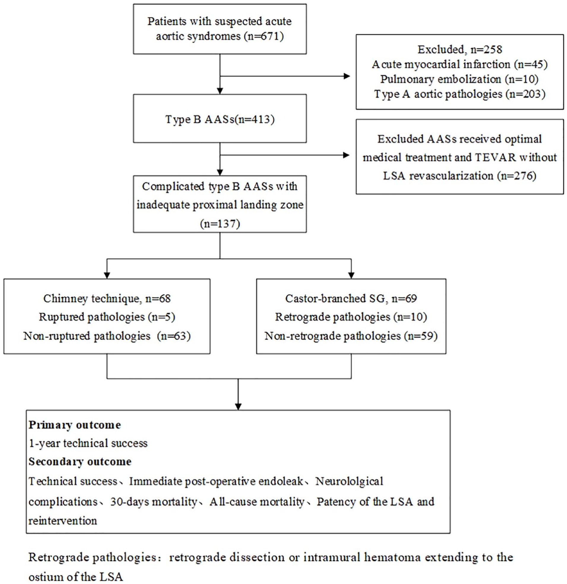
The flow chart for patient selection. AAS, acute aortic syndromes; TEVAR, thoracic endovascular aortic repair; LSA, left subclavian artery; SG, stent graft.
Definition and Outcome Variable Selection
Patients with the following characteristics were considered to be indicated for TEVAR: refractory symptoms and/or hypertension, aortic rupture, rapid aortic expansion, and visceral, renal, and/or limb malperfusion. Persistent pain was defined as refractory pain persisting for >12 hours despite adequate medical management, and rapid aortic expansion was defined as increase of the maximum diameter of the proximal descending thoracic aorta >4 mm from the first to the second computed tomography angiography (CTA) scan within 14 days of symptoms onset. The unfavorable proximal landing zone was defined as a primary entry tear located ≤15 mm distal to the LSA and/or retrograde dissection or intramural hematoma involving the ostium of the LSA. Primary technical success was defined as successful deployment of the device in the absence of conversion to open surgical repair, death within 24 hours, type Ia (antegrade perfusion of the false lumen) and III (modular disconnect, apposition failure or fabric tear induced leaks) endoleaks evidenced by procedural angiography, and stent graft and LSA occlusion. The threshold value for BBC was defined as a gap between the aortic wall and the sent graft with stent protrusion into the aortic lumen more than 5 mm (Figure 2). The 1-year technical success was defined as the absence of type I endoleak without the requirement of reintervention within 1 year of TEVAR.

The bird-beak configuration including the gap (arrowhead), the length of stent graft protrusion (line), and bare metal stent served as chimney stent (arrow).
CTA was performed for all patients before discharge, at postoperative 3 and 6 months, then annually.
Preprocedural Evaluation
With the help of EndoSize (Therenva SAS, France), all aortic pathologies were analyzed by 2 experts with >10 years of experience in the preoperative evaluation. No greater than 10% oversizing was applied. The deployment of the stent graft was finally determined according to intraoperative aortography.
Endovascular Procedure
All procedures were performed by the same 2 operators with >10 years of experience in TEVAR, and all patients received general anesthesia with tracheal intubation.
Chimney Technique
The left brachial artery (LBA) access was obtained percutaneously for deployment of a 6F sheath (Terumo Corporation, Tokyo, Japan). Then, a 5F pigtail catheter (Cook Medical, Bloomington, USA) was advanced over a guidewire into the ascending aorta for aortography. One side of the common femoral artery (CFA) was exposed. A 4F tapered catheter (Cordis, Miami, FL, USA) was advanced into the ascending aorta, and then the catheter was replaced with a super-stiff guidewire (Cook Medical). The stent graft (Life-tech Scientific, Shenzhen, China) was advanced into the aortic arch over the guidewire. With the administration of heparin (80 U/kg), the deployment of the stent graft (Life-tech Scientific) was performed under transient apnea with a systolic blood pressure ≤100 mmHg. Through the sheath in the LBA, a stiff guidewire (Abbott Medical) was advanced over the pigtail catheter (Cook Medical) into the ascending aorta. An 8 × 40/60 mm or 10 × 40/60 mm bare self-expanded stent (Zilver 635 Vascular Self-Expanding Stent, Cook Medical) was implanted to preserve the LSA. The pigtail catheter was advanced into the ascending aorta over the stiff guidewire, and immediate aortography was performed to confirm the exclusion of the entry tear and patency of the arch branch arteries.
Castor-Branched Stent Graft
Retrograde LBA access was obtained percutaneously, and one side of the CFA was exposed. Through the 6F sheath (Terumo) in the LBA, a 5F multipurpose angiographic (MPA) catheter (Cordis) was advanced over a guidewire into the exposed CFA and withdrawn from the arteriotomy site. Along with a guidewire, a 4F tapered catheter (Cordis) was delivered into the ascending aorta through the exposed CFA, and a super-stiff guidewire (Cook Medical) was exchanged. Through a 5F sheath (Terumo Corporation) inserted percutaneously into the contralateral CFA, a 5F pigtail catheter (Cook Medical) was delivered into the ascending aorta. The traction wire of the branch section was advanced into the MPA catheter and withdrawn from the tail of the catheter. The castor stent graft was delivered into the descending thoracic aorta over the super-stiff guidewire while simultaneously pulling the traction wire. With the outer sheath left in the descending aorta, the stent graft within the soft sheath was advanced into the aortic arch. The stent graft was advanced to the proper position after the soft sheath was removed, and the branch section was pulled into the LSA by drawing the traction wire. If the traction wire wrapped around the stent graft trunk, the stent graft was withdrawn into the outer sheath, rotated the main body to the correct orientation, and re-advanced into the arch. The stent graft trunk was quickly deployed by withdrawing the trigger wire followed by deployment of the branch section by withdrawing the traction wire. Immediate aortography was performed to confirm the exclusion of the dissection and patency of the LSA.
Statistical Analysis
Data are reported as the mean ± standard deviation and as percentages. Continuous variables were compared using the Student’s t test. Categorical variables were compared using the Pearson chi-square test, Fisher’s exact test, or Wilcoxon rank sum test. Kaplan-Meier curves were used to determine differences in all-cause mortality, patency of the LSA, and rate of reintervention between the 2 groups. Statistical significance was set at p<0.05. All statistical analyses were performed using SPSS software package 19 (IBM Corp, Armonk, NY, USA).
Results
Patient Characteristics
The demographics of the enrolled patients in both groups (Table 1) included the mean age of 54.25±13.21 years and 56.77±12.10 years in groups A and B, respectively. The majority of patients in both groups were male (51 in group A and 54 in group B, p=0.652). There was no significant difference in preoperative comorbidities between the 2 groups. One patient in group A underwent ascending aortic replacement due to an ascending aortic aneurysm 7 years prior. Trauma was the cause for 3 dissections in group A and 1 in group B. Except for the diameter of the distal landing zone, the parameters of the dissection also exhibited no significant difference between the 2 groups (Table 1).
Patients’ Characteristics.

Abbreviations: PAU, penetrating aortic ulcer.
Parameters of the Stent Grafts
Seventy-two and 74 stent grafts were deployed in group A and B, respectively (Table 2). The mean proximal diameter of the stent grafts was 32.54±3.37 mm in group A and 32.86±2.36 mm in group B, with no significant difference. The oversize and length of the aortic stent graft were greater in group B than in group A (204.64±17.54 mm vs 197.65±8.13 mm). The branch section had a larger diameter and shorter length than the chimney stent. The difference of the distance between the proximal end of the aortic graft and the branch between the 2 groups was negligible (7.72±3.01 mm vs 7.97±3.77 mm, p=0.668). A branch section was twisted and occluded in 1 patient in group B with a requirement of balloon dilation to maintain its patency, which was not observed in group A.
Parameters of the Stent Grafts.

Perioperative Outcome
All TEVARs were performed in the acute phase. Primary technical success was achieved in 66 and 67 patients with the duration of the procedure being 118.31±35.57 min and 119.93±25.87 min in groups A and B, respectively (Table 3). Four immediate postoperative endoleaks, including 2 type I endoleaks and 2 type II endoleaks, were detected in group A, and 2 immediate postoperative type I endoleaks were detected in group B. No further intervention was performed during the procedure, as the endoleaks were minor. One stroke and 1 acute myocardial infarction were confirmed in group B. Prolonged hypotension (>90 mmHg) and excessive device manipulation within the aortic arch was considered to be associated with the stroke. One patient in group A suffered from paralysis of the right upper extremity due to covering of the aberrant right subclavian artery during TEVAR, and 1 patient in group B suffered from paralysis of the left lower extremity due to extended coverage of thoracic aortic segment leading to spinal cord ischemia. There were 3 patients in group A and 2 patients in group B got pneumonia. No other major complications occurred in the perioperative period. The 30-day mortality was 4.4% (3/68) in group A and 2.9% (2/69) in group B, and the in-hospital mortality was 2.9% (2/68) in group A and 1.5% (1/69) in group B. One patient died of severe pneumonia and renal insufficiency. The cause was uncertain for the remaining deaths because the relatives refused autopsy. Patients in group A had significantly lower costs than those entailed by patients in group B (RMB 72.07 thousand vs 136.69 thousand during procedure; RMB 138.32 thousand vs 203.18 thousand in total). The incidence of BBC was significantly higher in group B than in group A (59.42% vs17.65%). The mean hospital length of stay was 16.35 days in group A and 17.26 days in group B, with a non-significant difference.
Perioperative Data and 1-Year Technical Success Rate.

Follow-up Outcome
Surveillance including clinical and imaging follow-up was performed prior to discharge, and then repeated postoperatively at 3 and 6 months, then annually. During the follow-up, both groups had similar 1-year technical success rates (98.4% in group A and 96.9% in group B).
The all-cause mortality in both groups during follow-up is shown in Figure 3. In group A, 1 sudden death occurred 13 months after TEVAR due to an uncertain cause, and 1 patient died of upper gastrointestinal hemorrhage 1 month after TEVAR. In group B, 1 patient died suddenly due to an uncertain cause 3 months after TEVAR, 1 died due to severe pneumonia 2 months after TEVAR, and 1 died of acute myocardial infarction 6 months after TEVAR. During follow-up, the mortality rates were 3.1% (n=2/65) and 4.5% (n=3/67) in groups A and B, respectively (p=0.654).

Freedom from all-cause mortality.
The patency of the LSA branches is shown in a figure (Figure 4). The last CTA revealed 2 and 3 occlusions of the LSA branches in groups A and B, respectively. No reintervention was performed because of absence of clinical symptoms. The occlusion rate of the LSA branches (3.2% in group A and 4.7% in group B) was not significantly different among groups during mid-term follow-up, and the patency was 96.8% and 95.3% in both groups, respectively.

Freedom from occlusion of the left subclavian artery (LSA).
The rate of reintervention is shown in a figure (Figure 5). Two proximal cuff stent grafts were deployed to eliminate type I endoleaks in group A at 14 months after TEVAR. Meanwhile, revascularization of the left common carotid artery and LSA was performed via the deployment of 2 chimney stents. A retrograde type A dissection was detected in 1 patient in group B 2 months after TEVAR, and he received open surgery. TEVAR assisted with double fenestration, to preserve the left common carotid artery and LSA, was performed to eliminate type I endoleak in 1 patient in group B 3 months after TEVAR. The reintervention rates were 1.6% and 3.1% in groups A and B, respectively.
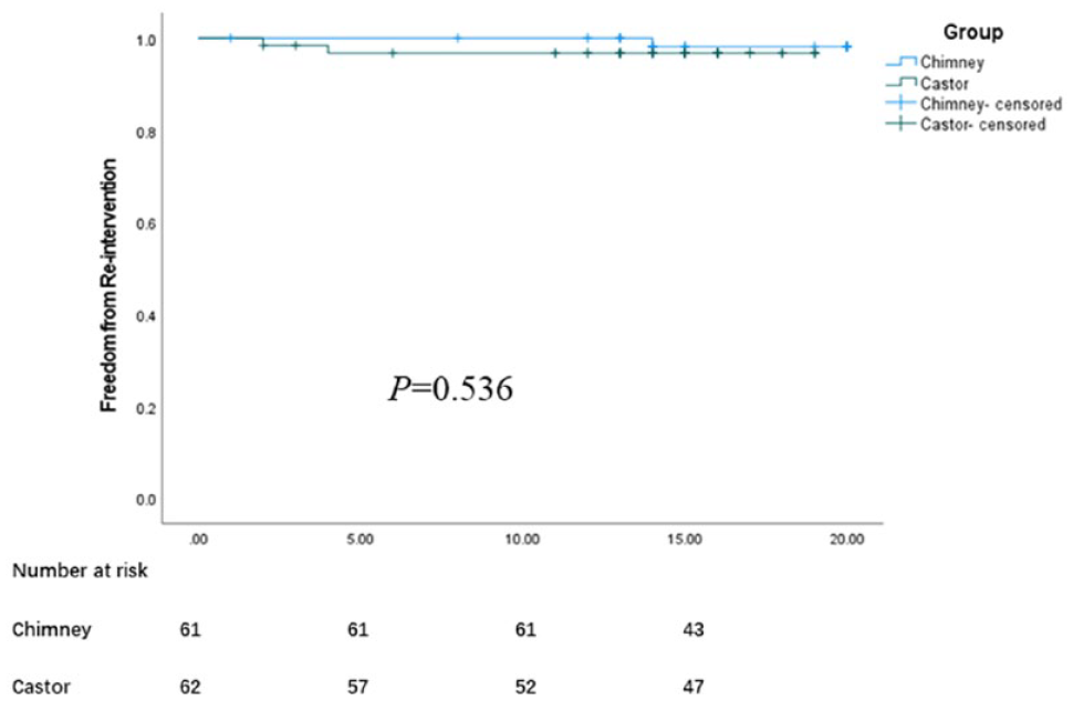
Freedom from reintervention.
Complications
A distal stent graft induced new entry at 4 months after TEVAR was confirmed in 1 patient in group B. No further intervention was performed in the absence of clinical symptoms. Twelve months after TEVAR, 1 patient in group A experienced cerebral hemorrhage. No other major complications were detected in either group during the follow-up.
Aortic Remodeling
The aortic remodeling is summarized in a table (Table 4). The last CTA confirmed the complete thrombosis of the false lumen in the distal aortic arch and stented segment of the thoracic aorta in all patients. Incomplete thrombosis or patency of the false lumen in the aorta distal to the stented segment was detected in 33 patients in group A and 34 in group B with the absence of excessive expansion (aortic growth >5 mm/year). Post-TEVAR changes of the maximum aortic diameter were analyzed at distal stent graft level and celiac trunk level in both groups, with a negligible difference at any level. During the mid-term follow-up, both techniques significantly contributed to favorable aortic remodeling.
Aorta Remodeling.

Abbreviations: FL, false lumen; SG, stent graft.
Pre-TEVAR vs Post-TEVAR. **Group A vs Group B.
Discussion
Although there is no randomized control trial, increasing evidence or general agreement demonstrated that TEVAR for complicated type B AAS is feasible and effective.1,6,7 The technical success rate ranged between 95% and 99%, and hospital mortality and neurological complications ranged between 2.6% and 9.8% and 0.6% and 7.5%, respectively.1,7,15 For patients with complicated type B AAS with suitable anatomy, TEVAR is an excellent alternative to open surgery with encouraging outcomes.
During TEVAR, a proximal landing zone ≥15 mm is a prerequisite for success.6,7 Intentional LSA coverage to extend the proximal landing zone increased the risk of stroke, spinal cord, and upper extremity ischemia.8,16 Therefore, for thoracic aortic pathologies involving the aortic arch or LSA, TEVAR combined with assistance techniques, including chimney and fenestration techniques, extends the proximal landing zone with preservation of the LSA.11,17–19 Branched devices were also available for preserving the LSA during zone 2 TEVAR.3,20,21 However, isolated LSA revascularization during zone 2 TEVAR using carotid-subclavian bypass or transposition is more likely to be accepted as the gold standard.12–14
Since the chimney technique was systemically introduced by Criado in 2007, 22 revascularization of the LSA using a chimney stent during zone 2 TEVAR was proven to be technically feasible and effective.17,23–25 However, the main concern of this technique is the risk of type I endoleak and the patency of the LSA.26,27 Previous studies have reported that the incidence of immediate postoperative type I endoleaks ranged from 0% to 17.6%.17,24,27,28 During the follow-up, the patency of the LSA branches ranged from 83% to 97%.23–25,27,29 In the present study, 68 patients received bare self-expanded chimney stent placement parallel to the main aortic stent graft, with a primary technical success rate of 97.06%, which is comparable to that of previous studies.17,23,25,28,30 Immediate postoperative type I endoleaks were detected in 2 cases with an entry tear or false lumen at the greater curvature. The chimney technique was considered to be associated with the high risk of endoleak for lesions located at the outer curve of the aortic arch. 24 During the follow-up, the patency of the chimney stent was high. We made the clinical decision to use a bare metal self-expanding stent as a chimney stent according to our experience as well as previous reports.9,10 The selection criteria for different types of chimney stents were not established because the covered stent was not employed in this study. No guideline or randomized control study has reported a protocol for choosing a chimney stent.
Currently, zone 2 TEVAR using branched stent grafts for preserving the LSA is considered to be safe and provide satisfactory perioperative outcomes.3,21,31 However, the type I endoleak and long-term patency of the LSA branch are still major concerns. As reported by previous studies, type I endoleak rate ranged from 1.4% to 18.2%,3,16,21 and the patency of the LSA ranged from 93% to 100%.3,21,31 For castor-treated patients, the primary technical success rate was 97.1%. Similar to that of previous reports, the incidence of immediate postoperative type I endoleak was 2.9%, and the patency of the LSA branches was 95.3% during mid-term follow-up. One type I endoleak resolved within 14 days without reintervention. Postprocedural hemodynamic changes sealed the entry tear and induced false lumen thrombosis. The other type I endoleak required reintervention because of persistent patency. For this patient, the entry tear at the lesser curvature was considered to be associated with the persistent type I endoleak.
Stent-related complications include endoleak, neurological complications, retrograde type A dissection, and distal stent-induced new entry. Two stent-induced new tears were found in group B during the follow-up, and none in group A. According to the European Society of Cardiology guidelines, almost no oversizing of the stent graft should be applied to patients with type B aortic dissections. 7 Greater oversize (4.93% and 7.69% in group A and B, p<0.001) was considered to be associated with an increased risk of stent-induced new entry tears in group B.
In accordance with previous studies, BBC appeared to be associated with an increased risk of type Ia endoleak, stent graft collapse, and distal migration of the stent graft.32–34 However, the relatively higher incidence of BBC in group B is discordant with the infrequency of perioperative complications. Statistical evidence for establishing the prognostic value of the BBC cannot be provided from the present study due to the limited number of complications. The stent graft with a proximal bare tip in group A ensures an active conformability by reducing the compression of the sealing stent and reduces interference with the bloodstream, and which was considered to be associated with the decreased risk of BBC. The castor-branched stent graft with absence of a proximal bare section is unable to conform to highly angulated arches completely due to the stiffness with long segment. According to previous reports, the conformable thoracic aortic graft exhibited high conformability (achieved in 95% of patients) by means of a telescoping segment of the inner curve of the stent graft, which can achieve the reduction in length of the inner curvature.34,35 Devices and their delivery systems engaged in group B should be modified to achieve improvement in conformability. A significant higher procedural cost was found in group B due to the high price of castor-branched stent grafts.
Carotid to subclavian bypass for the preservation of the LSA performed during TEVAR is feasible with a technical success rate of 100%. 13 The type I endoleak ranged from 2% to 2.5%, and the LSA patency ranged from 97% to 100%.13,36 However, a comparison between group A/B and carotid to subclavian bypass was not performed due to the limited number of patients undergoing carotid to subclavian bypass during TEVAR at our center.
The primary limitation of this study is that a comparison between the chimney technique or castor-branched stent graft and carotid to subclavian bypass was not performed. Indeed, carotid to subclavian bypass for the revascularization of the LSA should serve as the gold standard comparison data for other techniques and devices during zone 2 TEVAR. The inherent limitation of the retrospective analysis due to selection bias should also be considered when interpreting our findings. Furthermore, the experience of a single-center study with limited number of patients may not be applicable generally.
Conclusion
Both the chimney technique and castor-branched stent graft offer encouraging outcomes during mid-term follow-up and can serve as an alternative to limited surgical revascularization of the LSA during zone 2 TEVAR for type B AASs. Apart from the higher incidence of BBC for the castor-branched technique, the prognostic value of BBC has not been established due to the low incidence of complications in both groups. Further assessment with long-term follow-up is required for improving the treatment strategy and patient prognosis.
Footnotes
Declaration of Conflicting Interests
The author(s) declared no potential conflicts of interest with respect to the research, authorship, and/or publication of this article.
Funding
The author(s) disclosed receipt of the following financial support for the research, authorship, and/or publication of this article: This work was supported by the National Natural Science Foundation of China (11971269, 12171258), and the Key Technology Research and Development Program of Shandong Province (2019GSF108015).
