






Abstract
Purpose:
To report a successful revascularization case using the Rotarex™S atherothrombectomy system in a recent iliac limb thrombosis, and chronic hypogastric stent obstruction after previous aortoiliac aneurysm endovascular repair (EVAR).
Case Report:
A 72-year-old patient was treated for recent right iliac limb thrombosis and left iliac branch chronic hypogastric stent occlusion, 5 years after EVAR. A total endovascular approach, using both upper extremity and femoral vascular access, was settled with 2 Rotarex™S (6Fr and 10Fr) devices. The Rotarex™S catheters removed most of the intraluminal material, allowing additional endografts and bare metal stents to be deployed to support a new healthy lumen surface.
Conclusion:
The total endovascular approach provided by the Rotarex™S device appears to be safe and effective in treating aortoiliac endografts occlusions, both in subacute and chronic phases. Larger studies could highlight differences and eventual advantages compared with more traditional solutions.
Keywords
Introduction
Preservation of the pelvic vascular pattern during endovascular interventions performed for treatment of aortoiliac aneurysmal pathology (EVAR) is considered one of the main objectives to be achieved along with aneurysm sealing. The rationale behind this is to maintain antegrade flow into the pelvis, and to reduce the risk of ischemic complications associated with hypogastric occlusion, which may occur in up to 66% of the cases.1–5 Iliac branched devices (IBDs) demonstrated safety and effectiveness in the treatment of patient with aortoiliac aneurysms, with proven long-lasting hypogastric artery patency rates. 6
Nevertheless, during follow-up, as patency rate is never reported to be 100% over time, a percentage of patients receiving IBDs inevitably experience internal iliac artery occlusion, whether symptomatic or asymptomatic. There is not currently an indication for occluded hypogastric artery revascularization following endovascular aortoiliac treatments, except for cases associated with pelvic ischemia, bowel and/or spinal cord ischemia, with surgical reconstruction via hypogastric bypass resulting the most viable option in these cases.7,8
In this article, we describe an EVAR + IBD case with symptomatic late iliac limb and contralateral hypogastric branch occlusion successfully treated using rotational mechanical atherothrombectomy with the Rotarex™S (BD, Franklin Lakes, NJ, USA) catheters.
Case Report and Technique
Written informed consent was obtained from the patient for anonymous usage of data and images for scientific purposes.
We describe the case of a 72-year-old male who underwent EVAR and left IBD with Cook (Bloomington, IN, USA) endografts in 2016 for aortoiliac aneurysm repair. The postoperative course and early follow-up were uneventful until the 36 months scheduled visit when the left internal iliac artery stent was found occluded during duplex ultrasound evaluation. Since the patient presented with mild symptoms related to a non-disabling intermittent claudication (Rutherford classification category I),9,10 conservative management was given. Walking distance improved over time until February 2021 (59 months after EVAR), when the general practitioner requested an urgent vascular surgery assessment because of worsening, progressive bilateral claudication, following an episode of right limb acute pain onset which occurred 3 months before and resolved within few hours. Duplex scan and computed tomography angiography (CTA) revealed endograft right iliac limb occlusion with external and internal iliac artery patency in addition to the known left hypogastric stent occlusion (Figure 1).
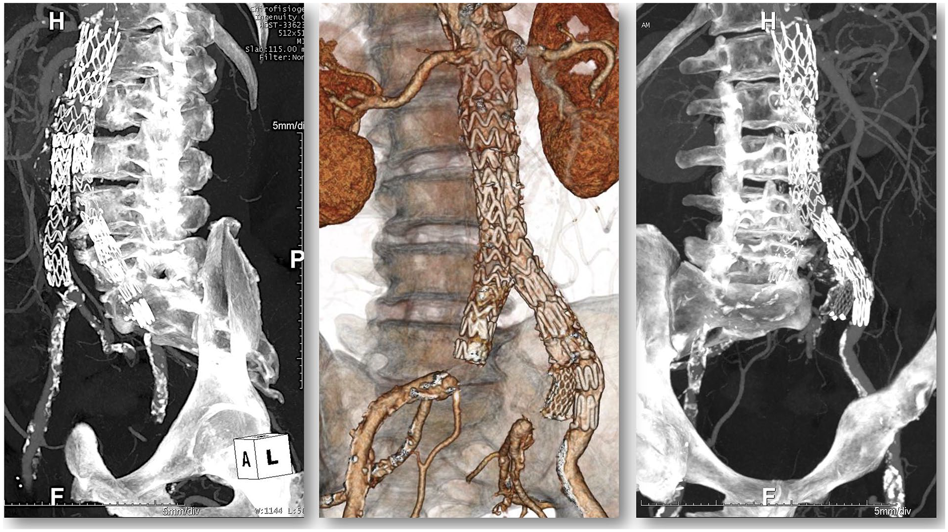
Preoperative computed tomography angiography (CTA) showing right iliac leg occlusion with external and internal iliac artery patency and left hypogastric stent inveterate occlusion.
On account of the extremely limited walking capacity (Rutherford classification category III), a total endovascular approach with rotational atherothrombectomy using the Rotarex™S catheters (BD, Franklin Lakes, NJ, USA) was planned.
The procedure was performed in a Hybrid Operating Theater equipped with a ceiling-mounted fixed angiography (Philips, Amsterdam, The Netherlands), under general anesthesia and systemic heparinization.
By direct right brachial artery surgical exposure, a 90-cm long Destination 8Fr introducer sheath (Terumo Corporation, Tokyo, Japan) was advanced through the arch, the descending aorta and inside the endograft until the left IBD lumen, just above the hypogastric branch origin.
Diagnostic angiogram confirmed complete hypogastric stent occlusion with late distal perfusion and adequate representation of the gluteal branches (Figure 2A). Using a 0.035″ hydrophilic stiff guide wire and a 6Fr Neuron catheter (Penumbra, Alameda, CA, USA), recanalization of the hypogastric branch was obtained, with control angiography revealing lumen recovery in the artery bifurcation site, associated with segmental dissection of the distal hypogastric and main gluteal branch first tract (Figure 2B and C).

Diagnostic arteriography confirming left hypogastric stent occlusion with late distal perfusion (A); intraoperative segmental dissection of the distal hypogastric (B and C); and gluteal dissection resolution with stent (D).
To solve the distal segmental dissection, 2 Bare Metal Stent X-position (Stentys, Paris, France) 3.5/4.5 mm × 27 mm were deployed, completely covering the gluteal artery dissecting flap (Figure 2D).
A 6Fr × 110 cm Rotarex™S rotational atherothrombectomy catheter was hence advanced from the brachial access, through the introducer sheath, and several device passages were executed inside the occluded hypogastric stent (Figure 3A).

Mechanical rotational thrombectomy with Rotarex system (A and B); hypogastric relining (C), angiographic control (D).
Control arteriography showed stent recanalization with some residual intraluminal material associated with focal residual dissection of the left internal iliac artery extending downstream to the gluteal branch (Figure 3B). Hypogastric relining was then carried out advancing, positioning, and deploying a balloon-expandable covered VBX Stent (W. L. Gore and Associates, Flagstaff, AZ, USA) 8 × 59 mm large, post-dilated with a 10 × 20 mm balloon. A Protegè (Medtronic, Minneapolis, MN, USA) bare metal stent (BMS) 12 to 40 mm was then deployed with its distal end proximal to the hypogastric bifurcation overlapped with the VBX, completing the distal dissection treatment (Figure 3C).
A new angiographic control demonstrated a satisfactory result with complete hypogastric recanalization, rapid contrast medium wash-out and maintained distal branches representation (Figure 3D).
Next, a 12Fr DrySeal (W. L. Gore and Associates, Flagstaff, AZ, USA) introducer sheath was advanced up to the iliac bifurcation from the right percutaneous femoral access. Selective arteriography confirmed right external and internal arteries patency with iliac endograft complete occlusion (Figure 4A).

Right-side diagnostic arteriography (A); Rotarex 10Fr inside iliac limb (B); control arteriography (C); and proximal ballooning after endograft relining (D).
Intraprosthetic recanalization was therefore obtained using a 0.035″ hydrophilic guide-wire and a Bernstein catheter (Cordis, Bridgewater, NJ, USA).
From the upper access, a 12 × 40 mm angioplasty balloon was positioned and inflated at the origin of the left iliac branch of the graft, for endoclamping purpose during Rotarex™S passages, avoiding risk of contralateral embolization (Figure 4B).
A 10Fr × 85 cm Rotarex™S rotational atherothrombectomy catheter was then advanced into the 12Fr introducer sheath and several passages performed inside the occluded graft segment. Control aortography documented treated axis recanalization with some intra-prosthetic endoluminal material persistence along the iliac limb (Figure 4C). Right iliac axis relining was then finalized by positioning and deploying a 16 × 100 mm Incraft (Cordis, Bridgewater, NJ, USA) iliac leg with the proximal landing zone at the main body flow divider and distal landing zone before the iliac bifurcation. A 12 × 40 mm Protegè BMS was then added distally, completing the intraluminal material exclusion (Figure 4D).
Finally, due to some signs of intraluminal material persistency inside the endograft’s main body (Figure 5A) at angiographic control, 2 additional Protegè 14 × 60 mm BMS were deployed and then dilated in kissing fashion with their proximal edge inside the graft’s main body and distal within the respective endoprosthetic legs (Figure 5B and C).

Residual thrombosis inside graft’s main body (A); endograft relining using kissing technique (B and C); completion angiography (D).
The final aortography showed excellent final morphological result with complete recanalization of the aorto-bi-iliac endoprosthesis and left IBD (Figure 5D). At the end of the procedure, both left pedal and right posterior tibial pulses were palpable. The patient was neurologically intact upon awakening.
The total amount of contrast media administrated during the procedure was 155 mL with a fluoroscopic time of 59′33″. The patient had a regular and uncomplicated postoperative course and was discharged on the third postoperative day, claudication free. The 30 days control CTA (Figure 6) and the 6-month duplex exam showed persisting patency of the revascularized segments.
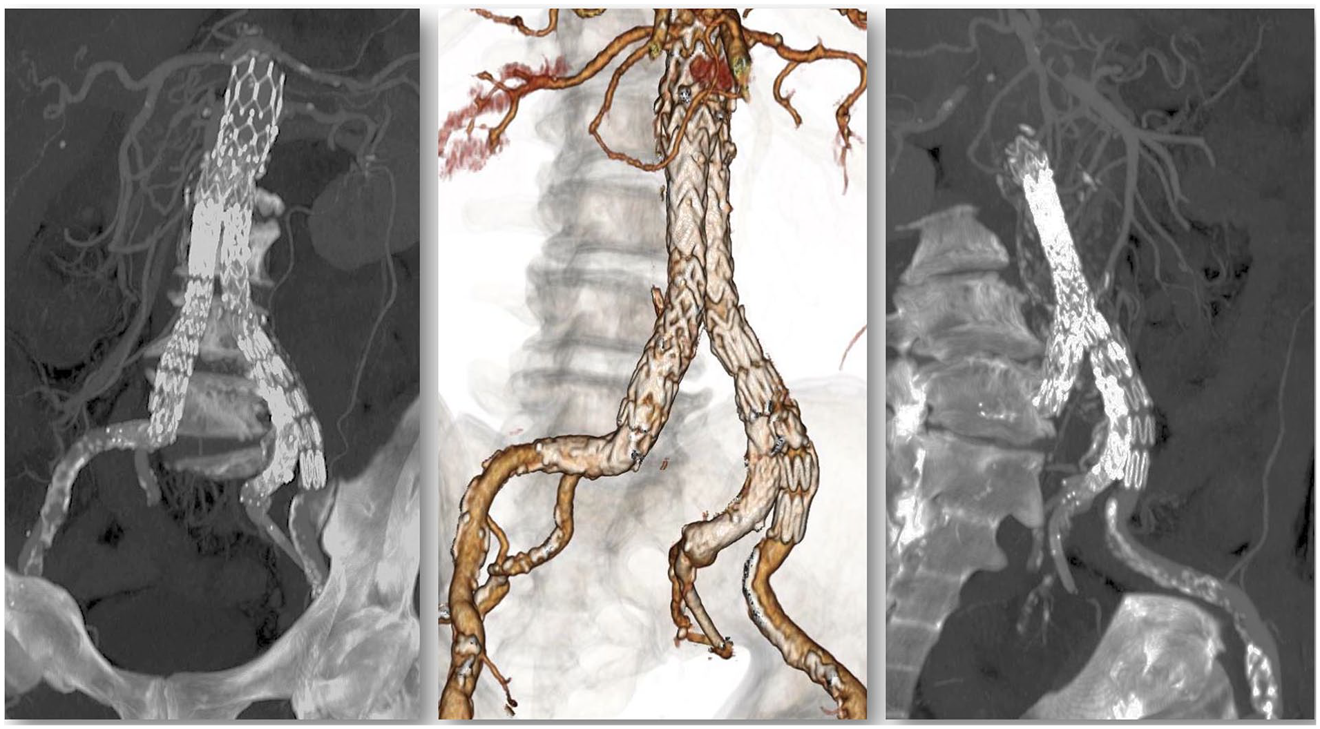
Control computed tomography angiography (CTA) at 1 month.
Discussion
Since the introduction of EVAR, stent graft design has continuously evolved, with the aim of improving outcomes in challenging anatomies. Considering the ever increasing number of patients receiving an aortic endograft, management of late device complications, such as limb occlusion, represents a current challenge. Despite the improved devices currently available, limb occlusion incidence rate can be as high as 8%,11,12 with limb graft occlusion being one of the major causes of re-hospitalization after EVAR. 13
According to a meta-analysis by Hammond et al, approximately 44% of patients with iliac occlusion after EVAR develop acute limb ischemia and nearly half (48%) of the graft thromboses are detected within 30 days from the index EVAR procedure. 11
Clinical presentation ranges from no symptoms to severe acute limb ischemia and may have a fatal outcome because of the risks involved with treatment. The 30-day mortality rate is estimated at 3.6% and the rate of limb loss within 30 days at 3.1%. 11
Revascularization is immediately required in acute limb ischemia, but may also be needed later in cases of ischemic symptoms worsening after chronic iliac occlusions.
Both surgical or endovascular and even hybrid approaches may be proposed, but limited evidence makes optimal management of iliac limb occlusion somewhat controversial. Consolidated treatment options include graft thrombectomy with eventual adjunctive balloon angioplasty or stenting, extra-anatomical bypass and thrombolytic infusion. 12 However, advanced total endovascular options are becoming widespread. Indeed, the most recent European Society for Vascular Surgery guidelines for the management of acute lower limb ischemia suggest to consider endovascular alternatives such as aspiration or endovascular thrombectomy in all patients with acute limb ischemia. 14
Technologic development has recently provided devices allowing minimally invasive treatment for acute as well as for chronic thrombosis, restoring the original endograft patency and avoiding open surgery. The Rotarex™S percutaneous mechanical thrombectomy system, although originally indicated for the treatment of acute, subacute, and chronic occlusions involving peripheral arteries, has been increasingly effectively adopted for this purpose even if no published reports are available as of today.
Providing a combined system, able to perform atherothrombectomy as well as aspiration with immediate blood flow restoration, the Rotarex™S device could overcome some of the limitations coming from loco-regional thrombolysis.
Intra-arterial thrombolytic infusion could be time-consuming, and at the same time, it determines a considerable risk of distal embolization and hemorrhage. 15
Moreover, in cases of progressive stratification and thrombus thickening over time, thrombolysis, thrombus aspiration, or balloon thrombectomy procedure effectiveness markedly decreases.
In these cases, open surgery is generally considered the first-line option, nevertheless a nonnegligible rate of surgical revascularization will eventually fail over time. 16 Furthermore, prosthetic infection is a possible complication which should always be considered. 17
To the author’s knowledge, a successful revascularization of a chronic hypogastric branch occlusion on previous IBD using the Rotarex™S system has never been reported before.
According to the IFU, the Rotarex™S catheter has applications in the treatment of arterial occlusions of native vessels, stents, prosthetic bypasses, and vascular accesses for dialysis.
The complete endovascular approach provided by the Rotarex™S system allows, once the target vessel has been recanalized, to perform adjunctive procedures such as percutaneous transluminal angioplasty/stenting or additional graft deployment to correct eventual intraluminal material persistence or the underlying pathology causing thrombosis.
In case of aortoiliac endograft recanalization, the authors suggest to always perform iliac limb relining in order to minimize the relapse risk.
Footnotes
Declaration of Conflicting Interests
The author(s) declared no potential conflicts of interest with respect to the research, authorship, and/or publication of this article.
Funding
The author(s) received no financial support for the research, authorship, and/or publication of this article.
