






Abstract
Purpose:
Takayasu arteritis is a large-vessel vasculitis in women of childbearing age that affects large vessels including the aorta and its main branches. Inflammation of arteries can produce lesions that lead to occlusion, stenosis, or aneurysms which can lead to complications. If signs of organ dysfunction are present, vascular intervention may be necessary.
Case Report:
In this article, we present a case of Takayasu arteritis with high-grade stenosis of all the great vessels of the thoracic aorta treated with drug-coated balloon (DCB) angioplasty and stenting requiring multiple follow-up interventions over a 1-year follow-up.
Conclusion:
The DCB angioplasty is a potential endovascular treatment for thoracic great artery stenosis in Takayasu arteritis that could be further explored.
Introduction
Takayasu arteritis is a rare large-vessel vasculitis most commonly occurring in young females that primarily affects the aorta and its main branches, although any large artery can be affected. 1 Onset of the disease usually occurs before age 50 and tends to have a subacute onset of symptoms which leads to a delay in diagnosis thus allowing the disease to progress and become symptomatic. 2 The mainstay of treatment is systemic glucocorticoids or glucocorticoid-sparring agents; however, vascular intervention may be necessary if organ ischemia is present. 3
Balloon angioplasty has been reported to have better long-term patency compared with stent placement for treatment of renal artery stenosis caused by Takayasu arteritis. 3 Drug-coated balloon (DCB) angioplasty has also been used to treat renal artery stenosis caused by Takayasu arteritis, 4 but to the best of our knowledge, the efficacy of DCB for treatment of stenosed thoracic arteries caused by Takayasu arteritis remains anecdotal. We present a case of a 24-year-old female with narrowing or occlusions of the innominate, right common carotid, left common carotid, left vertebral, left internal carotid, and left subclavian arteries who underwent vascular intervention by DCB angioplasty and stent placement.
Case Report
A 25-year-old white female presented with intermittent bilateral visual blurring, an occipital headache, and several syncopal episodes. Two months prior to admission at our facility, she was diagnosed with Takayasu arteritis, started on oral steroids, and was given a referral to a rheumatologist. She finished her course of steroids before she could get an appointment with a rheumatologist and started having recurrent syncopal episodes, so she presented to the emergency room with the above complaints.
Initial laboratory results were significant for an erythrocyte sedimentation rate (ESR) of 104 mm/h and C-reactive protein was 3.2 mg/dL. The patient’s 12-lead EKG showed sinus tachycardia with a rate of 120 beats/min. Cardiac troponin was nonelevated. Noncontrasted head computed tomography (CT) and CT angiogram of the head did not reveal any acute intracranial process. The CT angiogram of the neck showed extensive soft tissue thickening surrounding the aorta and origins of the great vessels extending proximally along their course in the neck consistent with Takayasu arteritis. Stenoses of the aortic artery branches were as follows: innominate 65%, left subclavian 90% proximal to the vertebral artery and occluded distal to the vertebral artery, left common carotid 80% right common carotid 80%, and left vertebral 30% (Figure 1). She was discharged home on high-dose glucocorticoid therapy as she had active acute vasculitis precluding vascular intervention. After she was discharged, her rheumatologist started her on mycophenolate mofetil for maintenance immunosuppression.
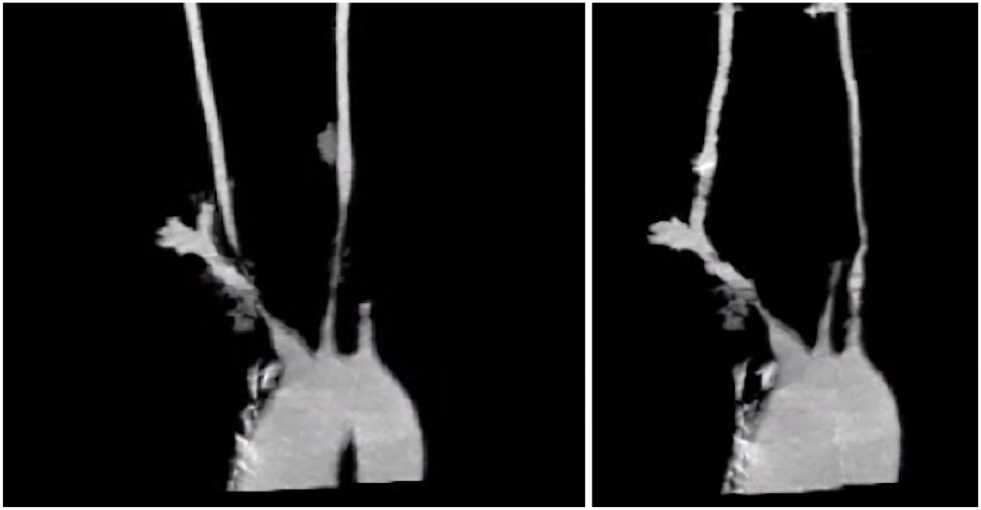
3D reconstruction of thoracic arterial vessels from CT angiogram of neck. Stenoses were measured at innominate 65%, left subclavian 100% occluded, left common carotid 80%, and right common carotid 80%.
DCB Angioplasty to Stenosed Thoracic Great Vessels
Twenty days later her ESR had normalized to 5 mm/h so she did not have active disease and could undergo intervention. Angiogram was performed showing stenosis of the innominate artery at 80% and the right common carotid artery showing 95% narrowing, left common carotid with 95%/subtotal occlusion, total occlusion of the left subclavian artery, and 90% narrowing of the left vertebral artery (Figure 2). Prior to intervention, the case was reviewed with multiple in-house and national consultants to discuss surgical versus percutaneous treatment options, but there was no consensus on best options because of the lack of data. Therefore, it was decided to attempt intervention with DCB angioplasty. Balloon and stent sizes were based on the carotid size, using a 1:1 sizing estimate. Angioplasty with a 7 mm × 60 mm IN.PACT Admiral DCB (Medtronic, Minneapolis, Minnesota) of the innominate artery was performed causing the patient to have momentary loss of consciousness with balloon inflations. Two further dilations of the innominate were performed with narrowing reduced to less than 30%. A SpiderFX distal protection device (Medtronic) was placed in the right internal carotid artery and the right common carotid artery was angioplastied with a 6 mm × 40 mm IN.PACT Admiral DCB, but a segment was poorly dilatable at 34 atm even after use of a cutting balloon, so 2 iCAST-covered stents (Atrium Medical Corp., Hudson, New Hampshire), 5 mm × 22 mm and 7 mm × 16 mm, were deployed with narrowing reduced to less than 40%. The use of covered stents was used to try to decrease restenosis because no large drug-eluting balloon–expandable stents were available in the United States and self-expanding drug-eluting stents were not chosen because of the lack of hoop strength. Her carotid stenosis was fibrotic, such that a balloon-expandable stent would be more likely to achieve larger lumen patency. Because of contrast limitations, further interventions were performed the following day. On the second day of intervention, a SpiderFX distal protection device was placed into the left internal carotid artery and the lesion was angioplastied with a 4 mm × 60 mm Sterling balloon (Boston Scientific Corp., Marlborough, Massachusetts) followed by a 5 mm × 60 mm IN.PACT Admiral DCB with narrowing reduced to 30%. After using multiple wires, catheters, and balloons, the occluded left subclavian artery was angioplastied with a 5 mm × 150 mm Lutonix DCB (Bard Peripheral Vascular Inc., Tempe, Arizona) with narrowing reduced to 25%. Angiography did not reveal any high-grade lesions in the thoracic aorta, abdominal aorta, celiac, or superior mesenteric arteries. She was discharged on clopidogrel, continued on mycophenolate mofetil and steroids, and started on metoprolol for persistent tachycardia.

Angiogram of thoracic great arteries prior to intervention. Stenoses were estimated at 80% for the innominate, 95% for the right common carotid, 95% for the left common carotid, total occlusion of the left subclavian artery, and 90% narrowing of the left vertebral artery.
Follow-up and Subsequent Interventions
By her 3-month follow-up, she noticed a return of her neck pain and tachycardia. Arterial duplex ultrasound showed recurrent innominate, bilateral carotid, and left subclavian artery stenosis. The decision was made to once again undergo intervention. The left common carotid had restenosis to 75% so was angioplastied with a 7 mm × 80 mm IN.PACT Admiral DCB, but still had intermediate stenosis so was further dilated with a cutting balloon which dissected the artery, so a 6 mm × 59 mm iCAST-covered stent was placed. The innominate artery had 50% stent restenosis, so a 7 mm × 22 mm iCAST-covered stent was deployed in the previous stent. The left subclavian had 60% restenosis so was angioplastied with a 6 mm × 150 mm IN.PACT Admiral DCB.
Two months after her second intervention, and 5 months after her index interventions, she developed right arm claudication. Angiogram showed that she had new stenosis of her right subclavian artery which was dilated with a 7 mm × 40 mm Lutonix DCB. The angiogram also showed patency of all the great thoracic vessels and no restenosis of previous lesions. At 9 months after her index interventions, arterial duplex ultrasound and CT angiogram of her chest did not show high-grade stenosis of her aortic branches.
At 1-year follow-up, she had increased velocities noted in the stents in both the right and left common carotid arteries on arterial duplex ultrasound. Aortic arch angiography showed persistent high-grade stenosis of the left vertebral artery for which intervention was previously deferred. Initially, a 4 mm × 32 mm Promus ELITE stent (Boston Scientific Corp.) was deployed in the left subclavian, but this caused plaque shift and jailing of the subclavian, so a 6 mm × 14 mm Express SD stent (Boston Scientific Corp.) was placed in the left subclavian at the level of the left vertebral artery. On intravascular ultrasound, there was a flap extending into the left subclavian artery below the origin of the left vertebral artery and a probable dissection with 2 lumens that likely developed as a result of prior intervention. Therefore, a second 4 mm × 20 mm Promus ELITE stent was placed in the left vertebral artery extending down into the left subclavian flap. The bilateral carotid arteries had stent restenosis and the right subclavian artery had intermediate stenosis, so the case was reviewed with multiple in-house and national consultants who recommended further carotid stenting rather than surgical intervention. Two weeks later, a 7 mm × 16 mm Express LD (Boston Scientific Corp.) was deployed in the right ostial subclavian artery. An iCAST-covered stent was deployed stent-in-stent in both the right (6 mm × 38 mm) and left (7 mm × 38 mm) common carotid arteries for stent-restenosis. Interestingly, the right common carotid was poorly dilatable during the initial intervention the previous year, but with 20 atm of pressure, the narrowing was reduced to less than 20%.
Discussion
Takayasu arteritis is a rare large-vessel vasculitis that typically affects the aorta and its branches. Endovascular treatment is indicated in patients with ischemic symptoms including syncope and transient ischemic attacks. In general, vascular interventions should not be performed in patients with active inflammation. 5 One study showed that patients who undergo endovascular repair have 10-year restenosis rates of 62% regardless of whether stents or nonstenting modalities were employed. 6 Another group that studied stenting versus percutaneous balloon angioplasty in hypertensive patients with renal artery stenosis due to Takayasu arteritis saw lower rates of restenosis in patients undergoing uncoated balloon angioplasty compared with patients who received stents. 7 Drug-coated balloon angioplasty has recently been used to treat stenotic lesions caused by Takayasu arteritis.4,8 To our knowledge, the use of DCBs to treat arterial stenosis involving all the major branches of the thoracic aorta due to Takayasu arteritis has not been reported.
In peripheral arterial disease, DCB has shown improved durability of treatment compared with uncoated balloon angioplasty. 9 In Takayasu arteritis, uncoated balloon angioplasty has been shown to be at least noninferior, and possibly superior, to stenting when it comes to restenosis of lesions. Therefore, we hypothesized that DCB angioplasty may even have lower restenosis rates than angioplasty with uncoated balloons. In this case, the patient underwent interventions that included DCB angioplasty as well as stenting.
Restenosis rates in Takayasu arteritis have been shown be high regardless of intervention modality, so it is likely she will need further interventions in the future. She had restenosis of lesions treated by both DCB and stenting, so it is difficult to determine whether her high restenosis rate was patient-specific, or whether DCB had any benefit. Therefore, any conclusions of the efficacy of intervention modality cannot be assessed. More studies are needed to assess whether DCB angioplasty has any advantage over uncoated balloon angioplasty as well as benefit over stenting in treatment of stenotic thoracic vessels due to Takayasu arteritis. Nonetheless, DCB angioplasty is a potential vascular intervention modality for stenosis and restenosis of aortic branches of the thoracic artery in Takayasu arteritis that could be explored in future studies.
Conclusion
We treated stenotic lesions, as well as restenosis of the same lesions, with DCB angioplasty of the great vessels of the thoracic aorta. In this case, it could not be determined if DCB had any benefit over stenting, but percutaneous treatment with DCBs could be explored as a treatment for thoracic arterial stenosis in Takayasu arteritis.
Footnotes
Authors’ Note
There has been no duplicate publication or submission of any part of the work. All authors have read and approved the manuscript.
Declaration of Conflicting Interests
The author(s) declared no potential conflicts of interest with respect to the research, authorship, and/or publication of this article.
Funding
The author(s) received no financial support for the research, authorship, and/or publication of this article.
