






Abstract
Background:
Vascular complications are a cause of increased morbidity and mortality when performing percutaneous procedures requiring large-bore arterial access. MANTA vascular closure device (VCD) is currently the only large-bore VCD using an intraluminal foot plate and an extraluminal collagen plug. The traditional depth locator approach might be compromised in; emergent cases without the required measurements, cases of hematoma formation, or other patient, procedure, or operator-specific variables. Furthermore, this technique can be used for postclosure in cases without the required measurement of depth. We describe vascular outcomes using fluoroscopy (fluoroscopic DOT technique) rather than traditional depth locator approach for vascular closure with the MANTA VCD.
Methods:
Fifty patients who underwent transcatheter aortic valve implantation (TAVI) were retrospectively analyzed using fluoroscopic DOT technique with 18F MANTA VCD between May and August 2021. All patients >18 years of age who qualified for transfemoral TAVI were included. Access was obtained with ultrasound guidance with vessel diameter of at least
Results:
In total, 50 patients were analyzed with a mean age of 81 years and majority were male (56%). Majority had comorbidities of hypertension (88%) and hyperlipidemia (94%), 24% had peripheral arterial disease, 38% coronary artery disease, and 58% were former smokers. Importantly, 40% were obese with an average body mass index (BMI) of 29 kg/m2. There were no bleeding or ischemic limb complications post MANTA VCD deployment using the fluoroscopic DOT technique. Furthermore, none of the patients required peripheral intervention from index procedure to 1 month post verified during their 1-month post TAVR follow-up.
Conclusion:
Fluoroscopic DOT technique using the MANTA VCD is highly reproducible and allows hemostasis in a predictable manner for procedures requiring large-bore arterial access in the absence of MANTA depth measurement.
Keywords
Introduction
Obtaining predictable and complete hemostasis is paramount to percutaneous procedures requiring large-bore arterial access such as transcatheter aortic valve replacement (TAVR), abdominal endovascular aneurysm repair (EVAR), mechanical circulatory support (MCS), or minimally invasive valve surgery. Bleeding complications related to large-bore arterial access is associated with increased in-hospital mortality, length of stay, and increased cost. 1 Transfemoral access is the preferred first-line approach for TAVR and is associated with 20% relative risk reduction in mortality in comparison with surgical aortic valve replacement. 2 Although the incidence of vascular complications and bleeding have decreased overtime from adoption of better practices, smaller delivery sheaths, and operator experience, they are still associated with increased length of stay and higher 1-year mortality (HR 2.31 [95% CI, 1.20–4.43] p=0.012).3–5 A recent observational study of 34 893 patients undergoing TAVR at 445 hospitals showed 9.3% of patients to have a vascular complication and 7.6% with in-hospital bleeding events. Vascular complications were associated with increased 30-day death (HR 2.23 [95% CI, 1.80–2.77]), death (adjusted HR 1.17 [95% CI, 1.05–1.30]) and rehospitalization (adjusted HR 1.14 [95% CI, 1.07–1.22]) at 1 year. Whereas, vascular bleeding events were also related to increased 30-day death (HR 3.71 [95% CI, 2.94–4.69]), death (HR 1.39 [95% CI, 1.23–1.56]), and hospital readmissions at 1 year (HR 1.19 [95% CI, 1.11–1.27]). 6 Hence, achieving adequate hemostasis free from vascular complications becomes paramount to improve both long- and short-term outcomes with large-bore arterial access related procedures.
MANTA VCD is a vascular closure device using an intraluminal footplate and extraluminal collagen plug (like ANGIO-SEAL) approved for large-bore vascular closure in 2019. 7 This study’s institution has switched to 100% MANTA VCD use for transfemoral TAVR procedures with an average of 250 procedures performed annually. Rapid, predictable, and complete hemostasis can be accomplished in majority of the TAVR cases where MANTA VCD is used. The first step of MANTA VCD is use of the MANTA depth locater, marked by centimeters (cm), to be advanced into femoral arteriotomy site for measurement of appropriate vessel depth which will be used at the end of procedure for final deployment steps. However, certain anatomic and patient-related factors can produce significant variability in this crucial measurement step and may lead to deployment at an improper level and therefore associated vascular complications. The device encompasses a radiopaque marker which locks the sliding suture knot that initially compacts the extravascular collagen plug and is a landmark used for future interventions. From operator experience, the initial depth finder measurement can be significantly variable from the final depth measurement due to patient and procedure-related factors such as intraprocedural hematoma formation, patient obesity, ioban film causing device movement, or operator skin pressure during depth finder step. A retrospective single center analysis of the fluoroscopic DOT technique taking advantage of the radiopaque marker was synthesized to allow for more precise and predictable deployment than the depth locater method. An additional advantage of this technique is its ability to be employed as dry postclosure in cases where either suture-mediated VCD have failed or when emergent large-bore arterial access procedures are performed; ie, MCS for cardiogenic shock.
Methods
A single-center retrospective study was conducted of 50 patients, >18 years of age, who underwent transfemoral TAVR with either Medtronic Evolut PRO or Edwards Sapien 3 Ultra for whom hemostasis was achieved with 18F MANTA VCD using fluoroscopic DOT method alone without the depth locator between May 2021 and August 2021 with prospective data obtained. While at the time, these patients consented to the procedure, obtaining written informed consent from the patients whose chart will be reviewed was not required as this study met the FDA’s criteria for consent to be waived, according to its guidance document IRB Waiver or Alteration of Informed Consent for Clinical Investigations Involving No More than Minimal Risk to Human Subjects (July 2017). Common femoral artery access was obtained with ultrasound guidance with a minimum lumen diameter of
Procedure and Device Description
MANTA VCD is available in 2 sizes, 14F and 18F, and allows reduced time to hemostasis with use of 10F-20F devices or sheaths with an outer diameter (OD) between 12F and 25F. The device consists of an absorbable collagen pad, stainless-steel locking component, and an absorbable polymer anchor connected by a nonabsorbable suture.
The first step involves use of the MANTA depth locator to locate the common femoral artery (CFA) access site. After obtaining CFA access and placing a 6F or 8F sheath, the MANTA depth locater (8F size) is inserted until steady pulsatile arterial blood flow is noted. Next, keeping the skin at neural position, the depth locater is slowly withdrawn until blood flow stops taking note of the visible cm marker at which blood flow ceases and adding 1 cm. This measurement is the marketed instructions for use (IFU) for final MANTA VCD deployment. After completion of procedure, the large-bore arterial access sheath is removed, and the MANTA introducer and sheath assembly placed hubbed into the arteriotomy site. The introducer is removed and the delivery tube inserted until a click is audible. MANTA handle and sheath are withdrawn corresponding to the measurement as assessed by the depth locater during step 1. The handle on the level is rotated back until a click is audible and the device withdrawn at least 45 degrees until yellow/green is seen at tension window. The blue lock tube is advanced lightly allowing the radiopaque lock to secure implant. Tension is increased on the MANTA closure handle until an audible click is heard completing device deployment (Figure 1).
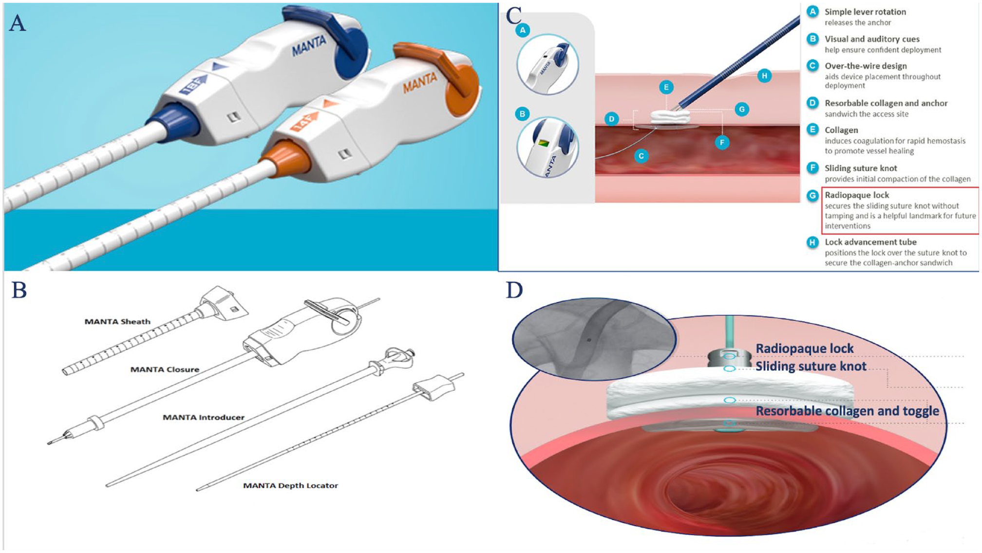
(A) 2 MANTA vascular closure device (VCD) sizes (14F and 18F) available currently in the market to achieve hemostasis for 10F-20F devices or sheaths. (B) Different components of MANTA VCD: MANTA depth locator, sheath, closure tube, and introducer. (C) Labeled different components of device while actively being deployed in target vessel for large-bore access closure. Note the “radiopaque lock” secures the sliding suture knot of the extra-luminal collagen plug prior to advancement of blue tamping tube and is a marker for future interventions. (D) Visualization of radiopaque lock on fluoroscopy.
Description of Fluoroscopic DOT Technique
Micropuncture arterial access was performed using modified Seldinger technique with ultrasound guidance for all 50 patients using 0.021-inch needle and fluoroscopic image saved with the 0.018-inch guidewire inserted at the CFA access level. Ultrasound guidance was used to ensure that no anterior CFA calcification was noted at needle site entry and that the CFA minimal lumen diameter was

MANTA VCD deployment using fluoroscopic DOT technique in a patient with groin hematoma. (A) MANTA depth locator measured vessel depth to be 5+1 cm = 6 cm. (B) Patient developed a hematoma during sheath and device exchanges. (C) Fluoroscopic image with micropuncture needle (white arrow) denoting exact arterial puncture site reviewed. (D) MANTA device fluoroscopic radiopaque DOT (white arrow) pulled back to level of micropuncture needle tip marking precise location of access site. (E) Due to the subcutaneous hematoma, measurement on MANTA sheath/tube assembly noted to be at 8 cm, instead of 6 cm as measured with the initial depth locator, with potential for too distal deployment and mechanism for device failure and related vascular complications if deployed exclusively based on initial depth locator measurement. (F) Digital subtraction angiography without any evidence of contrast extravasation or flow-limiting limb occlusion.
With standard IFU method, the MANTA introduction sheath is 1 cm inside the vessel from the arteriotomy site. Although, with fluoroscopic DOT technique, the introduction sheath is 2.5 cm inside the vessel from the arteriotomy site, thus is 1.5 cm deeper than IFU method.
Results
A total of 50 patients were analyzed retrospectively, with a mean age of 81.4 years, of which 28/50 (56%) were male. The mean BMI was 29 kg/m2, range (16.6–40.03 kg/m2), and obesity as identified by BMI of greater than 30 kg/m2 was present in 20/50 (40%) of patients. Majority had comorbidities of hypertension (88%), hyperlipidemia (94%), were former smokers (58%), and 17/50 (34%) had diabetes. Equal number of patients had chronic obstructive lung disease and chronic kidney disease (10/50, 20% each), and majority had either CAD with PCI or prior CABG (29/50 or 58% total). CHF was present in 30% of patients, while prior stroke history in 10% and concomitant PAD in 12/50 (24%) (Table 1). Primary outcome of major vascular complications (flow-limiting stenosis/dissection or active bleeding) requiring additional surgical or endovascular intervention as assessed by DSA from contralateral access site was seen in none of the patients using the fluoroscopic DOT technique (Table 1). Furthermore, there were no minor vascular complications requiring conservative treatment measures; ie, no additional manual compression performed after standard full dose protamine at the conclusion of procedure for bleeding, pseudoaneurysm or monitoring/presence of non-flow-limiting dissection or stenosis. Although DSA was performed till approximately 3 to 5 cm below the level of CFA bifurcation, there was no evidence of distal embolization assessed by patients’ symptoms or manual distal lower extremity pulse palpation or Doppler assessment pre- and postprocedure. Finally, none of the 50 patients required any peripheral angiogram or intervention from index procedure to 1-month post-TAVR follow up.
Baseline Demographics, Clinical Characteristics and Vascular Outcomes of Subjects (N=50).

Continuous data are presented as mean (range). Category data are given as frequency (percentage).
Abbreviations: CABG, coronary artery bypass graft; CAD, coronary artery disease; CHF, congestive heart failure; CKD, chronic kidney disease; COPD, chronic obstructive pulmonary disease; PCI, percutaneous coronary intervention.
As illustrated, with the fluoroscopic DOT technique, the foot plate is 1.5 cm deeper than the IFU method, and this did not correlate to any clinical adverse outcomes.
Discussion
Novel use of the fluoroscopic DOT technique for MANTA VCD deployment can allow precision and predictability in vascular closure, especially when procedure-, operator-, and patient-related factors can skew the MANTA depth finder measurements performed at the start of procedure. Outcomes of 50 patients undergoing transfemoral TAVR procedure using this technique showed no vascular complications of flow-limiting dissection or bleeding requiring endovascular or surgical intervention. Evidence from this observational study shows that a high level of predictability and MANTA VCD deployment can be achieved while maintaining a low rate of vascular complications. With the fluoroscopic DOT technique, the foot plate is 1.5 cm deeper within the vessel than with using manufacturers IFU method, and this did not correlate with any clinical adverse outcomes. On the contrary, this method can be advantageous in obese patients, those with intraprocedural hematomas or unexpected device movement; ie, ioban film, operator hand pressure, or combination of obesity and hematoma. Furthermore, while none of the patients in this study had external iliac artery stents or heavily calcified CFA vessels, knowledge of this technique and depth difference can allow the operator to deploy the vascular closure carefully under fluoroscopy taking account of the relevant patient-specific anatomy or the fluoroscopic dot can be placed 1 to 1.5 cm distal to the arteriotomy site. This approach allows more operator control and precision in deployment based on patient anatomy and overcomes other unexpected procedure related factors.
Evidence for MANTA VCD
The SAFE MANTA pivotal clinical study was a prospective, single-arm, multi-center investigational study of 341 patients where MANTA VCD was used for TAVR and large-bore endovascular procedures with mean effective sheath OD of 22F (7.3 mm). Mean time to hemostasis was only 65
MANTA VCD has been extended in application to obtain hemostasis for percutaneous axillary TAVR implantation, decannulation of venoarterial extracorporeal membrane oxygenation (VA-ECMO), in addition to minimally invasive valve surgery.12–15 However, it is important to note that the SAFE MANTA study selected patients without morbid obesity, severe calcification, or severely scarred femoral access area. While femoral artery calcification is best avoided with careful assessment by computed tomographic angiography during preplanning stage for TAVR, patient-related factors such as obesity and access vessel depth are difficult to preplan and navigate intraprocedurally. Thus, techniques to bring precision and predictability to unavoidable patient-related factors can be paramount to reduce vascular complications.
Benefits for Use of Fluoroscopic DOT Technique With MANTA VCD
Obesity and small femoral artery diameter with deep arteriotomy or greater vessel depth from skin puncture site appear to be strong patient-related variables associated with large-bore arterial access VCD failures.16,17 The initial MANTA depth locator measurement can be significantly different than the measurement toward the end of the procedure where this depth measurement is used for final deployment. A deeper arteriotomy site has been associated with increased MANTA-related vascular access site complications. 16 Additionally, anterior CFA calcification, 10 peripheral artery disease and vessel diameter < 6 mm, 18 low or high arteriotomies, 19 female gender, left CFA access, vessel angulation, and at least moderate anterior vessel calcification16,20 have all been independently associated with manta related vascular complications.
Operator- or procedure-related factors that lead to variability in MANTA depth finder measurement from start of procedure to the end include unintentional compression of skin by operator during depth locator measurement or during deployment process, iatrogenic hematoma accumulation during procedure related to device and sheath exchanges increasing skin to vessel depth distance (as shown in Figure 2), and sheath or depth locator entrapment or compression related to ioban film at the groin site which is commonly employed in OR setting to improve sterility. The fluoroscopic DOT technique allows precise and predictable MANTA VCD deployment at the large-bore arteriotomy site potentially overcoming these patient-, operator-, and procedure-related unpredictable factors. The operator theoretically has better control of the deployment process with knowledge of this technique and distance to the fluoroscopic dot to allow for more accurate deployment in setting of factors that can lead to variability in depth measurement. Although, with the technique, the foot plate is deployed 1.5 cm deeper than IFU method, this did not lead to any clinically significant adverse outcomes. The operators found the fluoroscopic DOT technique to be a reliable method to ensure proper MANTA VCD deployment as shown in these 50 patients who were free of any bleeding or ischemic limb complications and continue its use in daily practice.
Fluoroscopic DOT Technique for Dry/Postclosure Technique or in Cases of Failed Suture-Mediated VCD
Application of fluoroscopic DOT technique can be extended for use as dry field/postclosure during emergent cases or in cases where initial suture-mediated VCD fails. Dry field technique refers to emergency situations where emergent insertion of MCS is required without deployment of time consuming proglide sutures in preclose fashion. Postclosure options in these instances include balloon tamponade for 30 to 60 minutes or covered stent deployment which are both time and cost inefficient and commit a patient to unnecessary covered stent deployment at the CFA site. 20 While other techniques using either PVCD or ANGIO-SEAL (Terumo Interventional Systems, Somerset, NJ) of various size and numbers have been reported in case reports or small case series, they lack predictability and have not been performed in large numbers in a prospective manner to be considered a reliable method to achieve adequate hemostasis at a high success rate.21–24 Hence, this technique can also be successfully employed as postclosure in emergent MCS insertion cases such as Impella (Abiomed, Danvers, MA) or VA-ECMO as long as the initial arterial puncture site is known.
Limitations
Although the MANTA fluoroscopic DOT techniques appears to be a reliable technique to achieve hemostasis for large-bore arterial access in a predictable manner overcoming patient- and procedure-related variable factors, further validation in a randomized, prospective manner in a larger patient population is warranted. The fluoroscopic DOT technique was performed in the anterioposterior (AP) projection and was successful, using oblique perpendicular projections can improve outcomes with this technique, especially in patients with high or low CFA bifurcation. Additionally, validity of this technique as postclosure in emergent MCS or other emergent large-bore arterial access in larger series of patients is indicated as it can significantly improve vascular closure predictability and success rate in such cases.
Conclusion
The fluoroscopic DOT technique for MANTA VCD use after TAVR is a safe alternative to the standard application of the MANTA VCD. In addition, this technique can potentially be used for postclosure, for example, after emergent MCS insertion or failed suture mediated VCD. Further prospective studies using this technique in a larger patient population and validation of this technique as postclosure is warranted.
Footnotes
Acknowledgements
This is an original manuscript and has not been previously published or submitted to another journal.
Declaration of Conflicting Interests
The author(s) declared no potential conflicts of interest with respect to the research, authorship, and/or publication of this article.
Funding
The author(s) received no financial support for the research, authorship, and/or publication of this article.
