






Abstract
Purpose:
Common femoral artery (CFA)-occlusive disease has traditionally been treated with open surgery, yet nowadays the frailty of patients has induced to find new techniques of revascularisation by endovascular means. So far, intravascular lithotripsy (IVL) has shown promising results in several lower limbs arterial districts. The purpose of this article is to report our experience with IVL for severely calcified peripheral arterial disease (PAD) of the CFA.
Methods:
From November 2018 and October 2020, 10 consecutive patients (12 limbs) treated with IVL were prospectively enrolled in a dedicated database. Inclusion criteria were CFA localization of PAD, with a severe degree of calcification, a lesion length ≥10 mm, and a degree of stenosis ≥70% (severe). The only admitted adjunctive treatment was drug-coated balloon (DCB) angioplasty. Primary outcomes were technical and procedural success, clinical success, and target lesion revascularisation (TLR). Secondary outcomes were target extremity revascularisation (TER) and major adverse events (MAEs).
Results:
All patients underwent IVL with associated DCB angioplasty. The median percentage of achieved stenosis reduction was 55.5% (interquartile range [IQR] 50-60.75), with a technical and procedural success of 100%. Over the study period, TLR only occurred in one limb (8.3%), with a mean upgrade in Rutherford class of 2.7 ± 0.77. No target vessel and access site complications were reported, as well as no distal embolization. One death and one major amputation occurred over the follow-up period, both in the same patient.
Conclusions:
Based on our experience, IVL for selected cases of severely calcified CFA disease, associated with DCB angioplasty, may be considered a safe and effective technique. Of course, a long-term follow-up and a larger series of patients are needed to validate our results.
Keywords
Introduction
Peripheral arterial disease (PAD) of the common femoral artery (CFA) district has traditionally been treated with open revascularisation, particularly surgical endarterectomy. The multiple advantages of this technique are well-known: profunda femoris artery (PFA) branches preservation, which could be compromised by an endovascular procedure, 1 and compliance to hip flexion, which could ultimately lead to stent fracture; 2 moreover, good results and durability of the procedure 3 are well-established.
Endovascular repair has been continuously evolving during the last years, and revascularisations once only entrusted to open repair, are now mostly managed by endovascular means, such as the iliac axis treatment. 4 The fragility and the comorbidity rate of patients affected by PAD have stimulated the research for less-invasive revascularisation techniques. The CFA district is the extreme challenge for reasons discussed above, yet many options have been developed, such as plain old balloon angioplasty (POBA), drug-coated balloons (DCBs), cutting balloons, atherectomy, stenting, and drug-eluting stents (DESs). Endovascular options provide high rates of technical success, a shorter hospital length of stay and a low-rate of perioperative mortality and morbidity. 5 Yet, long-term patency appears to be not entirely satisfactory and reported target lesion revascularisation (TLR) rates are higher than open surgery. 6 Common femoral artery stenting has shown promising results, 7 yet hip flexion with the related possibility of stent fracture still remains an unresolved issue.
The main concern regarding CFA lesions is the frequent presence of calcifications, resulting in a reduction not only of the vessel lumen, but also of the arterial wall compliance, which could lead to dissection and/or thrombosis. Besides, after endovascular revascularisation of a severely calcified lesion, the postprocedural degree of stenosis tends to remain high, with poor procedural success and durability. 8 Intravascular lithotripsy (IVL) uses sonic pressure waves to modify the structure of intimal and medial calcium layers, producing shear stress that fractures calcium. 8 Many trials have been developed in the last 5 years8,9 regarding IVL applied to PAD also localized to the CFA district, and results are encouraging.
The aim of this article is to report our midterm results with IVL for severely calcified CFA-occlusive disease.
Materials and Methods
Study Design and Patients’ Demographics
From November 2018 to October 2020, all patients undergoing intervention for PAD were prospectively enrolled in a dedicated database. Of these, patients with a severely calcified CFA occlusive disease undergoing IVL associated with DCB angioplasty were retrospectively analyzed. All patients received exhaustive information about the procedure and all patients were consented for the surgical and endovascular procedures. Data were deidentified and collected in an anonymized database.
Inclusion criteria of this study were infrainguinal PAD localized in the CFA, with a severe degree of calcification, a lesion-length ≥10 mm, and a degree of stenosis ≥70% (severe). Patients with a previous aorto-iliac revascularisation, regardless of whether it was open or endovascular, were also included in the analysis. Patients selected for IVL had an American Society of Anaesthesiologists clinical status examination (ASA score) ≥3 and a Body Mass Index (BMI) ≥25 kg/m2.
We excluded patients with untreated PAD of the aorto-iliac segment and all cases where a simultaneous revascularisation of another arterial district of lower limbs was performed. Patients with no below-the-knee (BTK) patent axes were also excluded from the analysis.
We collected patients’ demographics, cardiovascular risk factors, ASA score, BMI values, preoperative antiaggregant or anticoagulant therapy, and months of survival. The lesions’ characteristics, such as length and percentage of stenosis, and the calcification degree were defined according to the Peripheral Academic Research Consortium (PARC). 10 We reported the duration of symptoms before the intervention, the preoperative clinical status according to Rutherford et al, 11 any previous endovascular treatments in the CFA region, previous aorto-iliac revascularisations, concomitant PAD localization in the superficial femoral artery (SFA) and PA and number of patent BTK axes. Intraoperative details were collected, such as the access type (percutaneous or surgical), the IVL pulse cycles performed in each intervention, and the duration of DCB application, as well as any intraoperative complication.
All lesions were preoperatively evaluated with duplex ultrasound (DUS) and computed tomography angiography (CTA), associated with an intraoperative evaluation with digital subtraction angiography (DSA), before and right after IVL.
Procedure Description
All procedures were performed with the Shockwave M5 Peripheral IVL system (Shockwave Medical Inc., Santa Clara, CA). This device has been described before,6,8 and it is composed of a generator, a cable connector, and a single-use over-the-wire catheter whose extremity contains lithotripsy emitters localized in an integrated balloon. The liquid contained in the balloon facilitates the sonic pressure-waves transmission to the calcified lesion with consequent fractures in the calcium.
After a percutaneous contralateral femoral access, a crossover was realized, and a 6 Fr-45 cm guiding sheath (Destination, Terumo Corporation, Tokyo, Japan) was placed in the homolateral external iliac artery. The lesion was crossed using a 0.014 Advantage (Terumo Corporation, Tokyo, Japan) or Regalia (Asahi Intecc, Japan) guidewire with a 4 Fr supporting catheter (Navicross, Terumo Corporation, Tokyo, Japan), and the Shockwave IVL system was placed at the level of the lesion. We used a 6 mm × 60 mm IVL system in every procedure with an average number of pulses of 150 (range: 120-180), the maximum pulses allowed being 300, and the system balloon was inflated at a subnominal pressure di 4 atm and subsequently 6 atm. After lithotripsy, we completed the lesion treatment with a DCB (Lutonix, Bard Peripheral Vascular, Inc. Tempe, AZ) inflated for 2 minutes. The final DSA allowed to verify the technical success of the procedure. All percutaneous accesses haemostasis have been achieved via manual compression.
One patient had both CFAs treated in the same session: an SFA surgical access was realized on one side, the first lesion was crossed with a 0.014 guidewire and a supporting catheter (Navicross, Terumo Corporation, Tokyo, Japan), and a crossover was realized to first treat the contralateral lesion.
Postoperative Management and Endpoints
All patients were given oral anticoagulation associated with a single antiplatelet therapy for at least 1 year after the procedure. Postoperative follow-up was primarily conducted with DUS every 3 months, reserving CTAs for cases of symptoms worsening, suspicion of recoiling of the lesion, or suspicion of PAD localization in another district.
Primary endpoints were technical success, procedural success, clinical success, and TLR rate. Technical and procedural success was defined according to Patel et al. 10 An improvement of at least one Rutherford category 11 without repeated treatments was considered as clinical success. Secondary endpoints were target extremity revascularisation (TER) and major adverse events (MAEs). 12 Major adverse events included all deaths, major amputations (both planned and unplanned), access site and target vessel complications (pseudoaneurysm, arteriovenous fistula, haematoma, dissection, and vessel thrombosis), distal embolization, and neurological, cardiac, gastrointestinal, and respiratory complications.
Target lesion revascularisation and TER rates indicate the rates of reintervention at the level of the lesion treated by IVL (including 1 cm above or below) and the rates of reintervention for a lesion localized far from the initial target site, respectively. 12 Diehm et al 12 define the subtraction of TLR and TER rates as an indicator for atherosclerosis progression. Restenosis was defined when an arterial lumen reduction ≥50% and a peak systolic velocity index ≥2.4 were found at DUS. 13
Statistical Analysis
Analysis were performed with R 4.0.3 (R Foundation for Statistical Computing, Vienna, Austria), and Prism GraphPad 9.0.2 (GraphPad Inc, San Diego, California). Patients’ baseline characteristics were reported as mean ± standard deviation (SD) or median + interquartile range (IQR), and frequency + percentage for continuous and categorical variables, respectively.
Results
From November 2018 to October 2020, 250 patients underwent PAD revascularization. Of these, 22 were operated of CFA revascularization and 13 patients (15 limbs) were subjected to IVL for PAD localized in the CFA. Ten patients (12 limbs) had a severely calcified CFA-occlusive disease and were therefore included in this analysis. Mean age was 75 ± 9.19 years old and most patients (70%) were males. Cardiovascular risk factors and comorbidities are summarized in Table 1: 50% of patients had coronary artery disease (CAD), all of them had underwent coronary revascularization, and 40% of patients as well had chronic obstructive pulmonary disease; chronic kidney disease was present in 30% of patients at a mild stage (stage II), 14 all patients had an ASA score of 3, and the mean BMI was 31 ± 4.3. All patients were on an antiplatelet medication, and in 7 cases (70%), this was associated with an anticoagulant medication.
Patients’ Demographics, Cardiovascular Risk Factors, and Preoperative Therapy. All Patients with Chronic Kidney Disease (CKD) Have 60< eGFR <90.

SD, standard deviation; IQR, interquartile range; NID, noninsulin dependent; ID, insulin dependent; CABG, coronary artery bypass grafting; PTCA, percutaneous transluminal coronary angioplasty; COPD, chronic obstructive pulmonary disease; BMI, body mass index; ASA, American Society of Anaesthesiologists.
The percentage is calculated over patients with CAD.
Preoperative characteristics are summarized in Table 2. All patients had a CFA localization of PAD, and in 58% and 42%, the disease was also localized in the SFA and the PA, respectively. Four patients had previous endovascular CFA revascularisation, consisting of POBA; 4 patients had a previous aorto-iliac revascularisation. About 50% of patients had critical limb-threatening ischemia (CLTI) (1 patient had a Rutherford grade 6), and the median duration of symptoms was 24 months (IQR: 12-24). Common femoral artery target lesion was severely calcified in 80% of patients, with a median degree of stenosis of 80% (IQR: 78.7-90) and a median length of 30 mm [20-34.5].
The Table Describes Preoperative Details on Clinical Status and Plaque Characteristics, as Long as the Postoperative Clinical Improvement and the Rate of Stenosis Reduction.

SD, standard deviation; CFA, common femoral artery; SFA, superficial femoral artery; PA, popliteal artery; PAD, peripheral arterial disease; BTK, below the knee.
Technical and procedural success was 100% with a median percentage of stenosis reduction of 55.5% (IQR: 50-60.75; Figures 1 and 2). Mean upgrade in Rutherford class was 2.7 ± 0.77. One patient had a repeated IVL procedure after 1 month because of restenosis (TLR = 8.3%). Over the study period, TER occurred in 2 limbs: 1 patient had a subsequent PAD localization in the SFA at the Hunter level, and 1 patient had a worsening of BTK axis disease. Median follow-up was 12 months (range: 6.75-13.50). Table 3 reports preoperative and postoperative Rutherford class and stenosis degree of all patients.
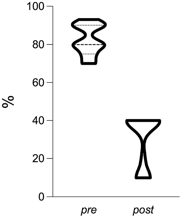
Preoperative and postoperative median (+IQR) common femoral artery stenosis rate.

Preprocedural and postprocedural digital subtraction angiography of a severely calcified common femoral artery stenosis. The final result did not require any stent implantation.
Overview of Preoperative and Postoperative Rutherford Class and Preoperative and Postoperative Degree of Stenosis, Focusing on Every Treated Limb.

There were no target vessel and access site complications, no distal embolization and no neurological, cardiac, gastrointestinal, and respiratory complications. Furthermore, PFA was patent both preoperatively and postoperatively, and no distal embolization was suspected nor evidenced at this level as well. One death and one major amputation occurred over the follow-up period in the same patient after 1 year from the IVL procedure, following a homolateral SFA recanalization and stenting.
Discussion
Open surgical revascularisation of the CFA district includes femoral endarterectomy, with or without patch angioplasty, and iliofemoral or femoro-femoral bypasses, using an autologous or a prosthetic vascular substitute, even without SFA reimplantation. These interventions allow to face multiple arterial lesion types, obtaining excellent technical success and long-term results.3,15 For a long time, CFA atherosclerotic disease has been considered a contraindication for endovascular repair because of hip mobility that could ultimately cause damages to implanted devices2,16 and because of the possibility of PFA branch loss 1 or complications of the femoral bifurcation, such as dissection or thrombosis. Yet, despite the fact that open revascularisations may also be performed avoiding general anesthesia in selected cases, in patients with important comorbidities, considered at high surgical risk, or in patients with a high BMI, the incidence of systemic and/or local complications is not neglectable. 17
The main challenge of endovascular procedures in the CFA territory is the presence of severely calcified plaques. Calcium distribution in the arterial wall varies according to patients’ comorbidities: the arteries calcifications can be limited to the intima in common atherosclerosis; the localization in the media, known as Mönckeberg sclerosis, with an associated circumferential distribution, is typically observed in patients with diabetes mellitus, end-stage renal disease (ESRD) under haemodialysis treatment, dysmetabolic conditions such as hyperparathyroidism and advanced age.
In the CFA district, POBA has obtained poor long-term results and high risk for adjunctive stenting and dissection. 18 Calcified plaques may reduce the efficiency of DCBs because of the difficulties in drug penetration due to the calcified barrier. Some authors19,20 have focused on CFA stenting, showing good early and long-term results with a freedom from TLR and TER at 5 years of 79% and 73%, respectively. A French multicentric prospective randomized controlled trial, the TECCO (Traitement des Lésions Athéromateuses de l’Artère Fémorale Commune par Technique Endovasculaire versus Chirurgie Ouverte), compared femoral endarterectomy to CFA stenting, showing comparable midterm results and lower complications of the endovascular treatment, 7 yet not specifying the prevalence and the degree of calcification in the stenosis. Furthermore, we believe that stent deployment should induce a more vigilant follow-up because of the risk of stent fracture.
Nowadays, the concept of vessel preparation in case of calcified lesion has become widely accepted. The analysis of the CONFIRM registry series21,22 reported that an orbital atherectomy (OA) realized before other endovascular treatments such as POBA, reduced the adverse events, which was not influenced by plaque characteristics. Yet, some authors 6 objected that atherectomy addresses only the calcium localized in the intimal tissue layer and does not differentiate between calcifications and soft tissue. Furthermore, there is a risk of distal embolization during the procedure.
By now, IVL appears to be the only technique focused on both intimal and medial build-ups of calcium, and the only technique capable of differentiation between calcium and soft tissue. 6 The DISRUPT PAD I 23 and II 8 trials evaluated the efficacy of IVL in femoro-popliteal diseases in patients with symptoms going from intermittent claudication to rest pain, excluding CLTI with associated ulcers. In the DISRUPT PAD I trial, 23 the obtained vessel patency was 100% and 82.1% at 30 days and 6 months, respectively, with no TLR and no MAEs in the follow-up period. In the DISRUPT PAD II trial, 8 freedom from TLR was 100%, 98.3%, and 79.3% at 1, 6, and 12 months, respectively. The DISRUPT PAD III observational study 9 included patients with Rutherford stages 5 and 6, and IVL was used as vessel preparation before other endovascular treatments such as POBA, DCB, stent deployment, or atherectomy: the technical success was 100% with a residual percentage of stenosis of 23.6 ± 9.7%.
The results of these trials are positive and encouraging, yet the main challenge of endovascular therapy is the CFA district, and unfortunately none of these trials was only focused on CFA lesions; furthermore, IVL preceded different types of adjunctive treatments. Brodmann et al 6 recently reported their experience on CFA with excellent results, with a postprocedural mean stenosis rate of 21.3%. Yet, no follow-up was specified and, even if the main adjunctive treatment was DCB, one patient received atherectomy and 2 patients only received IVL. Furthermore, the study included both moderate and severe degrees of calcifications.
Our goal was to concentrate on severely calcified stenosis of the CFA district treated with one single procedure type, that is IVL followed by DCB, and to report our midterm results. In fact, in our center, open repair has always been the technique of choice for calcified femoral bifurcation lesions. Yet, technical difficulties and the delay in inguinal wound healing in elderly patients and in those with high BMI and severe comorbidities, have forced us to consider other possibilities. Since 2014, we adopted the technique of vessel preparation with POBA before DCB application, with good long-term results, in line with literature findings. Yet, for calcified lesions results were not satisfactory, neither using atherectomy as vessel preparation technique. As for stent deployment in the CFA area, we do not agree with this technique because of reasons specified above and also because the stent presence may complicate any subsequent treatment in the same arterial district.
All patients in our cohort were treated with IVL, followed by DCB. The technique was satisfactory, with a procedural success rate of 100%. The calcified plaque becomes “cobblestone-like” at the end of the lithotripsy, and malleable enough to be remodeled with the DCB. In our experience, only one patient experienced reintervention in the same site, which was successfully performed again with IVL; this patient had a severely calcified preoperative plaque with a 90% degree of stenosis.
We believe that IVL presents many advantages, such as the action on both intimal and medial calcium layers and the absence of endothelial lesions, which prevents distal embolism and therefore does not require peripheral filters. Furthermore, no devices are left in the target lesion site, and the procedure is repeatable with a percutaneous access. In our experience, the IVL has proved to be a valid weapon in selected cases with severely calcified CFA-occlusive disease, such as patients with a high BMI and many comorbidities.
The study’s main limitations are the small number of patients, the short follow-up, and the absence of a control group. In fact, all patients have been treated with the same modality, that is IVL and DCB. Future studies should focus on the comparison between the solely use of IVL and IVL associated with other treatments. Because of the number of cases, we reported just a basic statistical analysis of data, and results were not stratified according to plaque characteristics, patients’ comorbidities, or preoperative medications.
Conclusions
Calcified arterial lesions represent a challenge, especially when calcifications are extended to the tunica media. Nowadays, a unanimous consent over the best endovascular technique to revascularize the CFA does not exist. We reported our experience using IVL associated with DCB in a cohort of patients with severely calcified CFA stenosis. Our results suggest that IVL is an effective and safe procedure, with no reported complications on the target lesion site, no distal embolization, and a satisfactory midterm patency. Larger series of patients and a longer follow-up are needed to validate our findings.
Footnotes
Declaration of Conflicting Interests
The author(s) declared no potential conflicts of interest with respect to the research, authorship, and/or publication of this article.
Funding
The author(s) received no financial support for the research, authorship, and/or publication of this article.
