






Abstract
Background:
To assess the clinical effects of diabetic peripheral neuropathy (DPN) in patients with chronic limb-threatening ischemia (CLTI) treated by primary infrapopliteal angioplasty for neuro-ischemic Rutherford 5, foot wounds.
Materials and methods:
Over a 10-year period (2009–2019), a series of 304 diabetic ischemic limbs adding or not evincible neuropathic affectation were treated by primary infrapopliteal angioplasty and their files were retrospectively reviewed. Mean length of treated arterial lesions was 6.1 cm (range 1–22 cm). Inferior limb vibration perception threshold diagnostic was performed for comparing and scoring detectable DPN in all studied diabetic patients (classed from 0 to 10 points). There were 19% limbs with normal (0–1 points) perception (group 1), 55% others with “mild” and “moderate” (2–6 points) neuropathic impairment (group 2), and 26% limbs showing “severe” (7–10 points) DPN (group 3).
Results:
Primary infrapopliteal angioplasty succeeded in 89% cases in group 1, in 82% in group 2, and in 68% of limbs in group 3. This latest group assembled the heaviest neuropathic affectation and arterial calcifications and proved the lowest clinical benefit at 36 months: 35% (95% confidence interval [CI]=22% to 48%) of primary patency, 36% (95% CI=22% to 50%) wound healing, and 54% (95% CI=39% to 69%) limb preservation rates. A comparison between groups 1 vs 3 and 2 vs 3 of primary patency (p=0.014 and p=0.043), tissue healing (p=0.049 and p=0.01), and limb salvage (p=0.006 and p=0.023) proved significant, yet without statistical weight for group 1 vs 2 (p>0.05). Overall survival was not significantly affected between groups (p=0.34).
Conclusion:
The presence of severe DPN may jeopardize the results of infrapopliteal angioplasty in terms of patency, tissue cicatrization, and limb preservation, yet without significance on survival of these patients. When present, DPN requires appropriate stratification as specific indicator in CLTI treatment.
Keywords
Introduction
It is estimated that approximately 15% of patients with diabetes will develop foot ulcers during their lifetime and, unfortunately, 14% to 43% will require a sort of amputation.1–3 Diabetic peripheral neuropathy (DPN), often accompanied by chronic limb-threatening ischemia (CLTI), 4 represents a current complication of diabetes mellitus affecting at least 60% of diabetic patients. 5 Up to 80% of diabetic foot ulcers are associated with varying stages of DPN, and approximately 37% exhibit additional latent or manifest ischemic features.1,5 Contemporary revascularization based on primary endovascular treatment (EVT) for CLTI has demonstrated encouraging feasibility, high reproducibility, low related complications,4,6 and limb salvage rates comparable with surgery.3,4,6–8 Although DPN itself has been recognized as a major risk factor for inferior limb amputation without additional threatening ischemia,1–6 only scarce information is available regarding its direct effect on the outcome(s) of primary endovascular arterial reconstruction. Despite a wide applicability, low invasiveness, and acceptable patency rates, infrapopliteal angioplasty is still limited by high restenosis outcomes.4,6 Possible influences of DPN on infrapopliteal arterial calcifications, on related occlusive disease, and concerning retarded wound cicatrization were stipulated yet, without pertinent clinical evidence until now.1,4,5 The aim of this study was to assess the possible influences of DPN (particularly its severe forms) as indicator of peripheral arterial disease (PAD) severity and the eventual related consequences in CLTI diabetic feet EVT. The present investigation directs a retrospective analysis of primary angioplasty outcomes in a homogeneous group of diabetic patients with CLTI, with or without concomitant neuropathic disease.
Materials and Methods
Patients
Between January 2009 and March 2020, a series of 304 ischemic limbs in 287 diabetic patients were treated with primary below-the-knee (BTK) angioplasty for CLTI, with or without evincible neuropathic symptoms. Medical files from 2 health institutions were retrospectively selected and analyzed.
Patient selection, a common interventional protocol, and follow-up were uniformly performed by multidisciplinary “diabetic foot” groups with approval from the local ethics committees. All studied limbs exhibited ischemic symptoms that were clinically assessed using duplex ultrasound and transcutaneous oximetry (TcPO2). In all selected cases of CLTI, punctual revascularization for tissue recovery2,3 and limb preservation were recommended.2,4,9
There were 195 (68%) males, and the mean age was 75.9 years (range 44–97 years). A total of 267 ischemic limbs (88%) were associated with type 2 diabetes, and 37 (12%) with type 1 diabetes (Table 1). A total of 228 patients (75%) were undergoing insulin-based treatment at the time of revascularization, and 237 interventions (78%) were performed in patients diagnosed with diabetes for >5 years. Patient demographic information and risk factors are summarized in Table 1. More than two-thirds of patients had been diagnosed with diabetes for >10 years, and 246 (81%) limbs exhibited different stages of peripheral neuropathy 10 coupled with CLTI features.1–4,9 Main features of endovascular procedures are summarized in Table 2.
Risk Factors and Angiographic and Clinical Characteristics of the Treated Limbs.

The bold values highlight significant statistical differences observed between groups, concerning: BTK >10 cm-length arterial disease, the presence of 2, or 3 tibial trunks having severe atherosclerosis, BTK-GLASS Grade 2 and 4, and Pedal-GLASS P1 and P2 lesions, and inferior limb types of wounds exhibiting Wagner grade 4, and ulcers >10 cm extent features.
Abbreviations: BTA, below-the-ankle; BTK, below-the-knee; GLASS, Global Limb Anatomic Staging System; TAP, target artery path; COPD, chronic obstructive pulmonary disease.
Main Locations of the Endovascular Treatment.

The bold values show significant deviations observed between groups, concerning the presence of 2, or 3 available tibial arteries run-off, alternatively of one single tibial artery run-off, and the feasibility of associated PTA in the pedal vessels.
Abbreviations: PTA, percutaneous transluminal angioplasty; TAP, target artery path; SFA, superficial femoral artery; PFA, profunda femoral artery.
Inclusion-exclusion criteria are presented in Table 3. The database used in this study primarily associated 457 initial inferior limb files, from which 365 (80%) were scheduled for primary EVT, 36 (8%) others for primary bypass, and 18 (4%) others for primary amputation. The remaining 38 (8%) files were excluded because of incomplete available clinical data. Among the initially EVT selected candidates, other 61 (17%) limbs were further eluded according to the exclusion criteria stated in the protocol (Table 3).
Patient’s Inclusion-Exclusion Criteria for Analysis.

Abbreviation: CLTI, chronic limb-threatening ischemia.
DPN Evaluation
The UK screening test score for DPN 10 represents an accessible, noninvasive, and highly reproducible method for screening and grading diabetic inferior limb neuropathic affectation. Based on vibration perception threshold, peripheral diabetic denervation can be easily detected, scored (0–10 points) assigning for normal (0–1), mild (2–4), moderate (5–6), and severe (7–10 points) presentations, and surveilled in direct relation with each patient’s glycemic control. 10
Following this stratification, 3 groups were further analyzed: without perceivable neuropathy (group 1), with “mild” to “moderate” neuropathic impairment (group 2), and having “severe” DPN (group 3).
Clinical presentations included 179 Wagner grade 2–3 (59%) and 125 (41%) Wagner grade 4 ischemic foot lesions 11 (Table 1).
Patients with end-stage renal disease and undergoing dialysis were excluded from analysis to avoid complementary etiologies generating tibial and pedal calcifications (other than neuropathic background) (Table 3).
Endpoints
Considering the homogeneous distribution of clinical presentations and atherosclerotic risk factors among all 3 patient groups (Table 1), the study was designed to compare postangioplasty results in terms of patency, tissue recovery (clinical success), and limb salvage rates (major endpoints) at specific time intervals from the primary endovascular approach. Patient survival was also evaluated as a secondary endpoint.
Preoperative assessment was performed in all cases, including the following: initial clinical evaluation of all neuro-ischemic limbs1–4,9 (arteriopathy and neuropathy), associated diabetic features,1,2,11 ankle-brachial index (ABI), ankle pressure (AP) or toe pressure (TP), and toe-brachial index (TBI) measurements (when technically applicable). The ABI evaluation was initially scheduled in all analyzed limbs. In cases in which ABI initially appeared irrelevant (>1.3) by the presence of arterial calcifications, or was technically unachievable, TBI was attempted instead. Regular duplex ultrasound scan and foot TcPO2 evaluation were indicated in all cases. These examinations were completed by computed tomographic angiography or by magnetic resonance imaging (in selected cases). In all cases, a consistent multidisciplinary “diabetic team” approach was implemented with similar perioperative and postoperative medications and wound care.
Endovascular interventions focused on opening a straight ilio-pedal arterial axis in technically affordable leg arteries as the “target artery path” (TAP), 4 with or without angiosomal orientation (optional wound targeted revascularization).4,8 Following systematic digital subtraction angiography, a plane of less resistance was used in all cases of chronic total occlusion (CTO) recanalization owing to endoluminal7,8,12 or extraluminal passages.13–15 The main locations of the staged angioplasties are summarized in Table 2. Multilevel arterial disease was a common finding among this cohort of diabetic patients with CLTI neuro-ischemia. Features of the targeted infragenicular atherosclerotic lesions (assessed according to the GLASS [Global Limb Anatomic Staging System] classification) 4 are summarized in Table 1.
Angioplasty was performed as a component of all interventions, with routine preoperative antiplatelet therapy consisting of daily administration of 160 mg aspirin or 75 mg clopidogrel. If absent, this treatment was started at least 72 hours before the procedure. Endovascular procedures (using intraluminal or extraluminal paths) were performed in the institutional operating room using a mobile C-arm device for fluoroscopic imaging while adhering to current EVT standards4,7,8,12,13 and previously described protocols from the authors’ interventional team.15,16 Vessel access was routinely achieved by ipsilateral antegrade femoral puncture and throughout the contralateral femoral route in selected cases. Popliteal or pedal retrograde punctures were not used in this cohort study. The target lesions were crossed using endoluminal or extraluminal methods involving hydrophilic 0.014 inch, 0.018 inch, or 0.035 inch guidewires (Cook Medical, Limerick, Ireland or Cordis, Santa Clara, California). Specifically, for BTK tibial and pedal arteries, low-profile 2 to 3 mm diameter balloons (from various companies), preferentially in an over-the-wire 0.014 inch system, sustained by a 55 or 70 cm 6F introducer sheath (Cook Medical or Cordis) were used. In 19 (6%) cases, cutting balloons (Boston Scientific, Marlborough, Massachusetts) were used for condensed or bulky calcifications in calf vessels. To maintain uniform results, patients who benefited from drug-coated devices (ie, balloons and stents) were not included in this study population. Associated bare metal stenting was performed in 64 instances (21%), specifically for multilevel iliac, superficial femoral artery, or popliteal (P1 segment) lesions. P2-P3, or infrapopliteal stenting, was not performed in the studied limbs. Synchronous angioplasties in the supragenicular arteries complementary to the tibial or pedal foot trunk trunks were applied punctually and detailed in Table 2. Groin hemostasis was currently performed using manual compression; closure devices were rarely used. In the postoperative period, all patients received aspirin (AAS, 80 mg/d) associated with clopidogrel (75 mg/d, except when contraindicated) during the first 3 months after revascularization, followed by AAS (80 mg/d) indefinitely.
Arterial calcifications were scored using a semiquantitative evaluation scale as follows: “spotted” (category 1), “moderate” (category 2, <50% of lesion length), and “severe” (category 3, >50% of vessel lesion length), also comprising annular and continuous calcifications (Table 1). When present (particularly for diabetic patients in group 3), category 3 calcification was associated with complex CTO of the tibial and pedal trunks. Concerning the infrapopliteal atherosclerotic occlusive disease, mean length of treated lesions was 8.9 cm (range 1–26 cm) (Table 1).
Concomitant Wound Approach
A uniform wound approach protocol was applied to all 3 patient groups, regardless of inclusion period. This multidisciplinary protocol included urgent debridement, local sepsis control, expeditious revascularization, adapted wound dressings, offloading devices, and eventual negative pressure wound therapy according to each clinical presentation.
Follow-up
All patients underwent surveillance by a multidisciplinary “diabetic foot team,” which included regular clinical and duplex ultrasound evaluation, current AP or TP, ABI, TBI, neuropathic Semmes-Weinstein monofilament evaluation, and periodic assessment of TcPO2.1,11 Follow-up was scheduled 1 month after discharge and every 6 months thereafter.
Definitions
Clinical grading of CLTI presentations was initially performed according to the revised Society of Vascular Surgery (SVS)/International Society for Cardio Vascular Surgery (ISCVS) standards, 9 the TransAtlantic InterSociety Consensus (ie, “TASC”) criteria,2,3 and further matched with the recent GVG (GLASS) recommendations. 4 The infrapopliteal angiographic classification of tibial-pedal atherosclerotic disease and appended calcification severity score (Table 1) was based on recent GVG/GLASS recommendations. 4 Parallel “diabetic foot” clinical features were graded based on the revised Wagner classification2,11 and the UK peripheral neuropathy screening score (0–1, normal; 2–4, mild; 5–6, moderate; and 7–9 points, severe peripheral neuropathy). 10 The severity of sensory neuropathy was evaluated using the Semmes-Weinstein monofilament test.1,10,11 Technical success was defined as patent revascularization of TAP 4 to allow direct arterial flow from the aortic level into the pedal arches. A maximal 30% residual stenosis was allowed on duplex control and angiography. Patency was verified using periodic duplex ultrasound scanning in all cases coupled with AP/TP, ABI, or TBI, and TcPO2 measurements.
Primary and secondary patency were defined following current available standards.2,4,9 Primary patency was defined following current available standards, as uninterrupted patency of the treated BTK arterial axis (at one or multiple levels) and free from iterative >30% restenosis, at specific time intervals in the follow-up. Secondary patency expressed regain of the initial permeability of one arterial axis, by iterative angioplasty without >30% remaining narrowing. The high probability to follow multiple target lesions simultaneously treated for distinct arterial axes in each patient with CLTI influenced the choice for analyzing secondary patency instead clinically-driven-target lesion revascularization (CD-TLR) as hemodynamic indicator in the present analysis. 4 Clinical success was defined as a postoperative ABI gain > 0.10 (when applicable), adding substantial improvement in wound recovery (at least 2 Rutherford categories)1,2,4,9 with or without minor amputations of the forefoot or toes.
Limb salvage implied no request for major amputation (calf or leg) and was disclosed since the functional autonomy of the patient was restored (walking or standing).2,4
Statistical Analysis
All data underwent “intention-to-treat” analysis. Data are presented as the mean ± standard deviation. The Kaplan-Meier life-table system was used to assess outcomes of primary and secondary patency, clinical success (wound healing), limb salvage, and survival proportions. 9 These parameters were further compared among groups using the log-rank (Mantel-Cox) test. The estimates are reported with the 95% confidence intervals (CI). The time-to-event data were studied between groups using Cox proportional hazards regression; the outcomes were reported as the hazard ratios (HR) and 95% CI. A p<0.05 was established as statistically significant. Main patient’s characteristics and individual risk factors were compared using the chi-square test. All analyses were performed using Prism (GraphPad, La Jolla, California) statistical software package.
Results
Primary infrapopliteal angioplasty was successful in 243 limbs (80%). In cases of unsuccessful EVT attempts, second-line surgical options were implemented. These patients were considered technical failures and were excluded from follow-up. Regarding each group, technical success was confirmed in 52/58 (89%) cases in group 1, 137/167 (82%) in group 2, and 54/79 (68%) in group 3. Following each TAP designation, 4 associated iliac, femoral, and/or popliteal angioplasties were performed in 93 (38%) of all successful infrapopliteal percutaneous transluminal angioplasties (PTAs; Table 2).
Among the initial 61 technical failures, 21 (35%) involved unsuccessful antegrade passage of the guidewire throughout highly-calcified CTOs and 7 (11%) were related to impossible balloon access over the positioned wire in similar densely-calcified environments. Six suboptimal balloon inflations (10%), 19 (32%) inappropriate residual stenoses (>30%), 4 tibial artery flow-limiting dissections and inability to pursue interventions (6%), and 4 (6%) other “elastic recoils” with collapsed lumen and TAP thrombosis were also encountered.
For all initially-failed angioplasty procedures, 19 alternative surgical revascularizations, 21 adjuvant endovascular interventions (endarterectomy or ultrasound recanalizations), 5 various-level venous arterializations, 2 complementary medical and wound approaches, and 14 inevitable major amputations were required.
An overall complication rate of 12% was observed. In 9 (3%) cases, major complications were noted: 2 limbs initially exhibited acute ischemia features requiring rapid surgical revascularization, 2 patients developed myocardial infarction, 3 patients experienced transient contrast-enhanced renal insufficiency with temporary dialysis, and 2 others developed groin hematomas requiring prompt surgical hemostasis. In the remaining 28 (9%) cases, minor complications with confined clinical repercussion were documented: 3 flow-restricted arterial perforations, 9 transitory arterial spasms, 2 distal embolisms solved with surgery and endo-aspiration, 6 superficial groin hematomas with spontaneous local resolution, 2 uncomplicated anginas, and 6 with self-limiting renal dysfunction.
The 30-day survival rate was 99% (one case of myocardial infarction). The mean follow-up was 11.8 ± 0.6 months (range 3–26.5 months), during which 27 (11%) limbs, grade 1, n=5 (2%); grade 2, n=13 (5%); and grade 3, n=9 (4%), were missed in deceased patients, and 12 other limbs were lost to follow-up before 12 months.
By initially applying the DPN/UK evaluation score in this study cohort, 10 58 limbs (19%) did not exhibit perceivable neuropathies (group 1), 167 (55%) exhibited (2–6 points) “mild” and “moderate” neuropathic impairment (group 2), and 79 (26%) remnant limbs exhibited (7–10 points) “severe” DPN (group 3).
The groups were compared regarding sex, age, and type and duration of common diabetic disease (Table 1).
During the follow-up period, ABI and TBI values showed significant (>0.10) gains in 172/243 (71%) limbs.
Over 1 year, 168 (69%) of the 243 successfully treated limbs exhibited wound healing: 39/52 (76%) in grade 1, 97/137 (71%) in grade 2, and 32/54 (59%) in grade 3. Overall, 31% (75/243) of wound relapses after initial healing were noted (grade 1, n=10; grade 2, n=38; and grade 3, n=27) during the first 2 years of follow-up. Interestingly, among the 27/grade 3 relapsed ulcers in 21 limbs, the initial arterial reconstruction was patent, and only complementary local ulcer treatment for severe neuropathy was administered.
At 1 year, the mean increases in TcPO2 were 28.7±2.6 mm Hg (range 23–44 mm Hg) in group 1, 23.1±3.0 mm Hg (range 21–39 mm Hg) in group 2, and 17±2.1 mm Hg (range 15–30 mm Hg) in group 3. When comparing these data as categorical variables, a significant difference was found between groups 1 and 2 (p=0.048) and groups 1 and 3 (p=0.012), although no meaningful clinical difference was found between groups 1 and 2. The ABI assessment was applicable in 220 (72%) of the studied limbs.
Primary patency rates (Figure 1) were 68% (95% CI=55% to 81%) and 56% (95% CI=41% to 71%) at 12 and 36 months for group 1; 64% (95% CI=56% to 72%) and 47% (95% CI=38% to 56%) for group 2; and 49% (95% CI=36% to 62%) and 35% (95% CI=22% to 48%) for group 3 (p=0.034), respectively, at the same time intervals. Separate log-rank (Mantel-Cox) test comparisons of primary patency values at 36 months between groups 1 and 3 (HR=0.51; 95% CI=0.302 to 0.884; p=0.014) and groups 2 and 3 (HR=0.63; 95% CI=0.404 to 0.996; p=0.043) were significant, but not between groups 1 and 2 (HR=0.77; 95% CI=0.478 to 1.268; p=0.330).
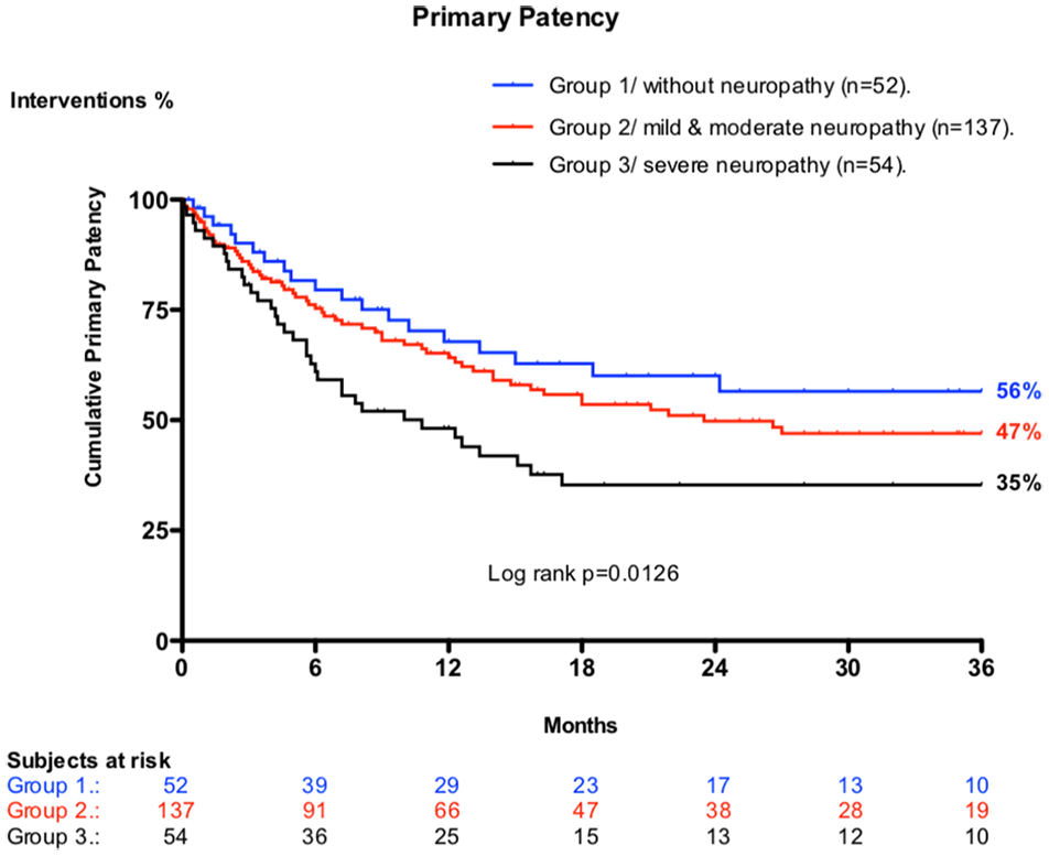
Comparison of primary patency between group 1 (lack of detectable diabetic peripheral neuropathy [DPN]) vs groups 2 (mild and moderate DPN) and 3 (severe DPN) that shows statistically significant differences (p=0.0126).
Secondary patency estimates (Figure 2) were 72% (95% CI=59% to 85%) and 60% (95% CI=45% to 75%) at 12 and 36 months for group 1; 66% (95% CI=58% to 74%) and 49% (95% CI=39% to 59%) for group 2; and 56% (95% CI=43% to 69%) and 45% (95% CI=31% to 59%) for group 3 (p=0.056), respectively, at identical time intervals. Correlation analysis of secondary patency rates at 36 months between groups 1 and 3 (HR=0.57; 95% CI=0.314 to 1.058; p=0.075), groups 2 and 3 (HR=0.78; 95% CI=0.484 to 1.274; p=0.240), and groups 1 and 2 (HR=0.71; 95% CI=0.426 to 1.184; p=0.147) demonstrated nonsignificant differences.

Comparison of secondary patency between group 1 (lack of diabetic peripheral neuropathy [DPN]) vs groups 2 (mild and moderate DPN) and 3 (severe DPN), demonstrating the absence of significant deviations (p=0.056).
Rates of freedom from amputation (ie, limb salvage, Figure 3) were estimated as 87% (95% CI=77% to 97%) and 73% (95% CI=58% to 88%) at 12 and 36 months for group 1; 79% (95% CI=72% to 88%) and 65% (95% CI=55% to 75%) for group 2; and 64% (95% CI=51% to 77%) and 54% (95% CI=39% to 69%) for group 3, respectively, at the same time periods. Independent comparison of limb salvage estimates between groups 1 and 3 at 36 months (HR=0.41; 95% CI=0.203 to 0.855; p=0.006) and groups 2 and 3 (HR=0.53; 95% CI=0.292 to 0.961; p=0.023) proved to be significant, but not between groups 1 and 2 (HR=0.689; 95% CI=0.352 to 1.347; p=0.200).

Similar comparison for limb salvage rates following endovascular treatment between group 1 (lack of diabetic peripheral neuropathy [DPN]) vs groups 2 (mild and moderate DPN) and 3 (severe DPN), showing meaningful statistical differences (p=0.0127).
Wound healing (ie, clinical success, Figure 4) estimates were 77% (95% CI=66% to 88%) and 61% (95% CI=43% to 79%) at 12 and 36 months for group 1; 72% (95% CI=64% to 80%) and 51% (95% CI=45% to 61%) for group 2; and 60% (95% CI=47% to 73%) and 36% (95% CI=22% to 50%) for group 3 (p=0.034), respectively, at the same time intervals. A similar comparison of wound healing between groups 1 and 3 at 36 months (HR=1.61; 95% CI=1.157–3.040; p=0.049) and groups 2 and 3 (HR=1.61; 95% CI=1.079 to 2.416; p=0.01) proved to be significant, but not between groups 1 and 2 (HR=0.14; 95% CI=0.785 to 1.677; p=0.680).

Comparison of clinical success (wound healing) after endovascular treatment between group 1 (lack of diabetic peripheral neuropathy [DPN]) vs groups 2 (mild and moderate DPN) and 3 (severe DPN), also proving significant differences (p=0.0120).
Among all patients included in this study, there were 36 (12%) major amputations (14 early failures and 22 others for disappointing evolution throughout the follow-up period).
In the whole, 29 patients (13%) died during this analysis. Among this contingent, 24 (83%) were lost from cardiac and other vascular causes, 2 (7%) from neoplastic disease, 2 (7%) from pulmonary infections and chronic obstructive pulmonary disease (COPD), and 1 (3%) following a generalized sepsis. Among the related cardiovascular deaths, 25% were noted in group 1, whereas 29% and 46% in groups 2 and 3, respectively.
Overall survival estimates in this study were 91% (95% CI=84% to 98%) and 56% (95% CI=36% to 76%) at 12 and 36 months for group 1; 88% (95% CI=62% to 96%) and 53% (95% CI=40% to 66%) for group 2; and 79% (95% CI=68% to 90%) and 49% (95% CI=33% to 55%) for group 3, respectively, at the same time intervals. Amputation-free survival (Figure 5) appeared not influenced at 3 years interval, related to the presence and severity of peripheral limb neuropathy (p=0.344).

No significant deviations were observed concerning amputation-free survival between same studied groups (p=0.3446).
Discussion
The present study revealed a significant difference in BTK primary angioplasty outcomes performed in diabetic patients with CLTI without DPN or with incipient-to-moderate peripheral neuropathic symptoms, compared with the severe DPN group. This latest group assembled the heaviest neuropathic affectation and arterial calcifications and proved the lowest clinical benefit at 36 months: 35% (95% CI=22% to 48%) of primary patency, 36% (95% CI=22% to 50%) wound healing, and 54% (95% CI=39% to 69%) limb preservation rates.
These findings appear to support the hypothesis that severe DPN (equivalent to group 3 of diabetic patients) may represent an independent indicator for poor technical and clinical outcomes of infrapopliteal angioplasty in diabetic patients with CLTI.
The primary PTA approach has been increasingly suggested as beneficial in BTK and below-ankle revascularization.1,4,8,14,17
The relevant literature reports that although most diabetic foot ulcers appear to be neuropathic1,5 in >60% of these presentations, ischemic involvement can be suspected.4,5,11 Although regarded as distinct pathologies, diabetic neuropathy and angiopathy share a common origin triggered by hyperglycemic-enhanced “vasa-vasorum” and “vasa-nervorum” arteriolar thickening. 5 This initial etiological process unfolds with parallel microcirculatory functional disorders in both pathologies, 5 worsened by advanced patient age and the duration of diabetic affectation itself.5,11
From this perspective, the “OPIDIA” study 18 observed that among 291 patients, up to 87% of diabetic foot wounds had a neuropathic background, whereas 62% were correlated with various degrees of chronic ischemic presentations. 18 Among the multifaceted pathologies comprising “diabetic foot syndrome” (DFS), peripheral neuropathy is currently believed to be associated with distal symmetrical sensorimotor neuropathy (DSN) and concomitant autonomic peripheral denervation (APN).5,18–20 The former has been considered to be the primary triggering factor for foot ulceration. 1,3,5,11,20 Unlike sensorimotor constituents, APN involves more challenging clinical diagnosis and stratification.5,18,20 Peripheral neuropathy and chronic ischemia are increasingly assimilated as “entangled” entities in the complex etiology of diabetic foot wounds.5,18,20,21 As mentioned, concomitant “vasa-vasorum” and “vasa-nervorum” thickening are interrelated within the wider scope of diabetic “functional microangiopathy,”5,18–21 which manifests at the systemic level.5,20–22 This simultaneous neurovascular entanglement varies, from subtle or subclinical forms to threatening inferior limb pathologies,5,20–24 that all define so-called diabetic “neuro-ischemic foot syndrome.”1,4,5,11,24 A few specific mechanisms have been proposed by which DPN independently hampers the peripheral macrovasculature and microvasculature of the foot, as well as the potential benefit of revascularization.4,11,21 Several authors have suggested that the main detrimental effect of DPN is the influence on lower limb arterial perfusion that includes a specific pro-inflammatory status of the neuropathic limb that harbors a more aggressive and distal peripheral atherosclerosis.5,11,22 Characteristic tibial and pedal tunica media arterial calcifications (“Monckeberg calcifications”)1,18,19 are present in >90% of DSN and APN inferior limbs.2,4,11,19,22,23 A parallel arterio-venular neuropathic shunting and the skin level as flow “steal” phenomenon5,11,24 are also specific to advanced DPN and trigger retarded tissue recovery independent of simultaneous ischemic affliction.1,5,11,22,24
Our study confirms previous clinical findings regarding lower feasibility of BTK angioplasty in heavily calcified tibial and pedal arteries.25,26 The present study reveals that technical feasibility of PTA, its primary patency, clinical success (ie, tissue recovery), and global limb preservation appeared all, directly, and significantly (p<0.50) to be influenced by the severity of concomitant DPN (clinical results in group 3). In our experience, group 3 (highest DPN affliction/UK 8–10 patients) gathered the highest percentage of category 3 calcifications, the lowest PTA technical feasibility (68%), worse primary patency (35%), poor wound healing (36%), and modest limb salvage rates (54%) at 3 years, despite uniform multidisciplinary follow-up.
The association between DPN and infragenicular medial arterial calcification is not a new concept. It was previously described by Edmonds et al 19 40 years ago in relation to “Monckeberg media-sclerosis,” primarily evinced in 1924. 19 These pioneering observations were further developed by the recent works of Lanzer et al 27 and Rocha-Singh et al 28 ; however, these authors did not directly correlate calcific presentations with the influence of DPN in CLTI and following specific revascularization approaches.
Diabetic neuropathy also impacts the cellular aspects of the tissue regeneration process itself (by lack of “neurotrophic tissue factor”),5,11 independently of eventual corrective revascularization.11,21,22,24,29
By studying cutaneous microvascular reactivity using laser Doppler in diabetic neuro-ischemic CLTI patients, Arora et al 21 observed that coexisting neuropathy negatively affects foot cutaneous microcirculation, even after successful lower extremity revascularization of large arterial trunks. 21 Alternatively, in a study assessing lower extremity bypasses in limbs affected by diabetic neuro-ischemia, Akbari et al 30 suggested that the severity of neuropathy only minimally affects the hemodynamic component of revascularization (patency vs occlusions). The authors also observed that the reversal of hypoxia appeared to stop the progression of peripheral neuropathy. 30 In a similar review, Boulton 31 emphasized the importance of neuropathy in the genesis of diabetic foot wounds, but also during recovery after revascularization. The simultaneous control of parallel risk factors, such as hyperglycemia, foot deformity, and infection, appears to be primordial in DFS treatment.1,29,31
Although without a precise etiological background, accumulating clinical data suggest a possible correlation between DPN (more specifically APN) severity19,29 and the extent of infragenicular calcifications inside DFS.19,28,29,32 Similar observations were also documented in a preliminary analysis published by our institutional diabetic team. 32
Interestingly, analogous observations are also available in the literature about abundant calcific deposits in CLTI leg arteries succeeding lumbar sympathectomy (intentional autonomic inferior limb denervation), 19 through a possible parallel mechanism that is not yet clearly understood.
A probably relevant clinical outcome of the present study is represented by the finding that survival rates demonstrated no significant difference at 36 months among the 3 groups of patients related to the presence and severity of peripheral limb neuropathy (p=0.344).
This aspect could be probably attributed to uniform multidisciplinary team surveillance (wounds, infection, glycemic levels, nutrition, etc) that sustained limb preservation, despite persistency of chronic neuro-ischemic factors. Additional markers of quality of life could be then documented in a future stage of this research.
Perspectives
Tibial-pedal arterial calcifications do not exhibit uniform characteristics, although all represent (including neuropathic calcifications) an important technical drawback for any type of revascularization for CLTI.1–4,12,22,26,33 Mustapha et al 33 recently described several distinct morphological types of infrainguinal arterial calcifications, of which diabetic medial arterial (ie, Monckeberg’s) sclerosis observed in neuropathic environments 19 represents but one among other specific tibial and pedal calcific patterns.19,32,33 As a result, using CT scan analysis, Tokuda et al 34 recommended the establishment of a specific inferior limb “calcific score” (CS) in patients with CLTI, undoubtedly, to provide better technical and clinical value. 34
In addition to these strictly morphological CS evaluations, a future functional stratification4,35 of each individual tibial/pedal hemodynamic flow features, peripheral vascular resistance at the foot level,4,35 and predicted patency can be highly beneficial in assessing these extensively calcified and stiffened arterial trunks connected to a distorted microvasculature, and to a current collateral loss.24,35 Increasing severity of DPN with increasing severity of CLTI represents most likely an association.
Probably, the global outcome of patients suffering from CLTI that adds DPN could be improved in the future only by applying standardized “diabetic team” cares in which vascular interventionists avail awareness of all complex vascular, neural, and tissue interactions casted inside myriad of clinical presentations enhanced by the vast “metabolic syndrome.”
Limitations
The present study was limited by the small number of enrolled cases and its retrospective design. It should be noted that technical skills and PTA technology characteristics could have undeniably improved over the 10-year observation period of this research, with plausible influence on overall technical success rates and other statistical data. We also acknowledge that specific strategies for BTK angioplasty, detailed anatomical characteristics of neuro-ischemic wounds, and other individual local and systemic risk factors could have additionally influenced patency, tissue recovery, and limb preservation rates, and that could not be extensively detailed in this observational DPN study.
Conclusion
In diabetic neuro-ischemic limbs, peripheral neuropathy evaluation appeared to be useful and requires independent screening and stratification, in parallel to that of CLTI. The presence of severe neuropathic affectation may jeopardize the clinical benefit of successful infrapopliteal angioplasty in terms of patency, tissue cicatrization, and limb preservation, but without significant survival gain in these patients.
When present, DPN necessitates appropriate multidisciplinary control and distinct stratification as independent indicator for tissue decay and limb loss, associated with current CLTI revascularization.
Footnotes
Acknowledgements
The authors acknowledge members of their institutional diabetic foot clinic for their unwavering support during enrollment, treatment, and follow-up of all patients included this study during the past decade. Special acknowledgment to the internal medicine and radiology departments for their effective assistance enabling this analysis. We gratefully acknowledge academic consultancy for the challenging endovascular applications and our institutional computing staff for data collection, processing, and statistical counseling during this research.

Declaration of Conflicting Interests
The author(s) declared no potential conflicts of interest with respect to the research, authorship, and/or publication of this article.
Funding
The author(s) received no financial support for the research, authorship, and/or publication of this article.