






Abstract
Purpose:
LncRNA-Atherosclerotic plaque pathogenesis-associated transcript (APPAT) could be detected in circulating blood and has been demonstrated to correlate with the development of atherosclerosis in our previous work. It could be a potential noninvasive biomarker for earlier diagnoses of clinical cardiovascular disease. Moreover, the expression of miR-647 increased in ox-LDL-treated vascular smooth muscle cells and peripheral blood of patients with coronary heart disease. A negative correlation between APPAT and miR-647 was confirmed, and FGF5 was screened as molecular target of miR-647. However, it is largely unclear how APPAT, miR-647, and FGF5 interact and function in disease development. Here, we aim to explore the underlying molecular mechanism in this progression.
Materials and Methods:
APPAT, miR-647, and FGF5 expression levels were detected by quantitative reverse transcription polymerase chain reaction; cell proliferation was detected by EdU incorporation assay; cell migration was detected by wound-healing assay; the molecular interaction of APPAT/FGF5 with miR-647 was verified by dual-luciferase reporter assay; the western blot was performed to determine the gene expression at protein levels; subcellular localizations of APPAT and miR-647 were observed by fluorescence in situ hybridization; cytosolic and nucleus fractionation assay was performed to further detect the distribution of miR-647.
Results:
APPAT and miR-647 have inverse effects on human aortic smooth muscle cells’ (HASMCs) proliferation and migration. APPAT negatively regulated the cell activity, whereas miR-647 did it in a positive way (p<0.05). Three pairs of molecular interplay were found: mutual negative regulation between APPAT and miR-647, APPAT downregulated FGF5, miR-647 regulation on FGF5 (p<0.05). Subcellular location assay confirmed the molecular interaction of APPAT and miR-647.
Conclusions:
APPAT could suppress the migration and proliferation of ox-LDL-treated HASMCs via interacting with miR-647 and FGF5. We revealed a nontypical competing endogenous RNA mechanism of long noncoding RNA in the progression of atherosclerosis.
Introduction
Atherosclerosis (AS) is a serious threat to human health worldwide which could cause cardiovascular and cerebrovascular diseases such as myocardial infarction, cerebral infarction, and coronary heart disease.1,2 Vascular smooth muscle cells (VSMCs) in arteries participate in the arterial contraction as well as the production of extracellular matrix in the healthy tissue. The abnormal states of VSMCs play an essential role in the process of AS. The aberrant proliferation and migration of VSMCs, which in response to different atherogenic factors such as smoking, drinking, obesity, and age, promote atherosclerotic plaque formation. 3 Comprehensive understanding of the molecular mechanism behind the proliferation and migration of VSMCs under certain risks will facilitate researches on the prevention and treatment of AS and related diseases.
Long noncoding RNAs (lncRNAs) are generally defined as noncoding transcripts with more than 200 nucleotides in length. Increasing evidence indicates that lncRNAs are related to many human diseases such as diabetes, cancer, and central nervous system diseases.4 –9 Current research has reported that many lncRNAs, for example, cyclin-dependent kinase inhibitor 2B antisense RNA 1 (ANRIL), 10 myocardial infarction–associated transcript (MIAT), 11 metastasis-associated lung adenocarcinoma transcript 1 (MALAT1), 12 and lncRNA-RNCR3, 13 played vital roles in cardiovascular disease. LncRNA-Atherosclerotic plaque pathogenesis-associated transcript (APPAT) could be detected in circulating blood and has been demonstrated to correlate with the development of AS in our previous work. 14 The APPAT were located in the cytoplasm of contractile VSMCs of artery tunica media and showed significant downregulation when VSMCs were treated with ox-LDL. Moreover, in human coronary artery tissue, APPAT significantly varied from healthy group to disease group. It could be used as a novel kind of noninvasive biomarker with the ability of monitor development of atherosclerotic plaque which is potentially important for earlier diagnoses of clinical cardiovascular disease. 15 It is known that during the development of AS plaque, the media-oriented VSMCs will accumulate in the intima by proliferation or directed migration. We questioned that APPAT may play roles in maintaining phenotype of VSMCs by affecting its proliferation and migration. However, the function and molecular mechanism of APPAT in the process of AS are not clear.
The microRNA (miRNA) is a subset of endogenous small ncRNAs approximately containing about 22 nucleotides.16,17 It primarily functions through post-transcriptional repression via binding to the 3′ untranslated region (3′-UTR) of target genes in the cytoplasm.18,19 Emerging evidences suggested that the miRNAs function not only in the cytoplasm but also in the nucleus,20,21 while the mechanism of nuclear miRNAs remains largely unclear. Most of the studies revealed that some miRNAs positively regulated the gene expression by targeting promoter elements.22 –26 For instance, the miR-589 activated cyclooxygenase-2 (COX-2) transcription via binding to the promoter of COX-2. 24 MiR-24-1 was considered as an unconventional mediator to active transcriptional gene through chromatin remodeling at enhancer regions. 25 In the previous study, miR-647 was considered as a biomarker of cancer.27 –29 The miR-647 could suppress the cell proliferation in non–small-cell lung cancer 30 and inhibit invasion and migration of gastric cancer cells.31,32 Moreover, our previous exploration revealed that the expression level of miR-647 increased in ox-LDL-treated VSMCs and peripheral blood of patients with coronary heart disease. And a negative correlation between APPAT and miR-647 was also confirmed. 14
In this study, we aimed to verify the molecular interaction of APPAT with miR-647 and their roles in the process of proliferation and migration in human aortic smooth muscle cells (HASMCs). We found that APPAT and miR-647 have reverse influence on the activity of HASMCs. Dual-luciferase reporter and subcellular location assay provided evidence of the possibility of molecular interaction. The FGF5 was screened as molecular target of miR-647 in our previous work and further confirmed in this article. The FGF5 was positively regulated by miR-647 but inversely regulated by APPAT. Our results revealed that APPAT could regulate the migration and proliferation of HASMCs via interplaying with miR-647 and FGF5, which is not consistent with the typical ceRNA loop interaction.
Materials and Methods
Cell Culture
The HASMCs were obtained from Saiqi Biological Engineering Co., Ltd (Shanghai) and were cultured in minimal medium (Gibco, New York) with 10% fetal calf serum (FBS; Gibco), 100 units/mL penicillin, and 100 µg/mL streptomycin. To construct the foam cell model in HASMCs, ox-LDL (Yiyuan Biotechnologies, Guangzhou) was used with a concentration of 80 µg/mL for 48 hours in complete medium.
Cell Transfection
The lentivirus, APPAT-overexpressing vector, and empty lentivirus, negative control, were synthesized by Obio Technology (Shanghai). The recombinant lentivirus was applied to the experiments with a titer of 4.8 × 10⁸ TU/mL for infection and the concentration of empty lentivirus used for infection was 2.04 × 10⁹ TU/mL. The HASMCs were infected with lentivirus for 12 hours for further experiments. The siRNA of APPAT and its corresponding negative control (siNC) were synthesized by RiboBio (Guangzhou). The miRNA oligonucleotides including miR-647 mimic, miR-647 inhibitor, and their corresponding negative control (mimic NC and inhibitor NC) were purchased from RiboBio. The HASMC transfection with miRNAs or siRNAs was executed by using the riboFECTCP Reagent (RiboBio) on the basis of the manufacturer’s protocol. After 48 hours transfection, the HASMCs were used for further experiments. The overexpression vector of FGF5 (Pexp-rb-Mam-EGFP-has-FGF5) and empty vector were obtained from RiboBio. Lipo3000 reagent (Invitrogen, Waltham, Massachusetts) was used in the HASMC transfection with plasmids, which is according to the manufacturer’s protocol for further experiments.
RNA Extraction and Quantitative Real-Time PCR
Total RNAs of HASMCs were isolated using Eastep Total RNA Extraction Kit (Promega, Madison, Wisconsin). GoScript Reverse Transcription System (Promega) and Bulge-Loop miRNA qRT-PCR Primer Kit (RiboBio) were used for reverse transcription of messenger RNA (mRNA) and miRNA, respectively. Quantitative PCR analysis was performed using Applied Biosystems 7500 Real-Time PCR System with SYBR green method (Applied Biosystems, Foster City, California). The primers used in amplification of targets sequences in this step were designed by Primer5 (online version) (lncRNA-APPAT: F5′-GCAGAGGCAGGTCACCAAC-3′, R5′-CAGGATGATTCGAGACCAGGA-3′; FGF5: F5′-AGCTTGTCCTTCCTCCTCCT-3′, R5′-ATAGCGCTACTGCTGCTCTG-3′). The primer of miR-647 was designed and manufactured by RiboBio (miR-647: R5′-GTGTTGGCCTGTGGCTG-3′, F5′-CTGACCCTCCCTCCTGC-3′). Glyceraldehyde phosphate dehydrogenase (GAPDH) (primer: F5′-GGGAGCCAAAAGGGTCAT-3,′ R5′-GAGTCCTTCCACGATACCAA-3′) was used as the endogenous control for mRNA expression and U6 (primer: F5′-TTATGGGTCCTAGCCTGAC-3,′ 5′-CACTATTGCGGGTCTGC-3′) for miRNA expression. The gene expression levels were described relative to endogenous control and the 2−∆∆Ct method was used to calculate relative expression level.
Cell Proliferation Assay
The 5-ethynyl-2′-deoxyuridine (EdU) incorporation assay was performed to examine the proliferation of HASMCs. For EdU incorporation assay, the staining procedure was performed using a Cell-Light EdU Apollo 488 In vitro Imaging Kit (RiboBio). The HASMCs were planked in 96-well plates (4 × 10³ cells/well). After that EdU working solution was added to the culture solution. Then, the cells were collected and fixed with 4% paraformaldehyde after 2 hours of incubation. The cells were stained by Apollo staining solution and Hoechst 33342. The EdU-positive cells were counted and normalized to the total number of Hoechst 33342–stained cells. The fluorescence signals were observed via fluorescence microscopy (ECLIPSE Ti; Nikon, Tokyo), and images were analyzed using ImageJ software (v1.52).
Wound-Healing Assay
The HASMCs were planked in 6-well culture plates (2 × 10⁵cells/well). Then, wound gap of the cell monolayer was created using a 200 μL pipette tip. The wound width was recorded at 0, 24, and 48 hours after scratching was created. The wound-healing images were captured in 4× fields using microscope (Motic, Xiamen) equipped with Image-pro Plus software (Media Cybernetics Inc., Rockville, Maryland). The ratio of the cell recovery area to the whole wound area was used to evaluate cell migration ability using ImageJ software (v1.52).
Dual-Luciferase Reporter Assays
Polymerase chain reaction was used to amplify the fragments of APPAT and FGF5 3′-UTR, and then the products were cloned into pGL3 report vector (Promega), respectively, to construct the wild-type reporter vector. To build the mutant reporter, corresponding mutated fragments were cloned into the pGL3 report vector (Promega). Reporter assays were performed using HASMCs. The HASMCs were planked in 96-well plates, and then co-transfected with luciferase reporter vector (pGL3-APPAT-WT, pGL3-APPAT-MUT, pGL3-FGF5-WT, and pGL3-FGF5-MUT) and miR-647 mimic/NC mimic. After 48 hours, luciferase activity was detected by the Dual-Luciferase Reporter Assay System (Promega).
Western Blot Analysis
Total protein was extracted from the HASMCs using RIPA (radioimmunoprecipitation assay) lysis buffer containing protease and phosphatase inhibitors (Solarbio, Beijing). Proteins were separated by 12% sodium dodecyl sulfate-polyacrylamide gel electrophoresis and transferred to polyvinylidene fluoride membranes (GE Healthcare, Buckinghamshire). The membranes with protein blot bands were blocked using 5% skimmed milk. Then the membranes were incubated with the antibodies containing anti-FGF5 (1:700 dilution, RefNum: 18171-1-AP; Proteintech, Rosemont, Illinois) and α-tubulin (1:1000 dilution, RefNum: 11224-1-AP; Proteintech) at 4°C overnight, followed by incubation with horseradish peroxidase–conjugated secondary antibodies (1:2000 dilution; Solarbio) for 2 hours at room temperature. The protein blot bands were visualized by an ECL Prime Western Blotting Detection Reagent (GE Healthcare) and Image Lab software (Bio-Rad, California).
Fluorescent In Situ Hybridization for Detection of APPAT and miR-647 in HASMCs
Ribo Fluorscent in Situ Hybridization Kit (RiboBio) and G-type m/miRNA in situ hybridization Kit (Foco, Guangzhou) were performed to detect the location of APPAT and miR-647 in HASMCs, respectively. The slides with HASMCs fixed by 4% paraformaldehyde were hybridized using CY3-labeled probe for APPAT and FITC probes specific for the miR-647 at 5 ng/μL concentration. Hybridization was performed at 37°C overnight, and slides were subsequently counterstained using DAPI, followed by visualization using fluorescence microscope (Leica, DMI6000B, Heerbrugg). The relative expression level was normalized (18s for APPAT and U6 for miR-647).
Cells’ Cytosolic and Nucleus Fractionation Assay
For the cells’ cytosolic and nucleus fractionation assay, the HASMCs were planked into 100 mm dish and collected into the Eppendorf tube. The cells were resuspended with TD solution and subsequently the RNA-EZ reagent, J Rnase-inhibitor-ribonucleoside-vanadyl complexes (RVC)/1% nonidetP40 (NP-40)/TD solution, was added into the Eppendorf tube. The process of cell lysis was performed on ice for 5 minutes and the mixture was centrifuged for 5 minutes. The cytoplasmic fraction was present in supernatant fraction and the nucleus fraction was contained in the precipitated part. The supernatant was removed to a new tube for further experiments. The precipitated part was resuspended in 0.5% NP-40/TD and then cleaved on ice for 5 minutes. After centrifugation for 5 minutes, the supernatant was removed, and then the precipitated part was resuspended in 0.5% NP-40/TD. The solution containing nucleus fraction of HASMCs was used in the further experiments.
Statistical Analysis
All data were analyzed by the GraphPad Prism Software (v5.0; GraphPad Software, California) and presented as the mean±SD. Statistical analysis among groups was determined by Student t test or 1-way analysis of variance followed by Bonferroni post hoc test. The level of statistical significance was considered at p<0.05.
Results
APPAT Inhibited the Migration and Proliferation of ox-LDL-Treated HASMCs
To explore the function of APPAT in HASMCs, the APPAT expression in HASMCs was interference by overexpression or knockdown. The transient overexpression of APPAT could remarkably increase the expression of APPAT compared with that of the negative control group (Mock) (p<0.01) (Figure S1A). Then, wound-healing assay revealed that enhanced expression of APPAT suppressed cell migration and attenuated the ox-LDL-induced cell migration (Figure 1A, p<0.05). Similarly, the effect of APPAT on the proliferation of HASMCs showed that APPAT overexpression attenuated the ox-LDL-induced cell proliferation (Figure 1B, p<0.001). Transfection with si-APPAT significantly reduced the expression level of APPAT in HASMCs (p<0.05) which verified the efficiency of designed interference of APPAT (Figure S1A and B). Then the wound-healing and EdU incorporation assay showed that knockdown of APPAT could aggravate the ox-LDL-induced increase of cell migration/proliferation (Figure 1C and D, p<0.01). Data of this step suggested that APPAT had inhibitive effects on migration and proliferation of ox-LDL-treated HASMCs.
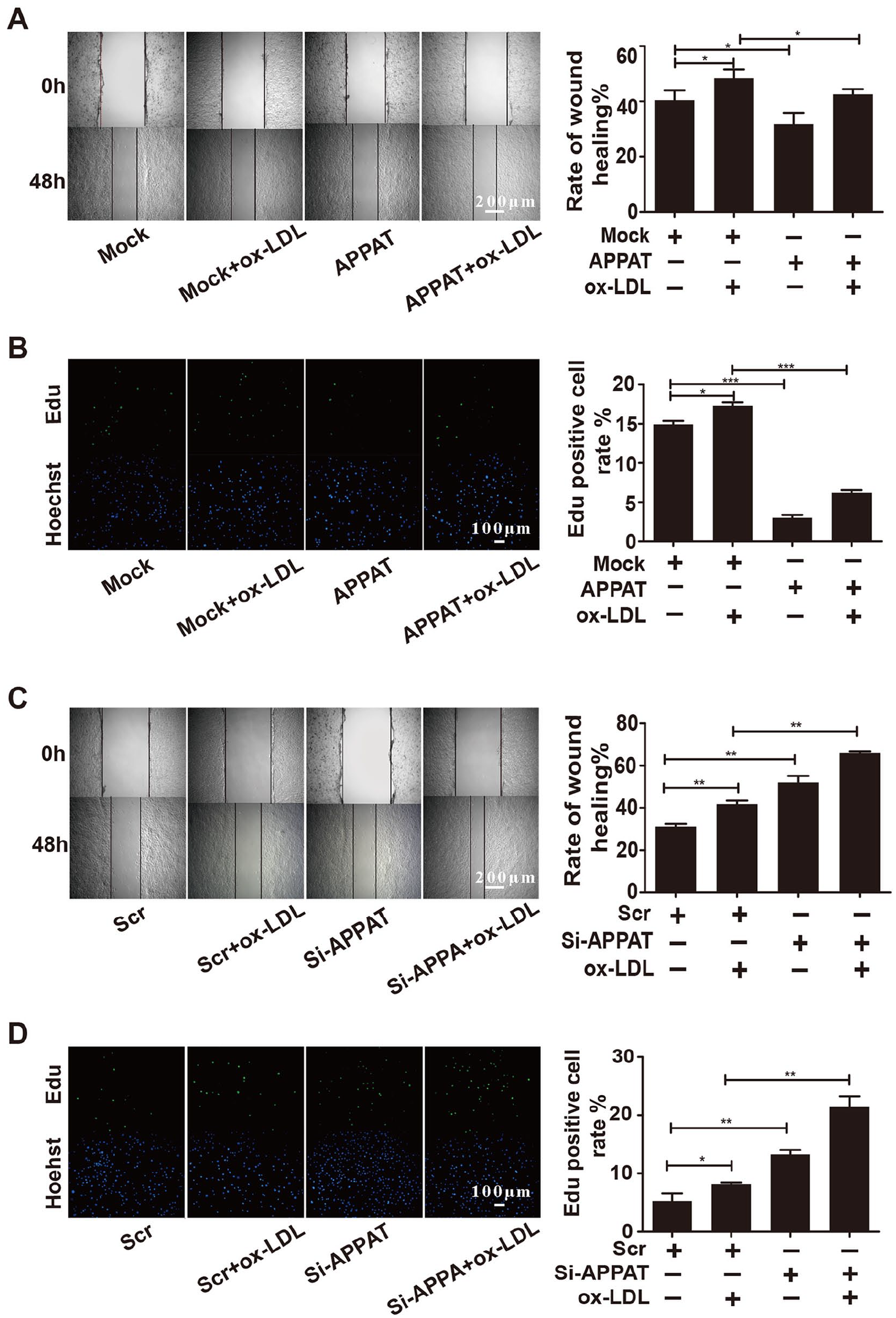
Proliferation and migration in response to APPAT overexpression/knockdown in ox-LDL-treated human aortic smooth muscle cells (HASMCs). Overexpression of APPAT on migration (A) and (B) proliferation (N=3). Knockdown of APPAT on (C) migration and (D) proliferation (N=3). The migration and proliferation were detected by wound-healing and EdU incorporation assay, respectively. In proliferation pictures, EdU: green, Hoechst: blue. (A) and (C)=40×, (B) and (D)=100×. Mock: control group with empty vehicle, APPAT: group of lentivirus vehicle of APPAT, Scr: control group with scramble siRNA of APPAT; si-APPAT: group of siRNA of APPAT. Concentration of ox-LDL is 80 µg/mL. APPAT, atherosclerotic plaque pathogenesis-associated transcript; EdU, 5-ethynyl-2′-deoxyuridine.
MiR-647 Improved the Migration and Proliferation of ox-LDL-Treated HASMCs
The miR-647 mimic and its negative control (NC mimic) were infected into ox-LDL-treated HASMCs to study the function of miR-647 in HASMCs, respectively. Pilot study confirmed the efficiency of mimic and inhibitor of miR-647 in HASMCs, respectively (Figure S2A and B, ***p<0.001, **p<0.01). Furthermore, the results of wound-healing assay revealed that enhanced expression of miR-647 facilitated the ox-LDL-induced increase of migration of HASMCs (Figure 2A, p<0.01). The EdU incorporation assay showed that overexpression of miR-647 has positive effect on cell proliferation (Figure 2B, p<0.05). Knockdown of miR-647 suppressed the ox-LDL-induced migration (Figure 2C, p<0.05) and proliferation (Figure 2D, p<0.05). These results indicated that miR-647 positively regulated the proliferation and migration of HASMCs.

Effects of miR-647 overexpression/knockdown on migration and proliferation in ox-LDL-treated human aortic smooth muscle cells (HASMCs). Overexpression of miR-647 on migration (A) and proliferation (B) (N=3). Knockdown of miR-647 on migration (C) and proliferation (D) (N=3). Migration and proliferation were detected by wound-healing assay and EdU incorporation assay, respectively. In proliferation pictures, EdU: green, Hoechst: blue. Mimic/inhibitor: synthesized mimic/inhibitor sequence of miR-647. Concentration of ox-LDL is 80 µg/mL. (A) and (C)=40×, (B) and (D)=100×. EdU, 5-ethynyl-2′-deoxyuridine.
APPAT Could Target miR-647
In the previous study, we found that the level of APPAT was inversely correlated with miR-647 in peripheral blood of patients with myocardial infraction. 14 To elucidate if APPAT interacts with miR-647 in HASMCs, a series of experiments were performed. Online bioinformatics tools (PicTar, miRDB, LncBase, miRWalk) suggested that APPAT could target miR-647 to form the complementary pairing (Figure 3A). Subsequently, the dual-luciferase reporter vectors including the wild-type (pGL3-APPAT-WT) and mutant binding sites (pGL3-APPAT-MUT) were constructed. And co-infection of miR-647mimics and pGL3-APPAT-WT led to a significant decrease in luciferase activity (p<0.001), whereas the luciferase activity of co-infection with miR-647 mimics and pGL3-APPAT-MUT exhibited no change (Figure 3B). The qPCR assay showed that overexpression of miR-647 decreased the expression of APPAT, and knockdown of miR-647 did in the reverse way (Figure 3C). These results demonstrated that miR-647 could bind with the APPAT.

MiR-647 is a target of APPAT. (A) The predicted binding site between APPAT and miR-647 (wild type=WT, mutant=MUT). (B) The luciferase constructs of pGL3-APPAT-WT or pGL3-miR-647-MUT were co-transfected with miR-647 mimic or NC mimic in human aortic smooth muscle cells (HASMCs) (N=4). Luciferase activity was detected by the Dual-Luciferase Reporter Assay System. (C) The expression levels of APPAT were determined in HASMCs after transfection with miR-647 mimic or inhibitor (N=3). Mimic/inhibitor: synthesized mimic/inhibitor sequence of miR-647. APPAT, atherosclerotic plaque pathogenesis-associated transcript; MUT, mutated-type; WT, wild-type.
FGF5 was a Predicting mRNA Target of miR-647
Online bioinformatics analysis found the potential putative binding sites between miR-647 and its potential targets FGF5. The FGF5 functions in human aortic endothelial cells and highly expressed in patients with hypertension33,34 and has a potential binding site with miR-647 (Figure 4A). Further tests confirmed that the expression levels of FGF5 significantly increased in ox-LDL-treated HASMCs (Figure 4B, p<0.05). To confirm the association between the expression levels of miR-647 and FGF5, the miR-647 was overexpressed/knockdown in HASMCs. The qPCR and western blot assay both showed that overexpression of miR-647 increased the expression of FGF5 (Figure 4C and D), and the knockdown of miR-647 led to a decrease of FGF5 (Figure 4E and F). The results suggested that FGF5 was a downstream target of miR-647 and been positively regulated by the latter in ox-LDL-treated HASMCs.

The interaction between miR-647 and FGF5. (A) Complementary sequences between miR-647 and FGF5 (wild type=WT, mutant=MUT). (B) The expression level of FGF5 in ox-LDL-treated human aortic smooth muscle cells (HASMCs) (N=3). The mRNA expression and protein expression levels of FGF5 were detected following transfecting miR-647 mimic (C, D) or miR-647 inhibitor (E, F) in ox-LDL-treated HASMCs by WB and qRT-PCR, respectively (N=3). Mimic/inhibitor: synthesized mimic/inhibitor sequence of miR-647. Concentration of ox-LDL is 80 µg/mL. FGF5, fibroblast growth factor 5; MUT, mutated type; qRT-PCR, quantitative reverse transcription polymerase chain reaction; WT, wild type.
APPAT Negatively Regulated the Expression of FGF5 in HASMCs
The expression pattern of FGF5 was further explored in HASMCs by transfection with lentiviruses and si-APPAT, respectively. Both qPCR and western blot assay revealed that overexpression of APPAT could decrease the FGF5 expression levels and then attenuate the ox-LDL-induced increase of FGF5 (Figure 5A and B). However, infection with si-APPAT could upregulate the FGF5 expression and aggravate the ox-LDL-induced increase of the latter (Figure 5C and D). The results suggested that APPAT could negatively regulate the FGF5 in ox-LDL-treated HASMCs.

The relation between APPAT and FGF5. The mRNA and protein levels of FGF5 in ox-LDL-treated human aortic smooth muscle cells (HASMCs) Overexpression of APPAT (A, B) (N=3). The mRNA and protein levels of FGF5 in ox-LDL-treated HASMCs after knockdown of APPAT (C, D) (N=3). The mRNA and protein levels were determined by qRT-PCR and Western Blot (WB), respectively. Mock: control group with empty vehicle, APPAT: group of lentivirus vehicle of APPAT, Scr: control group with scramble siRNA of APPAT, si-APPAT: group of siRNA of APPAT. Concentration of ox-LDL is 80 µg/mL. APPAT, atherosclerotic plaque pathogenesis-associated transcript; FGF5, fibroblast growth factor 5; qRT-PCR, quantitative reverse transcription polymerase chain reaction.
Subcellular Localization of APPAT and miR-647
Based on the above results, the possibility of molecular interaction was also explored by subcellular localizing test. Our previous study has described that the APPAT was located in the VSMCs of tunica media of coronary arteries and mainly enriched in the cytoplasm of VSMCs. 14 In this study, using CY3-labeled probe for APPAT, we specifically assessed the signal of APPAT in HASMCs and confirmed that APPAT mainly localized in the cytoplasm (Figure 6A). Given that cellular miRNAs existed in both nucleus and cytoplasm, in situ hybridization was performed to confirm the localization of miR-647. As shown in Figure 6B, the miR-647 existed simultaneously in cytoplasm and nucleus. Then, we performed the cells’ cytosolic and nucleus fractionation assay, which revealed that small ratios of miR-647 existed in the cytoplasm (Figure 6C).
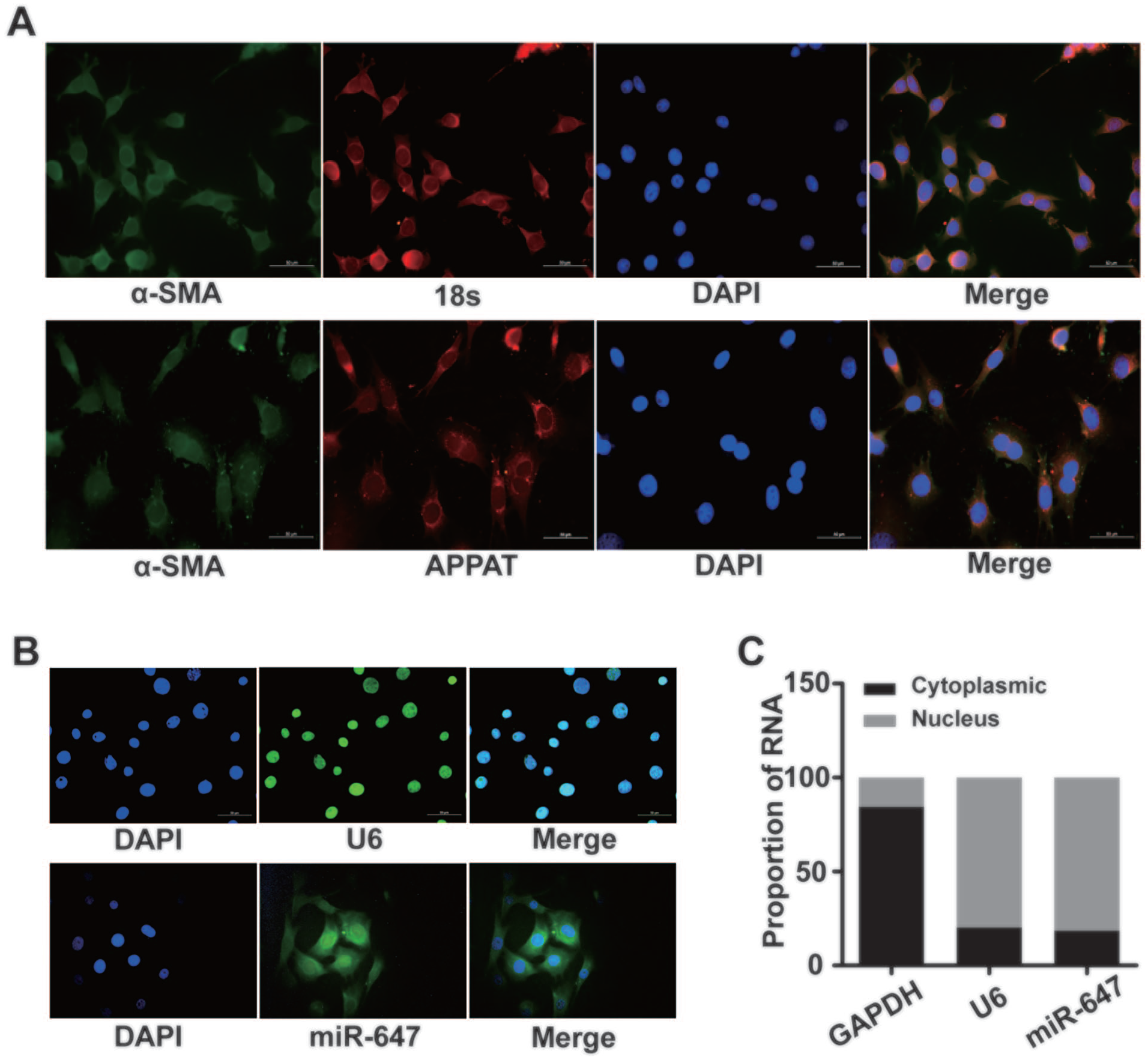
Subcellular location of APPAT and miR-647. (A) The location of APPAT was detected by fluorescent in situ hybridization (FISH) (N=3). DAPI: blue, α-smooth muscle actin (α-SMA): green, 18s RNA: red, and APPAT: red. Magnification 400×. (B) The distribution of miR-647 (N=3). FISH was performed on HASMCs, miR-647: green, U6: green, DAPI: blue. Magnification 400×. (C) The expression levels of miR-647 in the cytoplasm and nucleus in human aortic smooth muscle cells (HASMCs) were determined by qRT-PCR (N=4). U6: reference gene of nucleus, GAPDH: reference gene of cytoplasmic. APPAT, atherosclerotic plaque pathogenesis-associated transcript; GAPDH, glyceraldehyde phosphate dehydrogenase; qRT-PCR, quantitative reverse transcription polymerase chain reaction.
Molecular Interaction During the Proliferation and Migration of HASMCs
To explore if APPAT regulated the proliferation and migration of HASMCs via miR-647 and FGF5, a series of experiments were performed (Figure 7). First, overexpression and knockdown of APPAT in HASMCs confirmed its negative regulation on miR-647 expression (Figure 7A). The ox-LDL treatment exhibited promotion effect on expression of FGF5 (Figure 7B). But overexpression of APPAT attenuated those ox-LDL-induced effects.

APPAT regulated migration and proliferation of ox-LDL-treated human aortic smooth muscle cells (HASMCs) via interacting with miR-647 and FGF5. (A) The negative regulation of APPAT on miR-647 (N=4). (B) The expression of FGF5 in ox-LDL-treated HASMCs after transfection with lentivirus, miR-647 mimic, plasmid vector of FGF5 (N=3). The migration (C) and proliferation (D) were detected by wound-healing and EdU incorporation assay, respectively, in ox-LDL-treated HASMCs after transfection with lentivirus, miR-647 mimic, plasmid vector of FGF5 (N=3). Mock: control group with empty vehicle, APPAT: group of lentivirus vehicle of APPAT, Scr: control group with scramble siRNA of APPAT, si-APPAT: group of siRNA of APPAT, pcFGF5: plasmid vector of FGF5. Concentration of ox-LDL is 80 µg/mL. APPAT, atherosclerotic plaque pathogenesis-associated transcript; EdU, 5-ethynyl-2′-deoxyuridine; FGF5, fibroblast growth factor 5.
Moreover, the overexpressed miR-647 significantly impaired the regulating effects of APPAT on FGF5 and the influence of APPAT on ox-LDL-treated HASMCs. It also promoted the cell migration and proliferation (Figure 7C and D, p<0.01). In addition, the FGF5 showed an inverse effect to the regulation of APPAT on HASMCs’ activity. The above results suggested that APPAT regulated the HASMC proliferation and migration through interplay with the miR-647 and FGF5.
Discussion
A growing number of evidences showed that lncRNAs participate in the physiological and pathological processes of various diseases such as tumorigenesis, glaucoma, diabetes mellitus, and heterotopia endometriosis. 35 In addition, it has been confirmed that most of the lncRNAs play important roles in the occurrence and development of AS, particularly many circulating lncRNAs were suggested as markers for clinical diagnosis of cardiovascular diseases. 15 For instance, lncRNA SENCR and MALAT1 play roles in the migration of vascular cells and the growth of endothelial cells and participate in the pathogenesis of AS.36 –38 The etiology behind AS is complicated. And various components contribute to the plaque progression, among which the proliferation and migration of VSMCs play key role. The lncRNAs have been demonstrated to affect the migration of VSMCs from tunica media to intima during the development of AS.36,39,40 Here, we aimed to further explore the role of APPAT in the migration and proliferation of HASMCs and explore the molecular mechanism behind it, which may provide clues for understanding the development of AS.
In our previous studies, it is assumed that APPAT may function as a component of competing endogenous RNA (ceRNA) which regulates the expression of miR-647 in HASMCs and perform a vital role in the pathological progression of AS. 14 In this work, we first confirmed the suppression of APPAT on migration and proliferation in HASMCs. Then further tests verified that the APPAT could negatively regulate the expression of miR-647 in the HASMCs. The role of miR-647 in cell migration and proliferation has been verified in other studies.31,41 It targets SRF-MYH9 axis to inhibit the migration of gastric cancer cells. 41 However, we found miR-647 promoted the migration and proliferation of HASMCs, which was different from the previous study. The results of dual-luciferase reporter assay suggested that APPAT could target miR-647. Our previous study observed the expression level of miR-647 increased in peripheral blood of patients with coronary heart disease. And APPAT negatively correlated with miR-647. 14 Taken together, it was reasonable to conclude that the former could bind with the latter to conduct the regulating function in the progression of AS.
One unexpected result came from the subcellular distribution of APPAT and miR-647. The APPAT was enriched in cytoplasm while small ratios of the latter distributing in the cytoplasm. A large number of studies suggested that miRNA acted as negative regulators of gene expression via suppressing translation or directing sequence-specific complementary mRNA in the cytoplasm.17 –19 However, emerging evidences revealed that miRNAs could function not only in the cytoplasm, but also in nucleus.24 –26,42 –44 Mastui et al 24 showed that miR-589 could bind the promoter of COX-2 to activate COX-2 transcription which critically regulates the inflammation in disease. Li et al 26 showed that miR-320 acted in the nucleus and directly activated CD36 transcription in cardiomyocytes with diabetic conditions. Xiao et al 25 suggested that miRNA had a dual function, both to suppress and activate the transcriptional gene. For example, miR-24-1 in nucleus could function as unconventional mediators to activate transcriptional gene via chromatin remodeling at enhancer regions of the target gene. Meanwhile, it functions typically in cytoplasm as a repressor for its target gene. 25 The mechanism of nuclear miRNAs remains largely unclear. Interestingly, a new type of miRNAs named as nuclear-activating miRNAs (NamiRNAs) was defined in the nucleus recently, which could activate gene expression by binding to the enhancer and then play role in some diseases. 45 The NamiRNAs showed an miRNA-enhancer-target gene activation network, demonstrating their differential roles in the nucleus and cytoplasm. Of course, this potential mechanism should be confirmed further.45,46
Another interesting finding is that the results in our study were not in accordance with the classical function of miRNAs, of which it should negatively regulate the target gene in a ceRNA interaction loop. The FGF5 is a member of the family of fibroblast growth factors which play vital roles in regulating growth and invasion of tumor.47,48 It was identified as a downstream target of miR-647 and positively regulated. 14 It is possible that the positive regulatory role between miR-647 and FGF5 might be associated with other regulation mechanisms. The results of molecular interaction also provided evidences for the relationship of APPAT, miR-647, and FGF5 in normal physiology and disease from different aspects. Overexpression or knockdown on APPAT confirmed that it could negatively regulate the expression of FGF5 and miR-647 in ox-LDL-treated HASMCs. Rescue experiments further revealed that miR-647 and FGF5 could reverse the negative effects of APPAT on cell migration and proliferation. Combining the above results and the molecular basis of interaction, it could be suggested that APPAT regulated cell migration and proliferation via interplay with miR-647 and FGF5 in HASMCs and finally affected disease development.
The APPAT and miR-647 were located in HASMCs, and also detectable in peripheral blood as potential clinical markers for disease monitoring. How these molecules come to the blood, what is the relationship between blood concentration and cell expression of these molecules, what is the influence of coexistence of the cell and blood on the disease development, all these questions are planned to be answered in our following work based on well-designed animal model and clinical cohort sampling.
Conclusions
In summary, our findings revealed the role of APPAT in migration and proliferation of HASMCs, as well as a potential interacting network with miR-647 and FGF5 in AS, which could provide promising therapeutic targets for AS investigations (Figure 8). And the correlation between APPAT and miR-647 further confirm that their potential value as biomarker is monitoring the development of AS plaque which is potentially important for earlier diagnoses of clinical cardiovascular disease. This kind of molecular interacting model might be a specific angle to understand the lncRNA-miRNA-mRNA network in the disease progression.
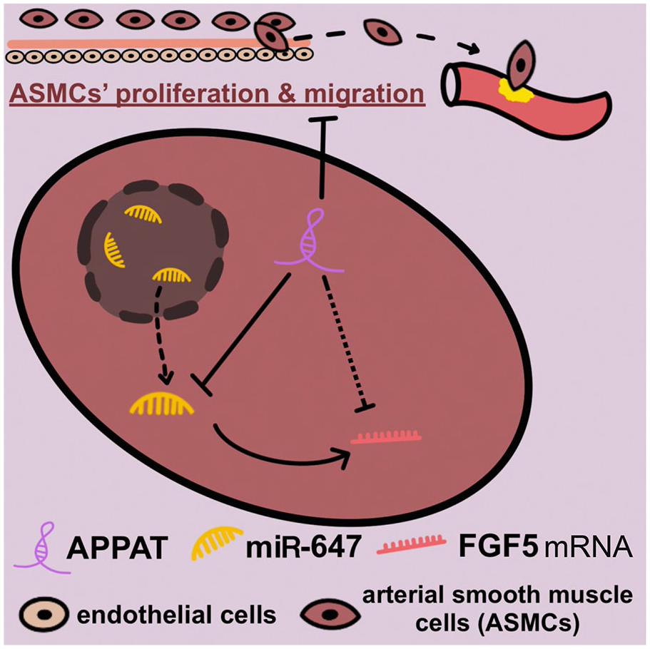
The interaction between APPAT, miR-647, and FGF5 and their role in the proliferation and migration of human aortic smooth muscle cells (HASMCs). The APPAT negatively regulated the expression of miR-647 and FGF5 and inhibited the migration of HASMCs. The miR-647 could function in the cytoplasm and increase the expression of FGF5. APPAT, atherosclerotic plaque pathogenesis-associated transcript; FGF5, fibroblast growth factor 5.
Supplemental Material
sj-tif-1-jet-10.1177_15266028221112247 – Supplemental material for The Lnc-RNA APPAT Suppresses Human Aortic Smooth Muscle Cell Proliferation and Migration by Interacting With MiR-647 and FGF5 in Atherosclerosis
Supplemental material, sj-tif-1-jet-10.1177_15266028221112247 for The Lnc-RNA APPAT Suppresses Human Aortic Smooth Muscle Cell Proliferation and Migration by Interacting With MiR-647 and FGF5 in Atherosclerosis by Fanming Meng, Luyang Han, Qin Liang, Shanshan Lu, Yanqing Huang and Junwen Liu in Journal of Endovascular Therapy
Supplemental Material
sj-tif-2-jet-10.1177_15266028221112247 – Supplemental material for The Lnc-RNA APPAT Suppresses Human Aortic Smooth Muscle Cell Proliferation and Migration by Interacting With MiR-647 and FGF5 in Atherosclerosis
Supplemental material, sj-tif-2-jet-10.1177_15266028221112247 for The Lnc-RNA APPAT Suppresses Human Aortic Smooth Muscle Cell Proliferation and Migration by Interacting With MiR-647 and FGF5 in Atherosclerosis by Fanming Meng, Luyang Han, Qin Liang, Shanshan Lu, Yanqing Huang and Junwen Liu in Journal of Endovascular Therapy
Supplemental Material
sj-tif-3-jet-10.1177_15266028221112247 – Supplemental material for The Lnc-RNA APPAT Suppresses Human Aortic Smooth Muscle Cell Proliferation and Migration by Interacting With MiR-647 and FGF5 in Atherosclerosis
Supplemental material, sj-tif-3-jet-10.1177_15266028221112247 for The Lnc-RNA APPAT Suppresses Human Aortic Smooth Muscle Cell Proliferation and Migration by Interacting With MiR-647 and FGF5 in Atherosclerosis by Fanming Meng, Luyang Han, Qin Liang, Shanshan Lu, Yanqing Huang and Junwen Liu in Journal of Endovascular Therapy
Footnotes
Author Contributions
FM and JL initially designed the present research. FM, HL, and QL performed the experiments and analyzed the results, were all major contributors in writing the manuscript. HY and LS provided the figures and mechanism drawing. JL supported the research and revised the manuscript. All authors read and approved the final manuscript.
Declaration of Conflicting Interests
The author(s) declared no potential conflicts of interest with respect to the research, authorship, and/or publication of this article.
Funding
The author(s) disclosed receipt of the following financial support for the research, authorship, and/or publication of this article: This work was supported by the National Natural Science Foundation of China (grant number 81770462, 81901923), Fundamental Research Funds for the Central Universities of Central South University (2020zzts781), and the Natural Science Foundation of Hunan Province (2018JJ3710).

Data Availability Statement
Data sharing is not applicable to this article as no new data were created or analyzed in this study.
Supplemental Material
Supplemental material for this article is available online.
References
Supplementary Material
Please find the following supplemental material available below.
For Open Access articles published under a Creative Commons License, all supplemental material carries the same license as the article it is associated with.
For non-Open Access articles published, all supplemental material carries a non-exclusive license, and permission requests for re-use of supplemental material or any part of supplemental material shall be sent directly to the copyright owner as specified in the copyright notice associated with the article.
