






Abstract
Deep venous arterialization (DVA) is a new and developing technique with promising outcomes. The DVA procedure can be performed surgically in a hybrid fashion or percutaneously. Over the last years, the hybrid and percutaneous techniques have been further developed and have become a focus of many DVA studies. Between 2017 and 2021, 5 different percutaneous deep venous arterialization (pDVA) techniques, and 2 hybrid procedures have been investigated. In total, 9 cohort studies and 2 case reports have been performed to evaluate their outcomes. Understandably, these studies mainly focused on the technique, patency, and outcomes after DVA. However, postprocedural management can be as challenging as the procedure itself but has not been a priority for further investigation. This review summarizes the different techniques proposed, and the follow-up care provided in literature. Follow-up care includes postoperative medication, edema occurrence and treatment, pain management, patency assessment, reintervention techniques, a staged amputation strategy if necessary, and appropriate wound care. Evidence from literature and own clinical experience was combined to provide recommendations for care after DVA.
Clinical Impact
After percutaneous deep venous arterialization (pDVA), the created arteriovenous circuit needs time to develop. Postprocedural care in patients after pDVA is essential in order to create optimal conditions for maturation of the circuit, and thus save the limb. However, current literature mainly focusses on the procedure itself, making postprocedural care an underexposed topic. Therefore, this study presents an overview of the available literature of postprocedural care of pDVA patients and provides recommendations based on expert opinion when current knowledge is limited.
Keywords
Introduction
Venous arterialization for the treatment of severe peripheral arterial disease was first postulated more than a century ago. 1 The rationale is to use the disease-free venous bed as an alternative conduit for distal perfusion with arterial blood. Many studies have been published, and the procedure has developed and evolved from a surgical approach to a hybrid and even entirely percutaneous procedure. 2
The surgical approach is becoming less popular because of the need to create surgical wounds in a malperfused area. In the hybrid approach, the anastomosis is performed in an open fashion, but valve disruption is established endovascularly.3,4 Usually, a great saphenous vein (GSV) or polytetrafluoroethylene (PTFE) graft is used to create a bypass between the popliteal artery and a tibial vein. Finally, venous collaterals are coiled or ligated to improve inflow to the foot. Limb salvages rates of 69% to 73% have been reported, but the hybrid procedure has not gained as much attention as the percutaneous techniques.3,4
Percutaneous approaches have the advantage of avoiding surgical wounds. Between 2017 and 2021, 5 different percutaneous deep venous arterialization (pDVA) techniques have been published.5–9 As the principles of these 5 approaches are similar (arteriovenous crossing, valve impairment, and distal flow achievement), the differences lie in the arteriovenous crossing point and, the devices and techniques used to cross from the artery into the vein and secure the arteriovenous fistula (AVF).
All approaches have their advantages and disadvantages, and a preferred method is primarily personally based then scientifically. Nevertheless, follow-up management may be similar, as a new conduit has been created using the venous system in all techniques. However, a comprehensive follow-up summary for post-DVA patients is lacking. Subsequently, this review aims to provide an overview of all aspects regarding postprocedural management and present an expert opinion based on clinical experience of the topics on which current knowledge is limited.
Literature Search
A search in EMBASE, Medline, and cross-references identified 14 studies to be included in this review to assess postprocedural management after deep venous arterialization (DVA). Two of the included studies investigated the results of a hybrid-DVA procedure,3,4 while the other 12 studies focused on a pDVA procedure.5,6,7–16 Surgical DVA procedures were excluded to reduce the clinical heterogeneity between the studies. Furthermore, letter to the editors,17–19 editorials, 20 commentaries, 21 and reviews2,22–24 were also excluded. However, although these studies were not included to evaluate postoperative care, they all have been read to gain more insights in the hypothesis, thoughts, and rationale of the authors performing DVA procedures. This information was taken into consideration for providing recommendations well thoroughly.
An overview of the included studies is presented in Table 1 and involves cohort studies (N = 9), technical notes (N = 3), and case reports (N = 2). The methodological quality of the cohort studies was assessed using the Methodological Index for Non-Randomized Studies (MINORS) score, with a global ideal score of 16. 25 For this study, a score of ≤8 was considered poor quality, 9–14 moderate quality, and ≥15 good quality. All included studies were of moderate quality (Figure 1A). The quality of case reports was assessed using the Joanna Briggs Institute Critical Appraisal Checklist. 26 The case reports were considered as sufficient to be included in this review (Figure 1B).

Quality assessment of included studies. (A) MINORS score of included cohort studies. (B) JBI Critical Appraisal Checklist for the case reports. MINORS, Methodological Index for Non-Randomized Studies; JBI, Joanna Briggs Institute.
Summary of Studies and Their Reported Postoperative Management Aspects.

Abbreviations: Amp, minor amputations; AV spear technique, simplified technique by Ichihashi; AVF, arteriovenous fistula; DVA, deep venous arterializations; FU, follow-up time in months; GSV, great saphenous vein; IVUS, intravascular ultrasound; Med, postoperative antiplatelet or anticoagulation medication; No., number of patients included in the studies; NR, not reported; pDVA, percutaneous deep venous arterialization; proximal, proximal part of the tibial vessels; PTFE, polytetrafluoroethylene; RI, reinterventions; VAST, venous arterialization simplified technique by Ysa; WC, wound care including vacuum-assisted therapy, split skin grafts, epidermal substitutes, and debridements; Y, yes, topics in follow-up care are reported/mentioned in the study.
Crossing point in hybrid-DVA studies specifies the location of the outflow AVF.
The 12 pDVA studies were performed using 5 different techniques and devices. Kum et al8,12 was the first to describe an entirely percutaneous technique, in which ultrasonic catheters (LimFlow, Paris, France) were used to create the AVF in the proximal part of the tibial vessels. Covered stents were used from the crossing point to the ankle to mature the AVF and redirecting flow distally. This technique is known as the LimFlow procedure.
An alternative technique was described by Gandini et al, 9 in which the AV crossover was created in the plantar vessels by a guidewire, while angioplasty of the arteriovenous anastomosis was performed to secure the AVF. Valve disruption was not required, and no stents were used as the AV fistula was created distally. 9
Migliara et al described another approach in which the Pioneer Plus IVUS-guided re-entry catheter (Philips, Amsterdam, The Netherlands) was used to cross at the level of proximal tibial vessels (PIPER technique).5,14,20 Valvulotomy was performed by a semicompliant balloon. Finally, covered stents were placed from the AVF to the foot. Venous collaterals were embolized 6 weeks later to focalize flow into the forefoot.
The venous arterialization simplified technique (VAST) was described by Ysa et al, 6 in which a low-profile balloon catheter and a snare were used to facilitate the AVF. The AVF was created in the distal tibial vessels and no stents were placed.
The simplified technique (AV spear technique) included crossing of the distal tibial vessels by direct percutaneous puncture under ultrasound guidance. A stent was used to secure the AVF. 7
The LimFlow device was used in 6 studies,8,11–13,15,16 the Pioneer Plus catheter with intravascular ultrasound (PIPER technique) in 3,5,10,14 a homemade device (VAST technique) in 1, 6 the AV spear technique in 1, 7 and the pDVA technique using regular devices in 1. 9 One cohort study 8 contained the same patient population as described in a technical note. 12 Therefore, these 2 studies were interpreted as one in follow-up care analysis further in this study.
Antiplatelet and Anticoagulation Medication
Postprocedural medication has been described in several pDVA studies, but consensus has not been achieved. Most studies used single antiplatelet therapy in combination with anticoagulation therapy for 3 to 6 months6,11,12,15,16 followed by indefinite single antiplatelet therapy.11,15,16 In 3 studies, dual antiplatelet therapy was used for at least 3 months,3,9,10 followed by lifelong aspirin (Table 2).3,10
Overview of Studies Reporting on Postprocedural Antiplatelet and Anticoagulation Medication and Their Outcomes on Reintervention Rates and Bleeding.

Abbreviations: AC, anticoagulation; DAPT, dual antiplatelet therapy; # Duplex, number of duplex measurements performed within 6 months of follow-up; DVA, deep venous arterialization; FU med, follow-up medication after 3 to 6 months postprocedural; IVUS, intravascular ultrasound; mo; months; No., number of patients included in the studies; NR, not reported; pDVA, percutaneous deep venous arterialization; PTFE, polytetrafluoroethylene; RR, Reintervention rates at 6 months of follow-up; SAPT, single antiplatelet therapy; VAST, venous arterialization simplified technique by Ysa.
Restenosis and Reinterventions
Primary patency rates were only reported in 2 studies,3,11 which was 66% at both 6 months follow-up. The reported reintervention rates for the studies using a combination of antiplatelet and anticoagulation therapy ranges from 25% to 86%, with a mean of 61% at 6 months follow-up. In the study which prescribed dual antiplatelet therapy and assessed the reintervention rate, the reintervention rate was 14% at 6 months follow-up. 9
Pharmacological Viewpoint
From a more pharmacological viewpoint, it is known that arterial thrombosis mainly consists of clotted thrombocytes due to activation by a damaged endothelial layer. 27 In patients with PAD, where the endothelial layer is often injured by a rupture of the atherosclerotic plaque, it is therefore necessary to use antiplatelet therapy to prevent the thrombocytes from clotting. 28 However, in post-pDVA patients, the venous system is also involved, and venous thrombosis typically exists from fibrin. 27 The formation of fibrin can effectively be prevented by anticoagulation medication as vitamin K antagonists or heparins. 29 However, it is uncertain if the occlusions that occur in post-pDVA patients can be attributed to arterial or venous thrombosis or a combination of both. In AVF studies for vascular access in patients with long-term kidney disease, the use of single or double antiplatelet therapy has not shown to reduce thrombosis rates or improve maturation. It is also the same for the use of anticoagulation therapy. 30 However, it is unknown if these results can be extrapolated to post-pDVA patients.
Covered Stents
Covered stents were used in most pDVA treatment techniques. When considering the appropriate medical treatment, the use of covered stents should also be taken into account. 31 A previous study evaluated the efficacy of various antiplatelet/anticoagulation regimes in patients with a Viabahn stent graft for femoropopliteal occlusive disease. 31 Three treatment groups were compared: (1) triple therapy including aspirin, clopidogrel, and warfarin, (2) indefinite dual antiplatelet therapy (aspirin and clopidogrel), and (3) indefinite aspirin with a temporarily 6 weeks clopidogrel. The triple therapy group showed the highest primary patency rates (68%, 56%, and 21%, for the 3 groups, respectively), but bleeding events also occurred more frequently (12%, 0%, and 0%, respectively). The indefinite dual antiplatelet therapy group showed similar outcomes as the triple therapy group with less bleeding events. The 12-month results from the temporarily dual therapy group were significantly worse compared to the other 2 treatment groups in all aspects including, freedom from reintervention (76%, 59%, and 21%), freedom from major adverse limb events (75%, 74%, and 62%), and freedom from thrombolysis (85%, 78% and, 63% for group 1, 2, and 3, respectively).
Expert Opinion
Based on existing literature on various domains, it is difficult to draw firm conclusions about the optimal medical treatment regime in post-pDVA patients. We consider low-molecular-weight heparin (LMWH) 2 times daily 0.6 to 0.8 mL depending on body weight, and clopidogrel 75 mg daily for at least 3 months, followed by lifelong dual antiplatelet therapy (clopidogrel 75 mg and Aspirin 100 mg daily). Therapeutic anticoagulation can be continued for other indications if necessary.
Edema, Cyanosis, and Necrosis
The occurrence of edema was described in 9 articles,3,4,8–10,13–15 of which 2 used a hybrid-DVA approach3,4 and 7 used a percutaneous DVA technique.8–10,13–15 Out of the 9 studies only 3 studies4,8,15 reported the treatment given, which included elevation of the leg. In one paper, diuretics were prescribed occasionally. 8 In addition, off-loading and hanging the legs down was advised to allow the hydrostatic pressure to encourage further formation of venous collaterals. 8
Purple Coloring of the Foot
Beside edema, cyanosis, or purple coloring of the forefoot was described by 2 studies9,14 and was suggested to be a result of venous hypertension. 14 The purple coloring disappeared within the first week postprocedural. 9
Necrosis
Superficial necrosis has been mentioned to become apparent in the first postprocedural period.3,4,14 Intentional demarcation of the affected toes was the preferred treatment option.3,4
Clinical Experience and Expert Opinion
In our experience with a cohort of 23 post-pDVA patients treated with the LimFlow system in Singapore and The Netherlands, edema occurred in all patients. Edema was mild and effectively treated by leg elevation, which is in line with previous literature. Also, to prevent excessive edema, we advised to elevate the leg for at least 24 hours postprocedural and off-loading of the heel was ensured to prevent pressure related wounds. In case of minor edema, the patient was allowed to hang the foot down for 2 hours followed by elevation for another 2 hours to encourage hydrostatic pressurization of the AV-circuit.
No studies mentioned the use of stockings to treat edema. It is uncertain if they were not used or not reported as only 3 studies reported their edema treatments given. In our cohort, thromboembolic foot pump and thromboembolic stockings were avoided as a precaution for the concern of compromising the venous outflow. Also, compressive bandages for wound care were discouraged and stocking net dressings like Tubifast (Mölnlycke Health Care, Gothenburg, Sweden) were used instead, fixated with tape.
Purple coloring of the foot was also noticed by our team and was deemed as a normal reaction on the pDVA procedure.
Progression of gangrene occurred in 13 patients (57%). Similar to the options discussed in the other studies,3,4 progressive necrotic tissue was left to demarcate and further assessed during follow-up visits in the outpatient clinic. Eventually, minor amputations were performed in case of wet gangrene.
Based on published literature and own experience, swelling and coloring of the foot is regularly seen after a pDVA and is a positive sign of venous perfusion. Keep the leg elevated for 24 hours can be considered and after this on indication. Progression of necrosis can occur due to steal from existing arterial collaterals. Demarcation of necrotic tissue should be strived for to wait with amputation until the AV-circuit has matured. Necrotectomy or a minor amputation can be considered in case of wet gangrene.
Pain Management
Only 3 articles reported the occurrence of postprocedural pain.4,13,14 However, none of the articles mentioned management of postprocedural pain. One study reported an increase in pain when the AV-circuit was occluded. 4 The other studies described pain as a postprocedural effect.13,14
Clinical Experience and Expert Opinion
In all patients in our cohort, the character of the pain changed from the typical ischemic pain type to a more engorgement type of pain. Pain management was handled by the primary team, and in case of more severe pain, pain specialists were consulted. Consultation of the APS was necessary in 5 patients (22%). In addition, patients were examined to identify the cause of the pain. Inflammation, reperfusion, ulceration, persistent ischemia, neuropathy, and a combination of the previously mentioned causes were considered and examined. Examination included clinical evaluation of the necrotic tissue or ulcer, TcPO2 measurements and DUS measurements. As ischemic pain post-pDVA may result from threatened patency of the circuit, examination could also include repeat angiography to accurately determine lack of distal perfusion and to assess if there was evidence of excessive shunting. In 3 patients, the pain was deemed related to an occlusion of the AV-circuit. Further causes were infection (N = 1) and a stealing collateral (N = 1).
We believe that the change of postprocedural pain from ischemic to engorgement is due to venous pressure and can be considered a result of the procedure. However, persisting, or an increase pain has in our experience always been a sign of malperfusion of the foot. Therefore, we recommend analyzing the AV-circuit with duplex ultrasound to detect stealing side branches or flow-limiting stenosis, in combination with a TcPO2 measurement of the foot when the pain is persistent or severe. An angiogram can be considered to resolve the cause and create a better forefoot perfusion.
Patency Assessment
Duplex Ultrasound
Patency by duplex ultrasound was assessed in 9 studies.3,4,6,8–11,15,16 Patency evaluations varied between the studies. Three studies4,10,16 evaluated patency frequently by duplex which was generally performed immediately after the procedure, at 1 week, 4 weeks and 3-, 6-, and 12-months postprocedural (Table 3).
Studies Reporting on Duplex Measurements and Their Outcomes on Reintervention Rates, Major Amputation, and Wound Healing.

The study by Schmidt et al did not perform duplex measurements in a regular basis (when necessary) and were therefore not included in the Table.
Abbreviations: # duplex, number of duplex measurements within 6 months of follow-up; DVA, deep venous arterialization; GSV, great saphenous vein; IVUS, intravascular ultrasound; MA, major amputation at 6 months of follow-up; NR, not reported; pDVA, percutaneous deep venous arterialization; PTFE, polytetrafluoroethylene; RR, reintervention rates; VAST, venous arterialization simplified technique by Ysa; WH, wound healing rate at 6 months of follow-up.
One study determined optimal threshold selection for peak systolic velocity (PSV) values in cm/s and volume flow (VF) values in ml/min for patent and AV-circuits at risk. Measurements were performed at 5 different points: (1) at the inflow arteries, (2) proximal 1/3 segment of the stented vein, (3) mid-segment of the stented vein, (4) distal 1/3 segment, and (5) the outflow vein. The PSV and VF measurements in the mid-segment were favorable of both reliability and diagnostical accuracy. PSV ≤ 55 cm/s and VF ≤ 195 mL/min measured mid stent were found predictive of failure. 16 Of note, the authors do warrant that side branches can evolve that do not necessarily influence the flow in the AV-circuit and could therefore be missed by duplex examination.
TcPO2 Measurements
TcPO2 was used to evaluate distal perfusion in 8 studies perioperatively but also including interval measurements.3,4,7,9–11,15 Intervals varied from every 2 weeks for the first 2 months and monthly after until wound healing was achieved or a major amputation was performed8,10,15 to 1-, 6- and 12-months postprocedural.3,11 All studies reported that a mean increase in oxygen pressure was noticed after the procedure. Generally, TcPO2 values ≥40 mm Hg were considered sufficient for wound healing.8,10 Details regarding treatment when a drop in oxygen pressure or stagnation of improvement was seen, were not reported in any of the studies.
Clinical Experience and Expert Opinion
A more liberal approach concerning duplex measurements was used in our clinic, especially the first 3 months postprocedural when most stenosis or occlusions were found. The median time to perform the first reintervention was 5 weeks (interquartile range [IQR], 3-9 weeks) and was performed for clinically relevant stenosis, occlusions, and stealing collaterals. The indication for the reinterventions was based on lesions and/or stealing collaterals found on duplex surveillance in 78%. Other indications included pain (11%) and persistent wounds (11%). Therefore, we would advise to assess patency frequently. At the first day postprocedural, patency of the AV-circuit can be evaluated by Doppler. The AV-circuit can be considered patent if a pulsating flow sound is heard on the plantar surface of the calcaneus. Duplex ultrasound measurements should be planned in the first week postprocedural in which the PSV and VF should be measured at least at mid stent and at more points of the AV-circuit, if possible. Further follow-up visits can be planned at 2-, 4-, 6-, 8-weeks, and 3-, 6-, 12-months postprocedural. In addition, TcPO2 measurements can be performed preprocedural, and at 1-, 3-, 6-, 12-months postprocedural to evaluate distal perfusion. This is of importance because duplex examination only is not sufficient to assess distal perfusion causing to potentially miss stealing side branches. In our case series, stealing side branches were noted because of a mismatch between the duplex ultrasound values and TcPO2 values. In these cases, there were high VF measurements of ±600 mL/min, but persistent low TcPO2 values of ±11 mm Hg laterally measured 4 weeks postprocedural. Therefore, a side branch was suspected, and a digital subtraction angiography (DSA) confirmed the diagnosis which was treated with embolization.
Reinterventions
Reinterventions after DVA are often necessary to re-establish patency. Almost all identified studies on DVA reported the necessity of reinterventions in one or multiple patients.4,6,8,10,11,13–16 In fact, reinterventions are mentioned in 2 studies as part of the procedure as maintenance of the AV-circuit.4,14 The hybrid-DVA procedure was described as a vascular procedure with multiple staged endovascular steps. After the initial bypass procedure, focalization of blood flow to the wound was performed 2 to 4 weeks later to create forward pressure. Additional procedures were performed as maintenance to re-establish patency when impairment was detected by duplex scan. 4 An angiography has been proposed at 4 to 6 weeks postprocedural to treat possible lesions and to focalize the arterial flow into the venous system, based on the venosome concept and wound lesion location. 14 In other studies,6,10,15 stealing collaterals were also embolized or ligated to increase forward pressure; however, it was not performed on a regular base4,14 In another study, embolization of stealing veins is performed during the primary procedure. 3
Reintervention Techniques
A total of 6 studies discussed the devices used during reinterventions. Thrombectomy for occlusion was necessary in 5 studies,8,10,11,15,16 of which 4 reported the device used, including AngioJet (N = 2)10,11 and Rotarex (N = 2).8,15 In 2 studies, additional medical thrombolysis was given.15,16 PTA for (re)stenosis was performed using a plain old balloon (POBA) or a drug-coated balloon (DCB).6,8,11,15,16 Stents were used in 2 studies in case of a suboptimal balloon angioplasty result.8,15 Reported indications to perform a reintervention were stenosis or occlusion as seen on duplex surveillance in most cases, and pain, a new wound or stagnant wound healing in the minority of cases.4,6,10,11,15,16
Clinical Experience and Expert Opinion
The venous arterialization seems to be a multiple staged procedure as the AV-circuit needs time to develop and reinterventions are often necessary to increase forward flow and pressure. Reintervention procedures performed in our clinics are similar as described in literature. Based on both, we advise the following: The indication for reintervention should be based on a combination of the TcPO2 measurements, DUS results and patient’s clinics. In case of an occlusion, percutaneous mechanical thrombectomy should be applied first, to create flow and unmask the cause of the occlusion followed by thrombolysis. When a stenosis is present, this can be treated by a high-pressure balloon or a cutting balloon, and DCBs or (drug eluting) stents, if necessary. Stealing side branches preventing distal pressurization can be ligated or coiled.
Amputation Strategy
Performing minor amputations is almost inescapable in the DVA patient population, in which many suffer from Rutherford 5 or 6 ischemia. This is also reflected in literature: a total of 8 studies reported the necessity of minor amputations. The minor amputation rate varied from 50% to 100% between the studies. Toes were amputated most often, followed by transmetatarsal amputations (TMAs),3,6,8,10,13 Lisfranc, and Chopart amputations.4,7 The technique and timing of the minor amputations has been discussed in 3 studies.4,12,14 Despite the different techniques used in the studies, their minor amputation approach is similar. A guillotine amputation to drain severe infection has been suggested before the DVA procedure. Definite amputation was considered after 6 to 8 weeks postprocedural. The rationale was that it takes time to develop retrograde tissue nutrition by veins and stimulation of the angiogenesis process that leads to a remodeling of the vascular distribution system of the foot. 12 Tension-free foot surgery has been suggested after the focalizing embolization procedure, so 6 weeks post-DVA. This was argued by the similar hypothesis that it takes time to arterialize the forefoot.4,14 Regarding skin closure, secondary healing was recommended, and sometimes a dermal substitute or dermo-epidermal graft was used to cover exposed bone and enhance wound healing.4,14
Clinical Experience and Expert Opinion
Minor amputations are crucial in the follow-up after DVA. As mentioned earlier, progression of ischemia can be seen in the first weeks postprocedural because the blood will flow into the veins and will directly return to the heart after DVA. During maturation of the AV-circuit, a distal perfusion of the forefoot arises through “new” vascular branches. This was also noticed in our cases, of which an example is shown in Figure 2. Therefore, we recommend to avoid definitive foot surgery at the initial period.
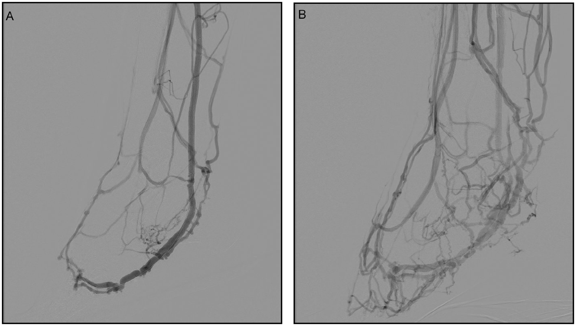
Image of 2 angiograms of the same patient. (A) Angiogram after the index procedure showing blood flow into the venous arch. (B) Angiogram 7 weeks later showing forward flow distal in the foot.
In addition, a staged amputation strategy is preferred to firstly drain the infection and secondly allow the AV-circuit to develop. A subsequent definitive amputation can be planned at least 6 weeks post-DVA. In our cohort, minor amputations deemed necessary in 13 patients (57%). An example of the staged amputation approach is shown in Figure 3. In one of our first cases, the skin was closed after a TMA. Wound dehiscence was seen a month later (Figure 4). In the following patients, the strategy was changed, and the skin was left open.
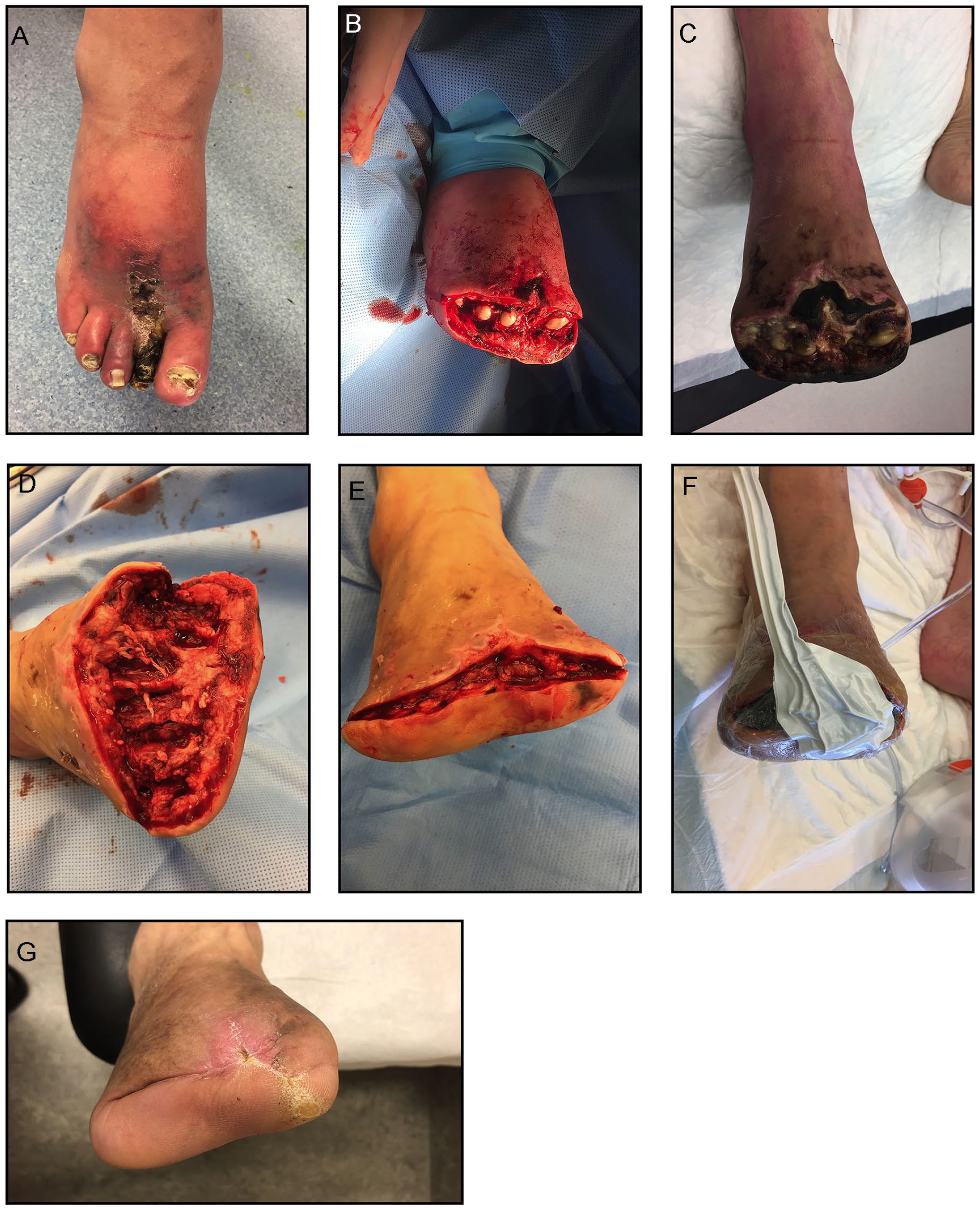
A staged amputation approach in a 63-year-old male. (A) Photograph showing affected tissue 6 weeks postpercutaneous deep venous arterialization. (B) Around 8 weeks postprocedural, the forefoot was amputated without removing the metatarsal cartilage. (C) Progression of necrosis was seen 2 weeks later. (D, E) Necrotic tissue and metatarsal heads were removed during the next amputation stage and the fascia was approximated. (F) Vacuum-assisted therapy was applied to accelerate wound healing. (G) Photograph of the fully healed wound.

A 28-year-old male in which a forefoot amputation was performed with primary closure. (A) Necrosis was left to demarcate after the percutaneous deep venous arterializations procedure. (B) A forefoot amputation was performed a month later, in which the skin was closed by sutures. (C) Necrosis started to develop at the site of the sutures a few days later, followed by wound dehiscence. Necrotectomy was performed in the operating theater to improve wound healing. (D) A split skin graft was placed after stagnant wound healing. (E) Wound healing was achieved at 8 months postprocedural.
Concerning the above, we advise to perform a staged amputation strategy as follows: the initial foot surgery should consist of necrotectomy of the gangrenous tissue and minor amputations in cases with a high risk of infection, leaving the skin open. When the amputation is through the metatarsal-phalangeal joint, the metatarsal heads should be left in place to prevent exposure of the cancellous bone. The second stage can include cleaning the wound surface, removing metatarsal heads and approximate the fascia to cover the underlying bone. Further wound care after DVA or minor amputations will be discussed in the following paragraph.
Wound Care
Wound care was briefly discussed in 5 studies.3,4,12,14 The use of vacuum-assisted therapy (VAC) was used in 23% to 100% of the cases.3,7,12 The use of split skin grafts was only described in one study 12 and was applied in 43% of the cases. Biosynthetic skin substitutes and rotational skin flaps were used in 7% and 3% of the cases, respectively. 3 Also dermal substitutes and dermo-epidermal grafts were used to enhance healing.4,14 Sole epithelialization-stimulating dressings were applied in 38% of the cases in one study. 3 Patients underwent debridements in 2 studies to improve healing potential.6,12
Clinical Experience and Expert Opinion
In our clinic, wound care was led by a multidisciplinary team including wound specialty nurses, physicians, and a podiatrist. We advise to treat dry gangrene conservatively with iodine in the initial 6 weeks. After a minor amputation, the wound can be covered with a gauze and netting. Compressive bandages should be discouraged. In case of wound debris during follow-up, special cleaning wound dressings as Prontosan® (B Braun, Melsungen, Germany) or Eusol can be used for debridement. Open wounds should be dressed with Hydrogels or Hydrofibre, for example, Aquacel to keep them moist. Vacuum-assisted therapy dressing can only be used when the wound bed is showing pink granulation and should generally be avoided in the first 6 weeks. In our first patient where VAC therapy was applied, the pressure was 120 mm Hg in a continuous setting. This was too intensive as the food colored white and the VAC was subsequently stopped after 3 days. In the following patients, the settings were changed to a pressure of 60 to 75 mm Hg with an intermittent suction frequency. This resulted in progression of wound healing in the following patients. Therefore, we recommend to apply VAC therapy intermittent with a low pressure of 60 to 70 mm Hg. In case of worsening infection or necrosis, the patient can be planned for surgical debridement in the operating theater. Necrotectomy can be performed using a knife and/or using the Versajet Hydrosurgery System (Smith & Nephew, London, United Kingdom).
Discussion
The advances in DVA over the last years have been impressive. Especially the percutaneous approach has shown most developments with promising results.6,10,11,13–15 The technique is becoming more accepted and applied in the treatment of patients with no-option CLTI, but there are still uncertainties on several aspects in the postprocedural management. A multimodal approach can be considered in the treatment of post-DVA patients.
Based on literature and clinical experience, we propose an algorithm for the challenges that interventionalists can face in the follow-up period after a DVA (Figure 5).

Timeline of postprocedural management. Six main subjects are shown. DVA, deep venous arterializations; APS, acute pain services; SAPT, single antiplatelet therapy; AC, anticoagulation; DAPT, dual antiplatelet therapy; TcPO2, transcutaneous oxygen pressure; DSA, digital subtraction angiography; MT heads, metatarsal heads; VAC, vacuum-assisted therapy; SSG, split skin grafts.
Edema and pain are the first events that can occur postprocedural and can be treated by elevation of the leg. A sudden increase in pain should be treated by pain specialists, and the cause should be identified. Postprocedural antiplatelet and anticoagulation therapy can include clopidogrel and heparin for the first 3 months postprocedural followed by lifelong dual antiplatelet therapy (clopidogrel and aspirin). Doppler can be performed in the first 48 hours postprocedural as this can be done at the ward and provides sufficient information about an open or occluded AV-circuit. Furthermore, we advise to evaluate patency by duplex ultrasound and assess forward pressure by TcPO2 measurements. Definitive foot surgery, debridements and wound enhancing therapies should be avoided within the first 6 weeks postprocedural as forward flow and pressure is unsufficient and evolving during that period.
With all developments in the technique and knowledge, the role of DVA for the treatment of no-option CLTI patients will become more important. The suggested recommendations were based on the reviewed studies and our experience with a cohort of 23 post-pDVA patients. The reviewed studies included cohort studies of moderate quality, case reports, and technical notes. Therefore, the level of evidence on which the recommendations were based can be considered moderate to low. In addition, the differences in pDVA techniques and procedures could affect postprocedural care and limit the generalizability of the propositions. Thus, we advocate for further research for this continuously developing revascularization method.
Conclusions
There are still many debated topics about postprocedural management in DVA patients. This review provides an overview of the current literature and suggests recommendations based on literature and clinical experience. Key elements in follow-up care are the acknowledgment that the DVA needs 6 weeks to develop before becoming effective, and a multimodal approach including surveillance, wound care, and a staged amputation strategy. The recommendations proposed may help future interventionalist in the present gap of knowledge in follow-up care of post-DVA patients. However, it is based on moderate to low quality evidence and therefore, further research is needed to draw firm conclusions.
Footnotes
Declaration of Conflicting Interests
The author(s) declared the following potential conflicts of interest with respect to the research, authorship, and/or publication of this article: Steven Kum is a paid consultant to LimFlow.
Funding
The author(s) received no financial support for the research, authorship, and/or publication of this article.
