






Abstract
An 81 year-old man presented with an asymptomatic juxtrarenal abdominal aortic aneurysm and was subsequently treated with a fenestrated endovascular Anaconda stent-graft. Surveillance imaging within the first postoperative year demonstrated a lower proximal sealing ring fracture. In the second postoperative surveillance year, the upper proximal sealing ring was also fractured with extension of the wire into the right paravertebral space. Despite these sealing ring fractures, there were no endoleak nor visceral stent complications and the patient continued on standard surveillance protocols. There are an increasing number of reports of fractured proximal sealing rings with the fenestrated Anaconda platform. Those analysing the surveillance scans of patients treated with this device should stay vigilant for the development of this complication.
Keywords
An 81 year-old man presented electively to the Vascular Surgery clinic with an asymptomatic 66 mm juxtarenal abdominal aortic aneurysm. A multidisciplinary team consensus was to undertake a fenestrated endovascular aneurysm repair (FEVAR) with a customized Anaconda stent-graft (Terumo Aortic, Inchinnan, Scotland, UK) incorporating 2 fenestrations for the renal arteries.
The Anaconda device is a self-expanding stent-graft incorporating fixation barbs with a proximal dual ring design that assumes a saddle shape with peaks and valleys when appropriately oversized and deployed in the aorta (Figure 1). The customized fenestrated platform is based on its more established standard infrarenal device design, except the peak and valley configuration can be rotated 90° to allow positioning of superior mesenteric or coeliac arteries in the anterior valley (the valleys in a standard infrarenal device are commonly lateral at the renal artery ostia). The 2 metal rings are nitinol wire bundles that form a single strand. The upper ring stent has a larger wire bundle diameter (higher number of wire turns) compared with the lower ring stent, resulting in higher radial strength. 1

The Anaconda fenestrated stent-graft. (A) The bifurcated piece comprising proximal sealing rings and nonmetallic fabric body housing multiple fenestrations. (B) Focus on the dual sealing ring configuration at the proximal stent-graft.
The procedure was undertaken without complication under general anesthetic, using percutaneous access, main body oversizing of 25% (the diameter of the visceral aortic segment was 24 mm and the stent-graft diameter was 30 mm), and sealing within both common iliac arteries. Postoperative surveillance imaging was undertaken with a computerized tomographic (CT) angiogram at 7 months post procedure. This demonstrated a fractured lower ring in the proximal sealing zone of the fenestrated graft with the free edge of the fractured wire abutting the adjacent lumbar vertebral body. However, there was no evidence of migration, endoleak, nor visceral stent complications. The fracture was stable in its appearances on repeat imaging with plain radiography (Figure 2B) and CT angiography 3 months later. A further year later (at a timepoint of 2 years post procedure), repeat plain radiography (Figure 2C) and CT angiography demonstrated an additional stent fracture of the upper sealing ring. The free end of this fractured upper sealing wire extended into the paravertebral space on the right side (Figure 3). Again, there was no evidence of migration, endoleak, nor visceral stent complications, and the residual sac had regressed in maximal diameter from 66 to 63 mm. A multidisciplinary team consensus was to manage the stent fracture conservatively and observe the aneurysm with standard surveillance protocols.
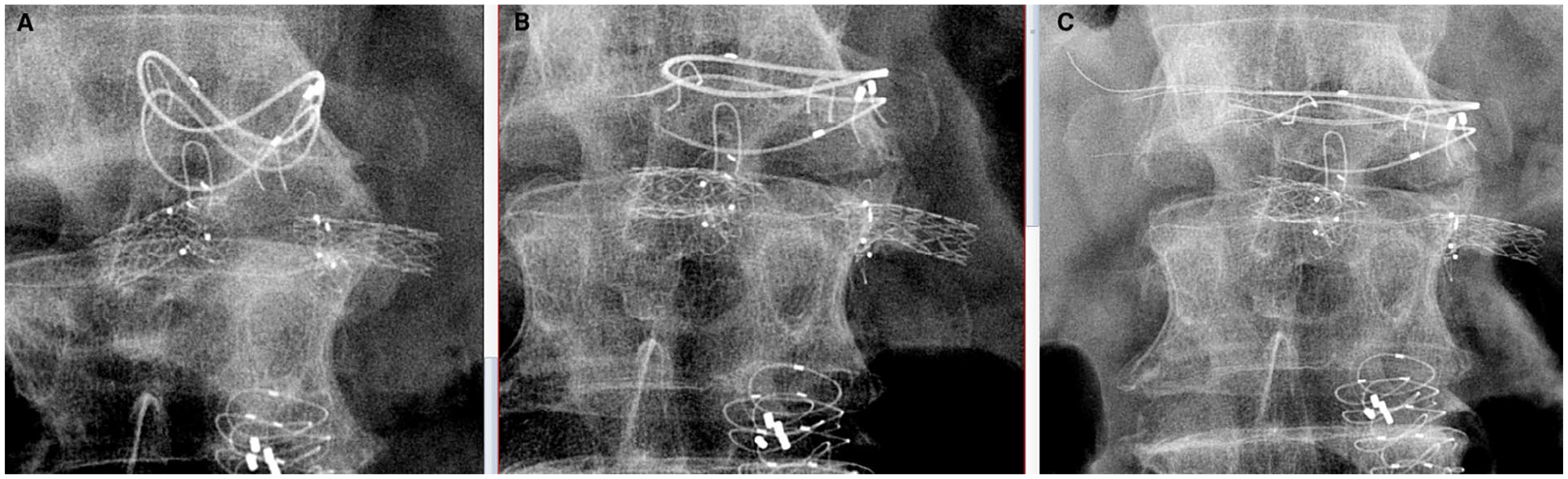
Plain radiographs of the dual ring system at the proximal sealing zone. (A) Normal configuration of the 2 “saddles” predischarge. (B) Flattening of both saddles with fracture of the lower ring at 1 year postoperative. (C) Fracture of both upper and lower rings at 2 years post procedure.

CT angiogram images at 2 years post procedure demonstrating the fractured top ring with displaced wire and the wire tip lying in the right paravertebral space. (A) Axial view and (B) 3D reconstructed view. CT, computerized tomographic; 3D, 3 dimensional.
This stent fracture is the first seen in our practice after implantation of 11 fenestrated Anaconda stent-grafts. There are very few Anaconda stent fractures reported in the literature and often none at all in published case series outcomes.2–4 However, recently, there has been unpublished data presented at a national vascular conference in the United Kingdom, where 15 of 253 (6%) patients treated with a customized Anaconda fenestrated stent-graft suffered stent fracture of the proximal ring. 5
It has been suggested that this specific graft design may be susceptible to complications from aggressive oversizing; the natural history of the device in situ involves continued expansion of the nitinol ring stents over time, and eventually the saddles of the rings flatten to become nearly circular by 2 years; excessive oversizing at the outset could lead to increased stress within the metal during this flattening process, potentially leading to fracture. 1 Those monitoring the surveillance scans of these patients should be vigilant for the development of this complication.
Footnotes
Declaration of Conflicting Interests
The author(s) declared no potential conflicts of interest with respect to the research, authorship, and/or publication of this article.
Funding
The author(s) received no financial support for the research, authorship, and/or publication of this article.
