






Abstract
Introduction:
En bloc kidney transplantation (EBKT) is a technique used to transplant pediatric kidneys to adult recipients, but can lead to certain complications seldom found in single-kidney transplantation. We report a case of renal artery embolization after EBKT due to intractable unilateral hydronephrosis and highlight the technical details and challenges of the procedure.
Case:
An 18-year-old female with MELAS syndrome underwent EBKT from a 10-month-old male baby. Two months later, the patient developed unilateral hydronephrosis and recurrent urinary tract infections, which was intractable to conventional therapy. Therefore, we underwent embolization of the problematic transplanted left kidney. Owing to the complicated anatomy and multiple angulations, multiple microcatheters, wires and support catheters were needed to select the renal arteries. Repeated procedures were required due to remnant flow from small branches and accessory renal arteries that were not easily visualized by conventional angiography, which were eventually detected by adjunctive use of 3-dimensional rotational angiography.
Conclusions:
Selective renal artery embolization after EBKT is challenging due to the short renal artery length and multiple angulations, yet it can still be performed safely and effectively by use of meticulous catheter-wire interactions and adjunctive intraoperative imaging techniques to delineate the precise anatomy of the target arteries.
Clinical Impact
Selective renal artery embolization, which is less invasive than nephrectomy, can be considered if the culprit kidney must inevitably be sacrificed in en bloc kidney transplantation.
Keywords
Introduction
En bloc kidney transplantation (EBKT) is a technique used to donate pediatric kidneys to adult recipients. It consists of procuring en bloc both kidneys together with the aorta and the inferior vena cava (IVC) and transplanting it into the same adult recipient, usually in the retroperitoneal space, by anastomosing the donor aorta and IVC to the recipient iliac vessels. Recent studies have demonstrated excellent graft function and outcomes after EBKT. As a result, EBKT is widely used as an approach to expand the donor pool and improve graft utilization.1 –3
Despite these promising results, EBKT is technically demanding, especially if the donor kidneys are small in size, and can lead to various complications that may occur less frequently during single-kidney transplantation. We report a case of an 18-year-old female who underwent EBKT and presented with recurrent urologic complications in one of the transplanted kidneys, which eventually required multiple embolization procedures to sacrifice the culprit kidney, with special emphasis on the endovascular techniques and advanced imaging modalities that were required to overcome the small and challenging anatomy.
Case
Patient History
The recipient was an 18-year-old female with congenital mitochondrial encephalomyopathy with lactic acidosis and stroke-like episodes (MELAS) syndrome who was on dialysis for end-stage renal disease (ESRD), secondary to focal segmental glomerulosclerosis. The donor was a 10-month-old male, who became brain dead after drowning in a bathtub. Both kidneys were retrieved from the donor en bloc with the aorta and inferior vena cava (IVC). However, injury to the left ureter of allograft occurred during the retrieval procedure, which was repaired during the bench procedure before transplanting it into the recipient. The suprarenal aorta and IVC were closed with 6–0 polypropylene running sutures. The caudal ends of the aorta and IVC were anastomosed with recipient’s right common iliac artery (CIA) and right external iliac vein (EIV), respectively. The ureters were anastomosed to the recipient bladder by an ureteroneocystostomy procedure, and ureteral double-J (DJ) catheters were inserted intraoperatively (Figure 1A). Postoperative recovery was uneventful. Immunosuppression induction was achieved using basiliximab and maintenance immunosuppression was done by means of a triple drug regimen consisting of tacrolimus, mycophenolate, and prednisolone. The serum creatinine level was 0.94 mg/dL on the fifth postoperative day and was since steadily maintained in the normal range. There were no specific findings regarding renal perfusion or complications evaluated by color Doppler ultrasound on the seventh postoperative day. The patient was discharged without complications after 2 weeks postoperatively.

En bloc kidney transplantation (EBKT), preoperative computed tomography (CT), and antegrade pyelography (AGP) findings. (A) EBKT was performed using both kidneys of a single donor. (B) Noncontrast CT shows hydronephrosis of the left transplanted kidney, (C) Follow-up CT performed after double-J (DJ) catheter removal showed mild aggravation of hydronephrosis. (D) Ureter outflow obstruction (arrow) as seen on AGP.
Eight weeks after EBKT, the patient presented to the emergency room with a fever of 38.3°C. Laboratory findings showed mild leukocytosis and elevated C-reactive protein (CRP), and bacteriuria was confirmed on urinalysis. We found severe hydronephrosis of the transplanted left kidney on noncontrast computed tomography (CT) (Figure 1B). Therefore, antibiotic therapy and DJ catheter removal were performed under the diagnosis of urinary tract infection (UTI) due to DJ catheter dysfunction. However, the fever did not subside despite continued therapy. Differential tests for the infection of BK virus and cytomegalovirus (CMV), and for acute rejection were performed, all of which came out negative. Follow-up contrast-enhanced CT was performed, which showed a slightly aggravated left hydronephrosis (Figure 1C). Ureter outflow obstruction was confirmed on antegrade pyelography (AGP), and a percutaneous nephrostomy (PCN) tube was inserted for the treatment of hydronephrosis (Figure 1D). To resolve the ureter outflow obstruction, balloon dilatation and DJ catheter reinsertion were performed. After 10 days of hospitalization, the patient recovered and was discharged with the DJ catheter in situ and the PCN removed. However, 5 weeks later, the patient returned to the hospital with the same symptoms and received the same treatment as the first hospitalization. The patient was hospitalized and discharged 5 times during a period of 10 months after EBKT, and obstructive uropathy did not resolve despite continuous antibiotic treatment and repeated radiologic interventions (PCN insertion, DJ catheterization and ureteral balloon dilatation). Eventually we decided to sacrifice the culprit transplanted left kidney by endovascular embolization.
Embolization Procedure
After 42 weeks of EBKT, embolization of the transplanted left kidney was performed through a left common femoral artery access with 5 Fr sheath. An arteriogram of the right common iliac artery showed the allograft aorta and both transplanted renal arteries on the right side of the patient (Figure 2A). The transplanted kidneys had normal renal artery anatomy and a left hydronephrosis was also visualized. Selective catheterization of the transplanted left renal artery was achieved using a 5 Fr Davis catheter (A&A M.D, SeongNam, Korea) and 1.7 to 2.8 Fr tapered Progreat Lambda 17 microcatheter (Terumo, Somerset, New Jersey, USA) (Figure 2B). Selective embolization for the renal artery of the transplanted left kidney was performed with gelatin (Nexsphere 100-300 um, Next Biomedical, Incheon, Korea), polyvinyl alcohol particles (PVA, COOK MEDICAL, Bloomington, Indiana, USA), and detachable coils (Concerto Coils, Medtronic, Minneapolis, MN, USA) (Figure 2C). The embolization procedure was considered to be completed successfully. Also, 100 cc/day of urine was continuously excreted through the PCN of transplanted left kidney after the procedure. One month after embolization, we decided to perform an additional embolization procedure since the culprit transplanted left kidney was still functional. Diagnostic arteriography was performed in the same way as the first embolization. Digital subtraction angiography (DSA) showed a small branch of transplanted left renal artery and an accessory left renal artery arising from the allograft aorta, which was not detected during the first embolization (Figure 2D). We tried to select this artery by exchanging various catheters with different shapes (Cobra, Davis, Simmons catheter) and wires. However, due to the multiple angulations and complicated anatomy to reach the desired branches, selection was unsuccessful. In addition, as the procedure time prolonged, the patient became irritable and did not cooperate, so the procedure was terminated. We tried a third embolization 2 weeks later, and this time the right common femoral artery was accessed. Conventional angiography with digital subtraction failed to demonstrate the target arteries, but we then performed a 3-dimensional rotational angiography (3D-RA) with low-contrast imaging (LCI) to visualize and reconstruct the anatomical configuration of the target arteries (Figure 3). Based on the exact information from the 3D reconstructed images, we selected the small branch arising from the transplanted left renal artery with a pre-shaped 90° bent 1.7 Fr microcatheter (Excelsior SL-10, Stryker Neurovascular, Freemont, California, USA) and wire (Synchro-14, Stryker Neurovascular, Freemont, California, USA). Selective embolization was then performed using detachable coils (Concerto coils, Medtronic, Minneapolis, MN, USA) (Figure 4A and B). For selection of the accessory left renal artery arising from the allograft aorta, we changed access to the left common femoral artery, and selective catheterization of the allograft aorta was performed using a 5 Fr Simmons catheter (A&A M.D, Seong Nam, Korea). A 1.7 to 2.8 Fr tapered Progreat Lambda 7 (Terumo, Somerset, New Jersey, USA) was successfully advanced into the accessory renal artery (Figure 4C), and embolization was performed using a mixture of histoacryl-lipiodol. A completion angiography showed total occlusion of the transplanted left renal arteries with no visible allograft parenchyme (Figure 4D). After embolization, urine was no longer excreted through the PCN, and a mild fever (up to 38°C) persisted for 2 days, but recovered with supportive care. Serum creatinine level was 0.83 mg/dL, similar to pre-embolization values. After 5 days of completion embolization, urine was no longer excreted through the inserted PCN, so it was removed. The patient was discharged on the seventh day after procedure and is currently under follow-up on an outpatient basis for 11 months.

The procedure of first and second embolization. (A) The digital subtraction angiography (DSA) through the right common iliac artery shows that transplanted kidneys have normal anatomy of the renal artery arising from the allograft aorta. (B) Selective catheterization of left transplanted kidney for embolization. (C) Final angiography shows successful embolization of the left transplanted kidney. (D) Angiography through allograft aorta demonstrated a small branch of the left transplanted renal artery and accessory renal artery (arrow) that was not detected at the time of the first embolization.
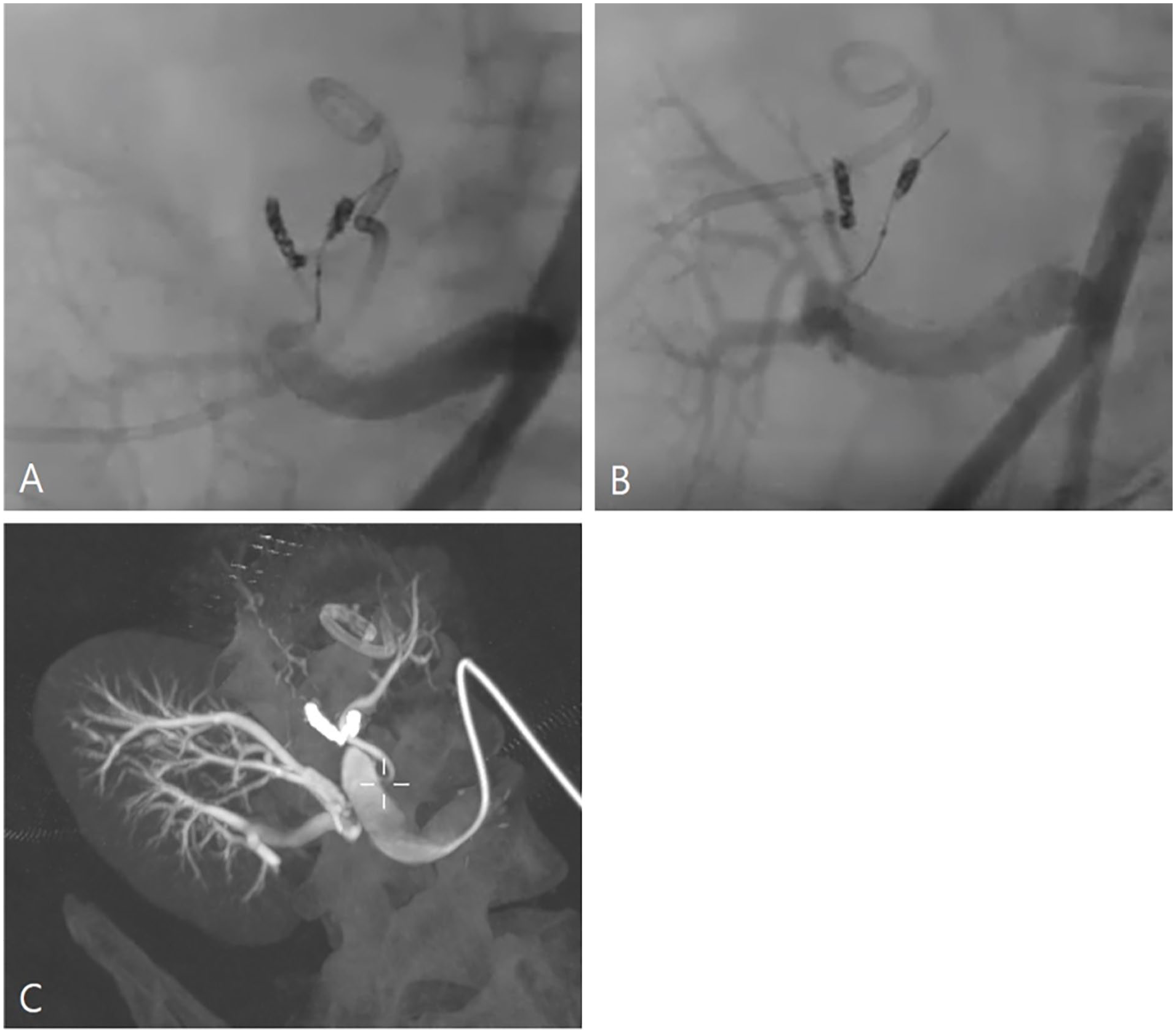
Low contrast imaging (LCI) and 3-dimensional rotational angiography (3D-RA) imaging. (A and B) Multi-angle imaging with 3D-RA can be used to structure the anatomical location of the renal artery of the transplanted kidney. (C) The LCI image shows the exact location of the origin of the accessory renal artery of the left transplanted kidney.
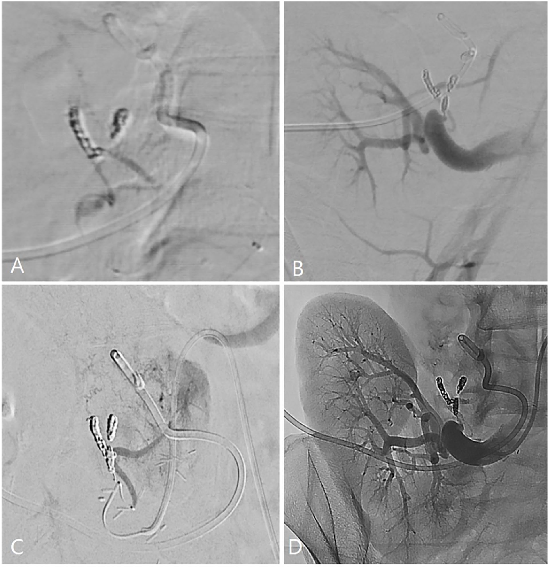
The procedure of third embolization. (A) Selective catheterization on the small branch of left transplanted renal artery using microcatheter and wire. (B) Successful coil embolization of small branch of the left transplanted kidney. (C) Selection and embolization of the accessory renal artery using histoacryl-lipiodol. (D) Final angiography show complete blockage of blood flow to the left transplanted kidney.
Discussion
We report a case of successful embolization of a culprit transplanted kidney after EBKT that was complicated by intractable hydronephrosis. Multiple embolization attempts were required due to the challenging anatomy arising from the small size of the kidneys leading to a short length of the renal arteries and branches, and the multiple angulations to reach the target vessels.
EBKT is a technique used to transplant pediatric kidneys into adults in order to overcome the size discrepancy. In the past, EBKT was seldom performed due to technical difficulties, leading to high urologic and vascular complications. However, with improved surgical skills and in order to overcome the disproportionate lack of donors, EBKT is nowadays being performed more often. Excellent graft function and long term outcomes have been reported for EBKT,1 –3 yet the incidence of urologic complication after EBKT is 55% to 33%, which is higher than standard kidney transplantation.1,3 –5 As shown in this case, ureter injury during donor kidney retrieval can occur, and can lead to postoperative urologic complications that may be intractable to medical therapy, eventually requiring the sacrifice of the transplanted kidney.
In this patient, during the 10 months from the initial transplantation to the timing of embolization, the transplanted kidney had significantly grown in size and the creatinine levels were in the normal range. The patient’s estimated glomerular filtration rate was 107.0, and the expected risk of ESRD after kidney sacrifice was 0.3, 1.9, 4.9, 10.1 per 10,000 at 5, 10, 15, 20 years after nephrectomy, respectively. 6 Based on these calculations, we judged that the culprit kidney could be sacrificed without causing significant renal function impairment. In this patient we decided to sacrifice the culprit kidney by endovascular means rather than open nephrectomy, since it is not only less invasive, but also to avoid possible injury to the normal functioning right transplanted kidney during surgery because it was expected that the surgical field would be complicated by repeated inflammation and fibrosis. Renal artery embolization is generally regarded as a safe and effective treatment method in various clinical situations and has been widely used as an alternative to nephrectomy. 7
During endovascular intervention, DSA is the gold standard procedure and widely used in many fields of endovascular treatment. However, DSA is limited with regard to visualizing the exact anatomy of vascular lesions with complex angioarchitecture.8,9 Therefore, 3D-RA was introduced to compensate for these shortcomings of DSA, and it is being used in the endovascular management of various vascular lesions such as cerebral and peripheral aneurysms, liver embolization, and vascular malformations.9 –14 In addition, 3D-RA has the advantage of reducing the amount of contrast medium compared to DSA because it acquires images with a single injection of contrast medium. Low-contrast imaging is a technology that can be performed immediately during angiography and is used for diagnosis of vascular lesions by providing CT-like images of various structures. 15 In this case, the use of 3D-RA and LCI allowed for accurate delineation of the complex arterial anatomy of EBKT, which may have not been easily visualized by conventional angiographic imaging. Based on this accurate anatomic information, meticulous planning, and selection of endovascular devices allowed for a technically successful embolization procedure.
Conclusion
Selective embolization of a transplanted kidney after EBKT is technically demanding and requires meticulous planning and device selection in order to overcome the challenging anatomy arising from the short and multiple angulations to select the renal arteries. However, it can be successfully performed with the guidance of advanced intraoperative imaging techniques such as 3D-RA and LCI, which delineate the precise anatomical location of the target vessels, allowing for accurate diagnosis and meticulous execution.
Footnotes
Declaration of Conflicting Interests
The author(s) declared no potential conflicts of interest with respect to the research, authorship, and/or publication of this article.
Funding
The author(s) received no financial support for the research, authorship, and/or publication of this article.
