






Abstract
Purpose:
To summarize experience with and the efficacy of fenestrated/branched thoracic endovascular repair (F/B-TEVAR) using physician-modified stent-grafts (PMSGs) under 3D printing guidance in triple aortic arch branch reconstruction.
Materials and Methods:
From February 2018 to April 2022, 14 cases of aortic arch aneurysms and 30 cases of aortic arch dissection (22 acute aortic arch dissection and 8 long-term aortic arch dissection)were treated by F/B-TEVAR in our department, including 34 males and 10 females, with an average age of 59.84 ± 11.72 years. Three aortic arch branches were affected in all patients. A 3D-printed model was made according to computed tomography angiography images and used to guide the fabrication of PMSGs. All patients were followed up.
Results:
A total of 132 branches were successfully reconstructed with no case of conversion to open surgery. The average operation time was 4.97 ± 1.40 hours, including a mean 44.05 ± 7.72 minutes for stent-graft customization, the mean postoperative hospitalization duration was 9.91 ± 4.47 days, the average intraoperative blood loss was 480.91 mL (100–2810 mL), and the mean postoperative intensive care unit monitoring duration was 1.02 days (0–5 days). No deaths occurred within 30 days of surgery. Postoperative neurological complications occurred in 1 case (2.3%), and retrograde type A dissection occurred in 1 case (2.3%).
Conclusion:
Compared with conventional surgery, triple aortic arch branch reconstruction under the guidance of 3D printing is a minimally invasive treatment method with the advantages of accurate positioning, rapid postoperative recovery, few complications, and reliable short- to mid-term effects.
Clinical Impact
At present the PMSG usually depend on imaging data and software calculation. With the guidance of 3D printing technology, image data could be transformed into 3D model, which has improved the accuracy of the positioning of the fenestrations. The diameter reduction technique and the internal mini cuff technique have made a complement to the slimed-down fenestration selection process and the low rate of endoleak. As reproducible study, our results may provide reference for TEVAR in different cases.
Keywords
Introduction
Thoracic endovascular repair (TEVAR) has been widely carried out in various countries and has become the first choice for the treatment of descending aortic lesions. 1 For lesions involving the aortic arch and ascending aorta, which endanger the important aortic arch branches, open surgery has traditionally been the best therapeutic method, even though it is traumatic and has high mortality and complication rates. In recent years, methods for the endovascular treatment of aortic arch and ascending aortic lesions have emerged. Despite the challenges of this technique, a number of studies have emerged recently, and more than acceptable results have been achieved especially in patients with high risk of traditional surgery.2,3
Three-dimensional (3D) printing technology has become a leading manufacturing technique in health care and medicine, it enables the production of anatomically matched and patient-specific devices and constructs with high tunability and complexity. 4 At our center, we used this technology to print anatomical models rather than medical devices, which allowed individualized and accurate positioning of the fenestrations for the aortic arch branches.
Materials and Methods
Study Design and Patient Population
The clinical data of 44 patients (34 males and 10 females, with a mean age of 59.84 ± 11.72 years) with aortic arch lesions who underwent treatment guided by 3D-printed technique in our Department from February 2018 to April 2022 were analyzed retrospectively. Among the 44 patients, there were 14 aortic arch aneurysms (aneurysm diameter: 49.55 ± 13.38 mm), 30 aortic arch dissections (3 cases of Ia endoleak and 2 cases of retrograde dissection after TEVAR of type B dissection, 2 cases after surgery of type A dissection, 9 cases of primary type A dissection, and 14 cases of primary type B dissection).
Indications for SG implantation included asymptomatic, degenerative, or traumatic aneurysms larger than 5 cm and all cystic aneurysms. In the setting of aortic arch dissection, the indications included a maximum dissecting aneurysm >50 mm, a false lumen diameter twice that of the true lumen and progressively enlarging (>10 mm/y), the pain in the chest or hoarseness of the voice, comorbid conditions, such as other ascending or descending aortic diseases, Marfan syndrome, family history of aortic dissection rupture.
A thin-slice enhanced computed tomography angiography from the carotid down to the femoral arteries was performed before the operation and formed the morphological basis of decision-making. Meanwhile we will consummate the decision-making process in 4 aspects:
Type of vascular lesion. For patients with aortic arch pathologies but a healthy ascending aorta, we will give priority to F/B TEVAR using physician-modified stent-graft (PMSG).
Patient’s physical condition. In the sitting of secondary surgery, elderly patients or patients with poor general conditions, we will give priority to F/B TEVAR using PMSG.
Degree of emergency. F/B TEVAR can be an alternative for emergency patients after failure of open surgery.
Patients’ preferences. The economic conditions of patients should be taken into consideration for the higher cost of SG.
This study was approved by the Medical Technology Ethics Committee, and the patients and their families signed the informed consent form.
3D-Printed Model Preparation
DICOM (Digital Imaging and Communications in Medicine) data files were imported into Mimics software for 3D reconstruction of the blood vessels in the aortic arch area, including the proximal and distal normal aorta, the diseased aorta and the branches of the arch. Accurate measurements of the normal aortic and triple-branched luminal longitudinal and transverse diameters near the distal end, as well as the lesion length, were obtained. The reconstructed 3D model data were further preprocessed using Geomagic Studio 2014 design software (Figure 1B). Nonparametric surface reconstruction of the blood vessels was performed using reverse engineering technology. Based on the mechanical characteristics of the aortic stent, blood vessel deformation after stent implantation was simulated, and the computer-aided design (CAD) mathematical model of the blood vessels was obtained. After the simulation analysis was performed using Geomagic Design Direct, the fenestrations for the 3 branches of the aortic arch were opened, and a 3D guide plate was designed (Figure 1C). The data were transmitted to a 3D printer to complete fabrication of the transparent hollow 3D aortic arch model. The 3D-printed model was disinfected with ethylene oxide and sealed for later use. The printing process took about 5 hours (1 hour for reconstruction, 3 hours for printing, and 1 hour for postprocessing) and the disinfection process took about 3 hours.
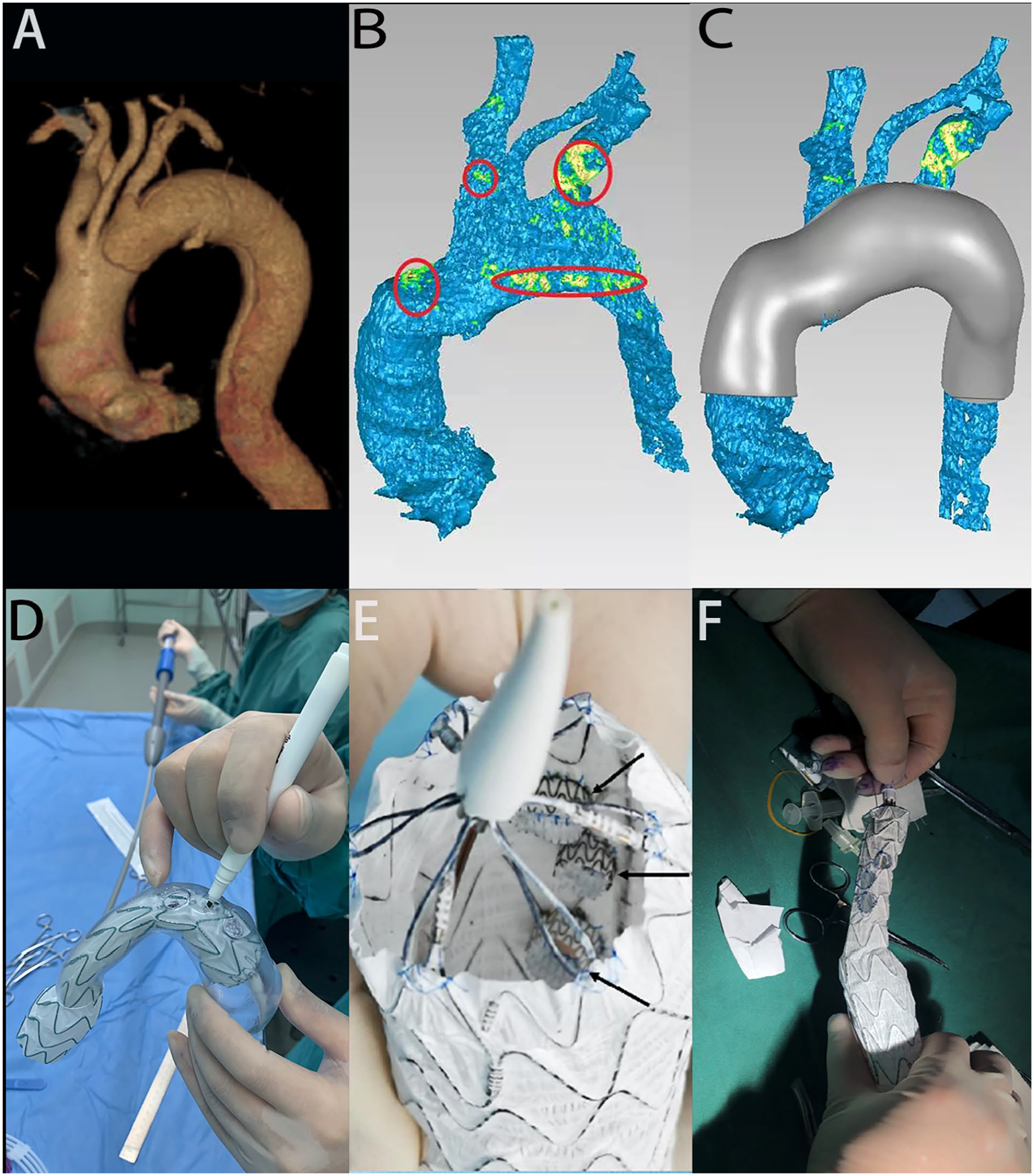
Repair of aortic arch aneurysm with triple prefenestrated stent-graft (SG) technique guided by 3D printing. (A) Preoperative 3D CT reconstruction. (B) Data preprocessing in Geomagic Studio 2014; red circles indicate an abnormal blood vessel or the site of possible intimal tear in the design of the 3D-printed model. (C) Design of the 3D-printed model. (D) Release of SG in the model to determine the fenestrations. (E) Internal mini cuffs (arrow). (F) Reduced diameter of SGt. CT, computed tomography.
Intraoperative PMSG Fabrication
According to the measurement results obtained using 3Mensio or EndoSize software, a suitable thoracic aortic stent-graft (SG) with a posterior release system (Ankura, China Lifetech Company; Captivia, Medtronic, USA) and branch artery SG (Endurant, iliac extension, Medtronic, USA; Fluency, Bard Company, USA; Viabahn, Gore Company, USA) were selected, and then the surgical scheme was designed according to the anatomical situation. Generally, for fenestrations where endoleak may occur (such as the branch artery from the aneurysm or the branch artery opening near the intimal tear), the internal mini cuff technique was adopted; and for fenestrations not affected by the lesion, the traditional fenestration technique without cuff was adopted. At the same time, the corresponding guidewire catheter, arterial sheath and dilation balloon were prepared.
The thoracic aortic SG was implanted into the transparent 3D-printed model, with the proximal end of the SG located at the planned anchoring site, slowly and completely released. During the release process, attention was given to adjusting the stent position at any time to ensure that the stent skeleton did not overlap the fenestration. Through the arterial branch fenestrations in the 3D model, the fenestration positions were marked on the SG membrane with a sterilized marker (Figures 1D and 2B).

Treatment of aortic arch aneurysm with triple prefenestrated SG technique guided by 3D printing. (A) Prebending of the stent. (B) Release of the stent in the model to determine the fenestrations. (C) Preoperative angiography of the aortic arch. (D) System for delivering the fenestrated stent before expansion. (E) Postoperative angiography of the aortic arch. (F) CT reconstruction at 1 month after surgery. SG, stent-graft; CT, computed tomography.
The SG was then removed from the model, and holes were created at the marked sites with an electrocautery device. In cases where the lesion affected the fenestration, it was necessary to prevent endoleak from the junctions between SGs. Usually, the internal mini cuff technique was adopted; that is, a Viabahn SG(Gore Company, USA) with an appropriate diameter (1–2 mm less than that of the expected branch, e.g., the fenestration for the 8 mm left carotid artery, should be 7 mm in diameter) was trimmed to a length of 3 to 5 mm, the external opening was continuously sutured with 5–0 nonabsorbable sutures (Prolene, Johnson & Johnson Company, USA), and the internal cuff was placed inside the aortic SG to prevent endoleak. For fenestrations that did not involve pathological changes, the fenestration was simply opened; similarly, the diameter of the fenestration was approximately 1 to 2 mm smaller than the expected diameter of the branch. A dehaired Tornado Embolization Microcoil (Cook Co., USA) was sewn to the fenestration edge with nonabsorbable sutures to reinforce the fenestration edge and was used as an intraoperative landmark. For aortic stents without reinforcing ribs (such as the Medtronic Captivia), a radiopaque X-ray mark was sewn to the top of the large curved side of the thoracic aortic SG for convenient positioning during angiography. The 4–0 or 5–0 prolene sutures and V-18 guide wire (Boston Scientific Company, USA) were combined on the minor curvature side of the thoracic aortic SG, and a detachable suture method was used to reduce the diameter of the anterior segment of the thoracic aortic SG by 30% to 50%. The segment of diameter reduction is from proximal bare stent to the left subclavian artery (LSA) fenestration, the total length was generally 80 to 100 mm. If the SG needed to enter the ascending aorta further, the tip of the SG could be trimmed to a shorter length to avoid damaging the heart valve. Finally, the thoracic aortic SG was placed into the delivery sheath again, and the stent delivery sheath was prebent in vitro again along the curvature under the guidance of the 3D model (Figure 2A).
Placement of Aortic and Branch Stents
At the same time as PMSG preparation, another group of surgeons also prepared the surgical approach. Usually, unilateral femoral access was established for aortic SG delivery, while the left brachial artery, left common carotid artery, and right axillary artery were used to access the aortic arch branches and deliver the corresponding covered stents. The catheter was placed at the level of the coronary artery, and then, digital subtraction angiography was performed for localization of the aorta (Figure 2C). Heparin (1 mg/kg) was injected intravenously. Then, the sheath used to deliver the aortic stent was inserted through the femoral artery until the stent reached the position on the aortic arch corresponding to the preshaped surface. The orientation of the fenestration was confirmed to be accurate according to the mark, the stent was slowly expanded, and the guide wire and catheter were inserted into their respective fenestrations from the right upper limb, left carotid artery, and left upper limb. At this time, the SG could be moved back and forth slightly to facilitate fenestration selection. Once the catheter entered the fenestration, the hard guide wire was replaced, and the appropriate long vascular sheath was inserted along the new guide wire (with the Viabahn stent and branch of Endurant iliac extension) or directly using the branch stent delivery system (with the Fluency stent) (Figure 2). After pulling out the fettered guide wire (V-18 guide wire, Boston Scientific Company, USA) and completely releasing the thoracic aortic stent, the stent in each arterial branch was released, and the balloon was expanded, such that, the stents completely fit the vessels. Owing to the reduced diameter of the main stent and fenestrations, the blood supply to the brain was sufficient, and no additional bypass technique was required during the operation. Angiography of the ascending aorta and aortic arch was performed to confirm the patency of each arterial branch and the absence of endoleakage, after which fenestrated/branched thoracic endovascular repair (F/B-TEVAR) was performed (Figure 2E). Patients were required to undergo postoperative follow-up computed tomography (CT) examinations (Figure 2F). In this group, 13 patients used aspirin enteric-coated tablets postoperatively, and 31 patients were treated aspirin enteric-coated tablets and rivaroxaban tablets. Except for patients allergic to rivaroxaban or any excipient in tablets, patients with clinically active bleeding, patients with liver disease or abnormal blood coagulation and clinically related bleeding risk, we asked all postoperative patients to take rivaroxaban for half a year or more, with a dose of 1 tablet (10 mg) per day.
Results
Under the guidance of 3D printing, 44 patients with aortic arch lesions were treated by total endovascular therapy with PMSGs; 29 patients were routinely treated with the internal mini cuff technique, while 15 patients (treated before 2019) were not (Table 1). A total of 132 branches were reconstructed, with a technical success rate of 100%. All branches were successfully reconstructed once without any cases of intraoperative mortality. The mean operation time was 4.97 ± 1.40 hours, including the mean duration required for custom stent preparation (44.05 ± 7.72 minutes) and the mean duration of the endovascular operation (79.64 ± 18.10 minutes). The average intraoperative blood loss was 480.91 mL (100–2810 mL), and the transfusion volume was 250.57 mL (0–1700 mL). The mean contrast agent volume was 134.59 ± 24.24 mL, and the mean radiation dose was 1508.73 ± 498.07 mGy. The mean postoperative intensive care unit (ICU) monitoring time was 1.02 days (0–5 days), and the mean postoperative hospitalization duration was 9.91 ± 4.47 days (Table 2). Postoperative neurological complications occurred in 1 case (2.3%) during hospitalization, and cured by medication. The patient presented headaches, dizziness, nausea, vomiting, numbness, and weakness in the extremities, and was diagnosed with ischemic stroke on head CT. Aspirin enteric-coated tablets and rivaroxaban tablets were used for anticoagulant therapy.
General Information of 44 Patients. a

Abbreviations: BMI, Body mass index; CHF, congestive heart failure.
Continuous data are presented as the mean ± standard deviation (range); categorical data are given as the number (percentage).
Procedure Details of 44 Patients.a

Abbreviation: ICU, intensive care unit.
Continuous data are presented as the mean ± standard deviation (range); categorical data are given as the number (percentage).
The mean follow-up time was 22.3 months (1–34 months). The aortic computed tomography angiography (CTA) follow-up examinations at 3 months, 6 months, and every year after the operation indicated that all 3 branches of the aortic arch were unobstructed, 42 patients reached 6 months of CTA follow-up, and 35 patients reached 12 months of CTA follow-up. One case (2.3%) of retrograde type A dissection occurred 2 months after the operation, which was treated with emergency open surgery and ascending aorta replacement at another hospital. During the follow-up, no other serious complications, such as cerebral infarction or paraplegia occurred, the mortality was 4.5% and the aorta-specific mortality was 0% (1 patient died of kidney failure and 1 patient died of lung cancer). There was 1 (2.3%) cases of type Ic endoleak among 29 (65.9%) patients treated using the internal mini cuff technique, considering the size of the branched stent is too small and regular follow-up was recommended; 1 type Ia (the insufficient size of the main aortic stent may be the explanation) and 2 type IIIc (occurred at the site of fenestrations stent junctions) endoleaks occurred in 3 (6.8%) of the other 15 (34.1%) patients without internal mini cuffs after the operation. The endoleaks occurred in the patients without internal mini cuffs (1 type Ia and 2 type IIIc endoleak) were cured after embolization, and the follow-up CTA showed good curative effect. The remaining case (1 type Ic endoleak) was small and only required long-term follow-up, the 6 months and 12 months follow-up CTA showed a slowly progressive course of endoleak. Typical CTA images are shown in Figures 2 and 3.

Treatment of aortic arch dissection with triple prefenestrated SG technique with internal mini cuffs guided by 3D printing. (A) Preoperative 3D CT reconstruction. (B) Release of the stent in the model to determine the fenestrations and reduced proximal SG diameter. (C) Preoperative angiography of the aortic arch. (D) Placement of sheathes into fenestrations. (E) Postoperative angiography of the aortic arch. (F) CT reconstruction at 3 months after surgery. CT, computed tomography; SG, stent-graft.
Discussion
At present, the endovascular treatment methods for aortic arch lesions mainly include chimney technique, standard TEVAR with hybrid adjuncts, including commercially available devices and PMSG.
The chimney technique, as a transitional technique for aortic arch lesions, with low technical difficulty and wide range of applications, is simple to perform. Studies have shown that the use of chimney SGs in the aortic arch can lead to an incidence of endoleak as high as 22%.5,6 At our center, the reverse retrograde type A dissection appeared in 3 of 12 patients who were implanted a chimney SGs into the ascending aorta within 1 year (3/12, 25%).
Commercially available devices have brought great convenience to the endovascular solutions, especially in emergency setting. However, among commercial branch SGs, only mature products with single branch can be purchased off the shelf; thus, aortic arch branch reconstruction methods still need to be combined with open surgery in most cases.7,8 It made sense to find a solution to make up the lack of the commercial device. Fenestrated/branched thoracic endovascular repair, guided by 3D printing technology, is a promising approach for the PMSG can be quickly and inexpensively fabricated using existing commercial thoracic aortic stents.
TEVAR using a custom-made multibranch device has closed the therapeutic gap in a subgroup of patients where neither conventional surgical approaches nor combined vascular and endovascular approaches could have been regarded as the therapy of first choice. 9 In the literature-related, a total of 39 patients had treated by total endovascular aortic arch repair at 8 academic centers using 3-vessel inner branch SGs (William Cook Europe, Bjaeverskov, Denmark) from 2016 to 2019. The technical success rate was 100%. The combined mortality and any stroke rate was 8% (3/39). Endoleak in 6 (3 type II, 1 type Ic, 1 type Ia/Ib, and 1 type IIIa), and target vessel stenosis in 1 patient. 10
Given that other solutions need to address high incidence of mortality, cerebral complications, and endoleak, F/B-TEVAR using PMSGs under the guidance of 3D printing technology has the advantages follow.
Individualized and accurate positioning of the fenestrations for the aortic arch branches using 3D printing technology. The 3D-printed model can intuitively reveal the spatial relations between the aortic dissection or aortic aneurysm and aortic arch branches to provide guidance for TEVAR for the treatment of these life-threatening aortic diseases and guide clinical management. In addition to PMSG fabrication, this model can also be used to simulate stent release and fenestration positioning in the vascular lumen before the operation. 11 Using the 3D model, all 3 fenestrations can be adjusted to fall on the membranous part of the SG without a part of the skeleton; additionally, the fenestrations can be reinforced and sutured to effectively counter any adverse influence on the structural strength of the aortic stent.
Low incidence of cerebral complications. The SG diameter was temporarily reduced by 30% to 50% using the diameter reduction technique combined with the guide wire and sutures. After the PMSG was inserted into the aortic arch, due to the difference in diameter between the primary SG and the blood vessel wall, the blood supply to the brain remained sufficient, and no additional bypass was needed during the operation. Without vascular occlusion, the branches of the arch remained unobstructed, and cerebral complications rarely occurred. Besides, all patients were adequately anticoagulated preoperatively and the mean intraoperative arterial pressure was higher than 120 mmHg to preserve the blood flow to the brain. Among the 44 patients in this group, only 1 patient (2.3%) with an aneurysm experienced transient cerebral ischemia after the operation. This rate is lower than Asciutto et al.’s 12 report considering the serious calcification of lesions, plaque shedding, and neurological complications. Asciutto reported that no diameter reducing ties were placed on the SG and the devices were re-sheathed with the aid of nylon tape and plastic vessel loops to wrap and compress the SG. More comparative studies are warranted, the diameter reduction technique may account for the differences.
Endoleak rarely occurred. In view of type IIIc endoleak, which easily occurs at the site of fenestrations stent junctions,13,14 the internal mini cuff technique was routinely used in the latter 25 patients in this group. An internal mini cuff of an appropriate size was sewn inside the larger stent, which established surface-to-surface contact instead of line-to-surface contact, effectively preventing the occurrence of endoleak. Only 1 case of type Ic endoleak occurred among 29 patients treated using the internal mini cuff branch technique. The rate is similar to Zhu et al.’s 15 report that said they sewed the loop of a snare (Amplatz GooseNeck, ev3, Plymouth, MN, USA) onto the edge of each fenestration for reinforcement. Since only 9 cases were reported and had a technical success rate of 90%, their repeatability still needs to face up to a test.
Slimmed-down fenestration selection process. In clinical work, we found that most commercial aortic SG delivery systems exhibit a certain memory effect. After forming a certain shape in a 3D-printed model of the aortic arch, the stent expands according to the shape assumed in the 3D model. Therefore, after the PMSG was collected into the delivery sheath, we prebent the stent along the large curved side again. During the operation, the delivery sheath was inserted directly without manual adjustment of the orientation, and the 3 fenestrations on the arch of the PMSG roughly faced the large curved side. Combined with the stent diameter reduction technique, this technique greatly simplified the fenestration selection process.
Appropriate access routes. We usually use the left brachial artery, the left common carotid artery, and the right axillary artery as access routes for stent delivery to the superior arterial branches. The choice of the approach will affect the incidence of different postoperative complications. The previous experience and the literature show that the stroke rate is higher for bilateral carotid artery access than unilateral carotid artery access. 16 Because the innominate artery is often implanted with a thick vascular sheath, the risk of complications related to right brachial artery access is obviously increased, 17 and the right axillary artery is a better choice. In this case, the patient’s head position does not need to be changed, and the right radial artery can be tested for blood pressure, which simplifies the operation. In the initial stage of treating patients with this method, we used bilateral common carotid artery and left brachial artery access and found that it was necessary to adjust the patient’s head position repeatedly during the operation, which was inconvenient and even sometimes caused massive bleeding due to detachment of the vascular sheath.
Fenestrated/branched thoracic endovascular repair was performed with a modified thoracic aortic stent with a bare stent and postrelease structure, and the proximal stent needed to enter the ascending aorta. Similar to conventional TEVAR, in F/B-TEVAR, intraoperative and postoperative reverse retrograde type A dissection is a key issue. 18 In this study group, patients with a completely healthy ascending aorta or who had undergone ascending aorta replacement underwent triple aortic arch branch reconstruction, and the bare segment of the aortic stent was placed in the intact ascending aorta, which may avoid the occurrence of type A dissection. The results demonstrated that no patients in the group had ascending aorta involvement. Meanwhile, appropriate expansion of the aortic stent is also very important. At present, our experience tells us that according to the diameter of the ascending aorta, the selection of approximately 10% oversized aortic SG to prevented type Ia endoleak. In this study, a patient with arch dissection developed type A dissection 2 months after the operation, which was considered to be related to poor postoperative blood pressure control and vascular dysplasia, suggesting the importance of preoperative evaluation and postoperative blood pressure management. During the follow-up, we found that when the aortic dissection was sealed well, CTA showed complete thrombosis in the false lumen after the operation, and the false lumen was reduced or even completely disappeared. For patients with aneurysms, complete thrombosis occurred in the aneurysm lumen after the operation, and the aneurysm did not change or shrink. These 2 phenomena demonstrate the satisfactory efficacy of the operation and prevention of endoleak. For dissection patients, if the false lumen shows no obvious change or enlargement, the occurrence of endoleak should be highly suspected. However, for aneurysm patients, if the lumen of the aneurysm is enlarged, even the contrast agent does not overflow in the expected time, the occurrence of endoleak should also be suspected. We used 3D printing to guide the prefenestration process combined with the internal mini cuff technique, which effectively reduced the incidence of endoleak at the junction of the stent while allowing accurate positioning.
In triple-branch TEVAR, once a significant arch arterial branch is occluded, serious cerebral complications may occur. Increasing the fenestration may reduce the difficulty of alignment but may also cause endoleak and neurological stroke. However, in our group, postoperative neurological complications occurred in 1 case (2.3%) during hospitalization, which is superior comparing other endovascular treatment methods.19,20 Thirty (68%) of 44 patients with aortic dissection instead of degenerative aneurysm may be the possible explanation. Besides, the stent diameter reduction technique, internal mini cuff technique and prebending technique can greatly reduce the incidence of cerebral complications.
From February 2018 to April 2022, a total number of 1464 aortic arch procedure were performed in our cardiac vascular institution, including 235 endovascular aortic arch repair, 97 hybrid repair, and 1132 open surgery. For lesions involving the aortic arch, open surgery is the optimal selection in most cases, such as young patients, patients with connective tissue disease, patients with unclear distinction between true and false lumen, and patients with severely distorted artery and structurally abnormal. Fenestrated/branched thoracic endovascular repair is an alternative to open surgery in cases that lesions involving the aortic arch expect the ascending aorta, secondary surgery, elderly patients or patients with poor general conditions. Since reported that the incidence of cerebral complications and endoleak remained high, hybrid surgery was rarely as priority procedure performed in our institution.
Conclusion
Fenestrated/branched thoracic endovascular repair under the guidance of 3D printing has demonstrated excellent clinical efficacy in the treatment of complex aortic arch lesions and triple aortic arch branch reconstruction on medium- and short-term follow-ups, but further observation of the long-term results is needed. Compared with existing endovascular treatments, including chimney technique, standard TEVAR with hybrid adjuncts, off-the-shelf and custom-made devices, this treatment strategy has several advantages:(1) individualized and accurate positioning of the fenestrations, (2) low incidence of cerebral complications, (3) endoleak rarely occurred, (4) slimmed-down fenestration selection process, and (5) appropriate access routes.
Footnotes
Declaration of Conflicting Interests
The author(s) declared no potential conflicts of interest with respect to the research, authorship, and/or publication of this article.
Funding
The author(s) disclosed receipt of the following financial support for the research, authorship, and/or publication of this article: This work was supported by the Outstanding Youth Project of Nanjing (JQX17003), Jiangsu Provincial Medical Youth Talent (QXRC201621), and Social Development Program of Jiangsu Province (BE2019604).
