






Abstract
Objectives:
To report the method and curative effect of using a physician modified Y-type iliac branch device (PMYIBD) to preserve the internal iliac artery during the endovascular repair of abdominal aortic aneurysms.
Methods:
From September 2018 to April 2022, 24 patients with abdominal aortic aneurysms or dissecting aneurysms, including 19 true aneurysms and 5 dissecting aneurysms involving the common iliac artery were treated in our department. The average age was (65.3 ± 7.6) years. All patients underwent preoperative evaluation by enhanced computed tomography. Combined with intraoperative angiography, a Y-type stent graft was prepared during surgery to perform endovascular repair of abdominal aortic aneurysms.
Results:
All operations were successfully completed. The average operative duration was (224.8 ± 44.1) minutes. A total of 24 internal iliac arteries were reconstructed. The average follow-up time was (27.1 ± 13.5) months. During the follow-up, there was no expansion of aneurysm cavity, no endoleak or stent displacement, and no death occurred in all patients.
Discussion:
The physician-modified Y-type iliac branched device (PMYIBD) provides an effective method for full-cavity repair. It has a wide range of indications and convenience. According to follow-up results, the early and mid-term had good curative effects; however, the long-term effects require further follow-up.
Conclusion:
The modified Y-type IBD technique is safe and effective for aortic diseases involving internal iliac artery especially with complex anatomy.
Clinical Impact
It is meaningful to preserve the IIA during EVAR. The use of PMYIBD provides a simple and effective method for the total endovascular repair of aortic diseases involving the IIA. Several advantages such as minimal trauma, low mortality, low complication rates and perfect short- and medium-term effects emerge in clinical practice. PMYIBDs are good choices for clinicians before suitable commercial stents are available in markets.
Introduction
When performing endovascular aneurysm repair (EVAR) for abdominal aortic aneurysms (AAAs), the expansion of the common iliac artery (CIA) in the distal anchoring area has always been a clinically challenging problem. Embolizing the internal iliac artery (IIA) and extending the stent to the external iliac artery can completely seal the aneurysm. This is the most commonly used method in clinical practice, but it may cause some complications. 1 The literature shows that during EVAR, approximately 18%–40% of lesions involve at least one CIA, with bilateral CIA involvement in 12% of patients. In EVAR, several methods, such as sandwich, periscope, and hybrid surgical techniques, have been used to retain the IIA. 2 In recent years, the application of an iliac branch device (IBD) has been recognized as the best method to preserve blood flow in the IIA. 3 Although the devices are available in other countries or regions, there are no commercially available IBD in China of practice. The similar commercial devices that are widely used in Chinese market are not suitable for our patient population. Moreover, each patient’s anatomy poses certain requirements, which cannot be fully met by commercially available IBDs. Physician modified Y-type iliac branch device (PMYIBD) may be a feasible option.
Materials and Methods
General Information
From September 2018 to April 2022, a total of 24 patients underwent EVAR using the PMYIBDs in our department, including 4 patients with an AAA combined with a left iliac aneurysm and 6 patients with an AAA combined with a right iliac aneurysm. There were 7 patients with an AAA combined with a bilateral iliac aneurysm, 1 patient with an AAA combined with an iliac artery dissection, 1 patient with a thoracic-AAA, and 2 patients with a thoracic-abdominal aortic dissection (Table 1). The clinical trial was approved by the Medical Technology Ethics Committee of Nanjing Drum Tower Hospital, the Affiliated Hospital of Nanjing University Medical School. All patients signed informed consent before surgery. Patient inclusion criteria were as follows: AAA patients with iliac artery dilatation involving the ostia of the IIA as assessed by preoperative computed tomography angiography (CTA), patients with iliac aneurysms involving the IIA as assessed by preoperative CTA, patients with arterial dissection or dissecting aneurysm involving the IIA as assessed by preoperative CTA, and at least one IIA flow of including patient needs to be preserved. Exclusion criteria of patients: patients with poor general condition and unable to afford surgery, patients suffering from various concomitant diseases, and patients with a survival period of less than 2 years.
Demographics and Comorbidities of 24 Patients in the Study.

Continuous data are presented as the median (range); categorical data are given as the counts (percentage).
Preoperative Preparation
All patients underwent thin-slice enhanced computed tomography (CT) before surgery. The original data files were imported into 3mensio software for 3-dimensional reconstruction. The diameter of the abdominal aorta, CIA, external iliac artery, and IIA was measured, and the path length was calculated. According to the measurement results, an appropriate aortic stent and arterial branch stent were selected. In our group of patients, the abdominal aorta stent grafts used were the Ankura (Lifetech, China), Endurant (Medtronic, USA), and Zenith (Cook, USA) grafts. A VIABAHN stent graft (Gore, USA) was used to make Y-type legs. VIABAHN stent grafts or Fluency stent grafts (Bard, USA) were used for the IIA or external iliac artery stent. The diameter of the Y-type leg was chosen according to the caliber of the IIA. Generally, the caliber (r) of the Y-type leg was slightly smaller than the diameter of the IIA. The corresponding iliac branch distal port diameter (R) was calculated as R2=2r2. The details are shown in Figure 1C.
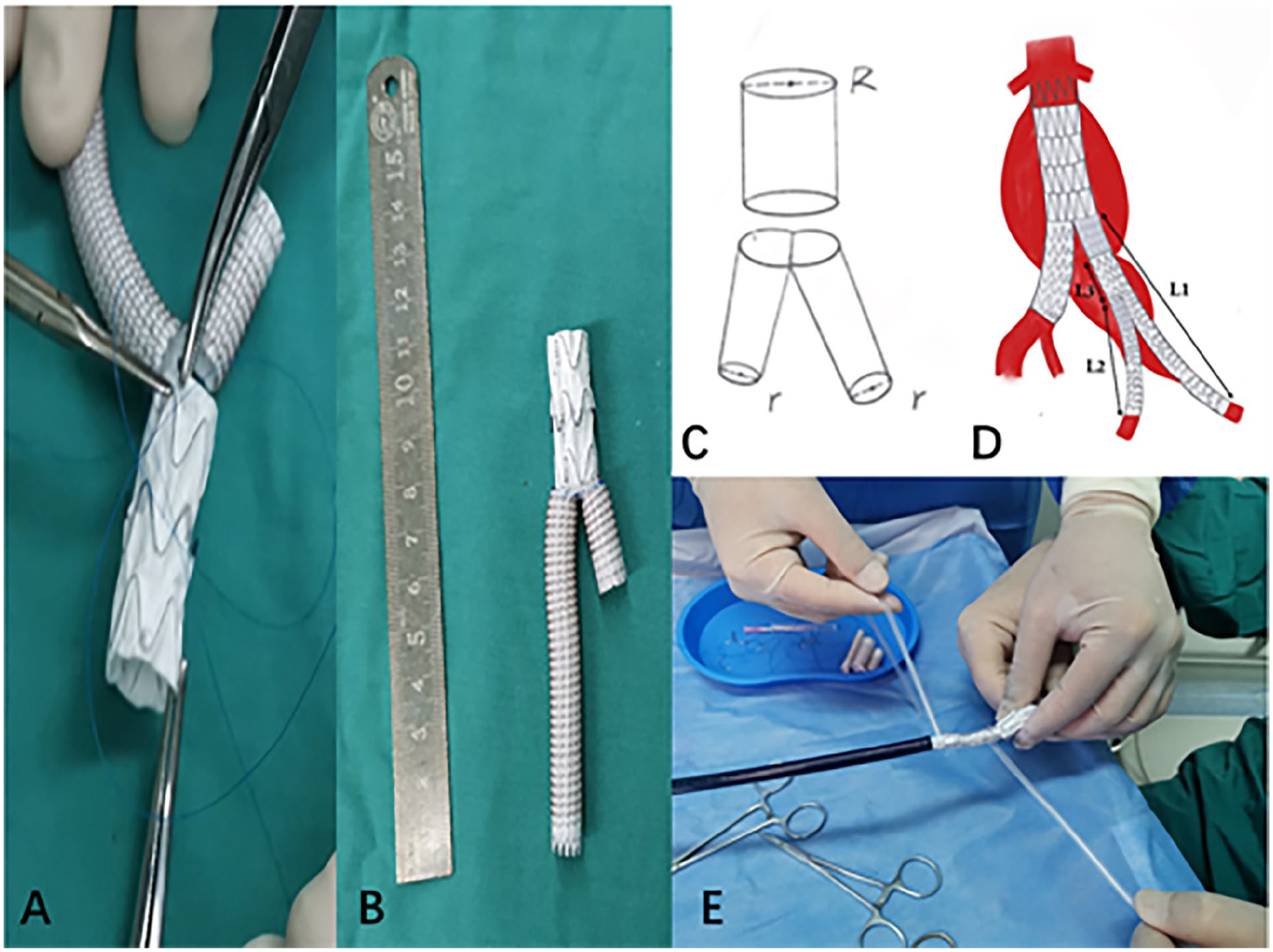
Physician modified Y-type iliac branch device (PMYIBD) production and reinstallment. (A) The VIABAHN (Gore, USA) stent was trimmed vertically, leaving approximately one-third of the graft connection; then, it was folded to fit the trimmed iliac artery stent end to end. (B) Prepared PMYIBD. (C) The iliac branch distal port diameter (R) and the caliber (r) of the Y-type leg. (D) The relationship of the distance from the end of the short leg of the aortic stent to the internal iliac opening (L1), 1 side of the VIABAHN stent (L2) and the iliac branch trimmed length (L3). (E) Using the infusion set strap to reload the PMYIBD into the delivery system.
Procedures and Intraoperative Stent Production
Generally, 3 approaches, 1 in the left upper limb and both lower limbs, are required. Because the branch stent delivery sheath is thick, a surgical incision is usually made in the upper limb to expose the left brachial artery. The bilateral femoral artery approach can be applied with percutaneous access with closure device or surgical incisions as an approach to the abdominal aorta.
During the operation, if 1 of the IIAs needed to be sacrificed, IIA embolization was performed first. First, the abdominal aortic stent body was fed from the femoral artery contralateral to the IA that needed to be preserved. The main stent body was completely released, and the delivery sheath was withdrawn (the main stent body delivery sheath needed to be temporarily retained at this time). The long leg of the stent was released or the iliac branches were connected to a healthy distal anchoring zone. Then, the guide wire catheter was fed into the short leg of the main stent body from the femoral artery on the side of the IA which needs to be preserved. The labeled catheter was exchanged for an aortic angiography device. According to angiography, the distance from the end of the short leg of the aortic stent to the internal iliac opening (L1) was measured.
According to the measured length, a VIABAHN stent graft with an iliac branch was selected to make the Y-type stent graft on table. The 10-cm-long VIABAHN stent was trimmed vertically, leaving approximately one-third of the covered segment connected (Figure 1A). One side of the stent was approximately 3~4 cm (L2), and the other side was approximately 4~7 cm. The iliac branch was trimmed to an appropriate length (L3). Then, the distal end of the iliac branch was anastomosed with the row end of the folded VIABAHN stent to make a PMYIBD. We used Johnson 5–0 Prolene suture for the anastomosis between the iliac branch and the VIABAHN stent graft. Make sure that the length L1>L2+L3 (Figure 1D). The short leg of the stent was confirmed to be located at the proximal end of the opening of the IIA, generally about 1~2 cm. The iliac branch mark was trimmed so that the proximal iliac branch had a visible mark in the direction of the short leg or the original stiffener of the iliac branch was aligned with the short-leg side (Figure 1). After the IBD was completed, it was reinstalled into the delivery sheath of the original abdominal aortic stent. The infusion set strap was used to reinstall the Y-type stent into the delivery system (Figure 1E). If the iliac branch delivery sheath was thin, the main stent delivery sheath which was large enough could be used to reinstall the manufactured IBD.
The IBD extension wire was fed into the iliac branch of the abdominal aorta stent and gradually released until the shorter branch opened. The long sheath was delivered from the left upper limb approach. The guide wire catheter was sent along the long sheath into the branch of the IIA, and the IIA is selected through the branch. The stent graft was sent to the IIA after confirming that it was located in the IIA. The PMYIBD was then completely released. If the longer branch could not extend to the external iliac artery, a suitable stent graft was implanted from the external iliac artery to the PMYIBD. Finally, release the IIA stent. A suitable balloon was used to expand the joints of each stent to completely repair the lesion. The patients were instructed to use enhanced CT for follow-up at the 3rd and 6th month after operation. Follow-up was kept annually thereafter.
Statistical Analyses
Continuous data are presented as the means ± standard deviation or the median (range); categorical data are shown as the counts (percentage). Statistical analysis was performed using Graph Pad Prism software (version 8.0.1; GraphPad Software, San Diego, CA, USA).
Results
From September 2018 to April 2022, the author used physician-modified iliac branched devices to treat 24 patients, without occlusion and obvious endoleaks in follow-up. The average follow-up time was (27.1±13.5) months. The average operative duration was (224.8±44.1) minutes, of which about 22 minutes were used for stent modification, and 50 to 70 minutes were used for endovascular procedure approximately. The average intraoperative blood loss was (110.4±125.0) ml. No blood transfusions were needed. The average volume of contrast agent used was (108.8±7.4) ml. The average hospital stay was (11.9±4.0) days. The intensive care unit (ICU) monitoring time was 1 day. A total of 24 IIAs were reconstructed, including 15 with Fluency stent grafts (Bard, USA) and 11 with VIABAHN stent grafts (Gore, USA). Among them, 22 patients received 1 stent graft and 2 patients received 2 stent grafts. The average diameter of the IIA was (9.1±1.2) cm. After operations, IIA patency was maintained in all 24 patients without IIA occlusion. Small endoleaks from the PMYIBD were observed in a half of patients during the operation. The endoleaks were type IV endoleak from the connect of the stent grafts, which were related to the pinholes at anastomosis. Moreover, the IBDs were stuck at the opening of the branch stent in 2 cases. The stent graft was delivered to the IIA only after exchanging the guide wire and the balloon for harder replacements, which greatly prolonged the operative duration. Two patients experienced intermittent claudication of the gluteal muscles after surgery, which improved after conservative medical treatment. The claudication occurred on which side the IIA was not reconstructed. Besides, iatrogenic type B aortic dissection occurred postoperatively in 1 case. All patients were asked to have regular follow-up with enhanced CT. Intermittent claudication of gluteal muscle on the side of IIA embolism occurred in 4 patients, which disappeared after half a year in follow-up. No endoleak was reported during the follow-up. No other complication, such as stent displacement or occlusion was found during the postoperative follow-up examination. No patient showed expansion of the aneurysm cavity or obvious symptoms during follow-up. Typical surgical procedures and imaging data are shown in Figures 2 and 3.

Endovascular treatment of abdominal aortic aneurysms (AAAs) involving bilateral common iliac arteries (CIAs). (A) Preoperative computed tomography (CT). (B) Preoperative angiography. (C) VIABAHN stent entering the left internal iliac artery (IIA) through the short leg of the Y-type stent. (D) Postoperative angiography. (E) Postoperative review, with no obstruction of the AAA or left IIA.

Thoracic-abdominal aortic dissection with internal iliac artery (IIA)-preserving full-cavity repair. (A) Dissecting aneurysm at the distal end after thoracic endovascular aneurysm repair (TEVAR). (B) Dissecting aneurysm of the abdominal aorta and iliac artery at the distal end after fenestration of the main thoracic-abdominal segment. (C) Preoperative angiography showing that the breach was in the bilateral iliac arterial opening. (D) Catheter entering the right IIA. (E) Postoperative angiography. (F) Postoperative angiography showing complete disappearance of the dissecting aneurysm and no obstruction of the right IIA.
Discussion
Since Roy K. Greenberg first reported physician-modified IBDs in 2006, 4 full-cavity treatments for iliac artery diseases have been widely performed abroad. At present, a large number of studies have demonstrated that IBDs are safe and effective. Iliac branch devices can preserve blood flow in at least one IIA in bilateral CIA expansion patients during EVAR. Compared with traditional open surgery, this approach has the same surgical effect. What’s more, IBD has the advantages of minimal invasion, short operation time and quick recovery, which is thus favored by clinicians.5,6 Numerous commercial stents have been developed. To date, there are 3 commercially available IBDs on the global market. 7 Related retrospective studies have revealed that the surgical success rate is high (80%–100%) with appropriate anatomical conditions. However, whether the stent is a side-branch IBD (eg, Cook’s ZBIS stent) or a parallel-branch IBD (eg, Gore’s IBE stent), there are strict requirements in terms of the patient’s anatomy, such as stents only being applicable to true iliac artery aneurysms, common iliac arteries (CIAs) > 18 mm in diameter, and CIAs>40 mm in length. 8
The anatomical features of Asian patients are slightly different from those of western patients. Therefore, commercial IBDs on the global market are not suitable for Chinese patient population. Moreover, there are no commercially available IBDs on the Chinese market. The treatment of such patients is greatly restricted. In the past, the only feasible method was embolization in China if patients could not tolerate open surgery. If both IIAs were sealed at the same time, intermittent claudication, sexual dysfunction, colonic ischemia, and perineal skin ulcers were likely to occur. 9
Since there are no commercially available iliac artery stents on the Chinese market, physician-modified IBDs have emerged in China for the treatment of AAAs involving the iliac artery or iliac aneurysms. One or both IIAs should be preserved during the operation to prevent related complications. At present, such physician-modified IBDs are similar in structure to Cook’s commercial stents. With the side-branch type, the iliac artery extension branch is used as the main body. The side of the stent is fenestrated, and the artificial blood vessel or stent graft is sutured at the fenestration. The suture needs to be marked with a developing marker to be led out from the side of the main stent body (Figure 4).

Iliac artery stent structure. (A) Schematic diagram of Cook’s commercial iliac artery stent. 10 (B) Branch stent with directional collaterals made by Greenberg et al. (C) Physician modified Y-type iliac branch device (PMYIBD) made by author.
Compared with embolization of the IIA, preservation of the IIA blood flow and avoidance of ischemic complications are important measures to improve the quality of life of patients. Iliac branch device implantation is a safe and effective method to preserve IIA blood flow, for patients with suitable anatomical conditions. Moreover, the technical success rate and mid-term prognosis of bilateral IBD implantation are similar to those of unilateral IBD implantation. 4 A multicenter study showed that the patency rate, endoleak rate, reintervention rate and other surgical effects and prognosis after IBD use are satisfactory. 11 It is demonstrated that IBD surgery has the advantage of fewer iliac complications than flared limbs, which is recommended for young patients. 12 In terms of preserving the blood flow of the IIA, the IBD and cross-chimney technique have similar surgical success rates and IIA patency rates, which are suitable for surgical patients who require IIA retention. 13 Due to the different anatomical conditions of different patients, the use of IBDs is subject to certain restrictions. Physician-modified IBDs break through the limitations of traditional IBDs, of which the feasibility and efficacy have been clinically verified.14,15
Compared with traditional IBDs, PMYIBDs described in this article possess the following advantages: (1) It is simple and convenient to prepare. It is not necessary for surgeons to make a hole in the side of the iliac artery stent and to make difficult end-to-side anastomosis. Only anastomosis of the end of the iliac artery stent to the folded VIABAHN stent is required. Additionally, developing markers are not needed. (2) The length and thickness of the stent can be varied. Physician modified Y-type iliac branch device of different sizes can be prepared through trimming or suturing iliac branches and VIABAHN stents of different sizes according to the anatomical conditions. (3) The brachial artery approach can be adopted in operations. Because the PMYIBD has a straight angle, the stent connection will not be stuck. The stent graft can easily enter the IIA and be released without the protection of long sheaths. Moreover, the angle of the stent graft is appropriate, and it will not become kinky. (4) Because the Y-type structure takes up little space, it can be applied not only for aneurysms but also for dissecting iliac artery aneurysms with a very small true lumen. The device can be used to repair the distal rupture of aortic dissection and preserve the IIA. (5) PMYIBDs are feasible options for all patients who are not suitable for commercial IBDs, such as iliac artery lumen stenosis, aortic dissection involvement, CIA too short. It is especially suitable when commercial IBDs are not inappropriate.
Generally, PMYIBDs have several major limitations: (1) PMYIBDs need long operation time and complicated device-modifying technique. Only experienced surgeons are able to perform the operation. (2) IBDs can only enter through an anterograde approach via brachial artery, which will lead to increasing surgical injury. (3) Because mental skeletons of VIABAHN are located on the outside, it is difficult to reload the PMYIBD into the delivery sheath. Larger delivery sheathes are always needed. (4) PMYIBD requires numerous stent grafts, which causes huge cost and high potential of endoleaks. Patients with poor financial situation barely accept the operation.
There are several reported complications after IBDs implantation including endoleaks, buttock claudication, and IBD-associated reintervention. 2 We observed complications such as intermittent claudication, type IV endoleak, and iatrogenic type B aortic dissection in minority of our patients postoperatively. Intermittent claudication occurred in patients who preserved 1 side of IIA. All type IV endoleaks disappeared at follow-up.
Conclusion
In short, how to preserve the IIA during EVAR has always been a hot topic in aortic surgery. The use of PMYIBD provides a simple and effective method for the total endovascular repair of aortic diseases involving the IIA. The surgical method has a wide range of indications and several advantages such as minimal trauma, low mortality, low complication rates, and perfect short- and medium-term effects. Physician modified Y-type iliac branch devices are effective for aortic diseases involving IIA before suitable commercial stents are available in markets. Further researches are required to investigate the long-term efficacy.
Footnotes
Declaration of Conflicting Interests
The author(s) declared no potential conflicts of interest with respect to the research, authorship, and/or publication of this article.
Funding
The author(s) disclosed receipt of the following financial support for the research, authorship, and/or publication of this article: This work was supported by the Outstanding Youth Project of Nanjing (JQX17003), Jiangsu Provincial Medical Youth Talent (QXRC201621), and Social Development Program of Jiangsu Province (BE2019604).
Informed Consent
Written informed consent was obtained from all participants for their clinical records to be used in this study.
