
Abstract
Background:
Varicose vein disease is characterized by dilated and tortuous lower extremity veins and affects approximately 37.25% of Indian population. Endovenous ablation is the first-line treatment approved for superficial venous reflux. A new technology recently available is a nontumescent and nonthermal method (VenaSeal) wherein aliquots of cyanoacrylate glue are delivered throughout the length of vein resulting in venous obliteration. The aim of this study was to evaluate the clinical outcomes of endovenous glue ablation for treatment of superficial venous reflux in patients with symptomatic varicose vein disease.
Materials and Methods:
This was a single-center retrospective observational study of 175 patients with 6, 12, and 18 months follow-up. The treatment success was measured in terms of vein occlusion rate on Duplex scan and clinical outcomes in terms of improvement in revised Venous Clinical Severity Score (VCSS).
Results:
A total of 176 limbs were treated over a study period of 1 year 5 months with 6-, 12-, and 18-month follow-up. The most common CEAP classification was C2 seen in 83/176 (47.2%) limbs. Incompetent great saphenous vein (GSV) was ablated in 170, incompetent short saphenous vein (SSV) in 5 and accessory GSV in 1 patient. The preprocedure GSV diameter (supine position) was 5.7±1.8 mm and the length of ablated vein was 32.9±7 cm. The technical success was 100% with no device related complications. The vein closure rate was 100% at 6 months and 99.4% at 12 and 18 months. The preprocedural VCSS score was 5.8±2.2. There was statistically significant improvement in VCSS score at 6 months (2.1±1.2; p<0.001) with sustained improvement at 12 months (1.7±0.99; p<0.001) and 18 months (1.4±0.67; p<0.001). The most common side effect was thrombophlebitis seen in 13/176 (7.4%) limbs. There was no serious adverse event and paraesthesia was not seen.
Conclusion:
This study has demonstrated high efficacy and safety of endovenous glue ablation in terms of truncal closure, technical and clinical success in symptomatic patients with chronic venous insufficiency in Indian population. However, complications like thrombophlebitis and hypersensitivity reactions are reported with cyanoacrylate ablation.
Clinical Impact
It will authenticate the safety and efficacy of glue based ablation for the treatment of lower limb varicose veins. Cyanoacrolate glue based non thermal Ablation of lower limb Varicose Veins has better efficacy, fewer side effects and practically painless compared to thermal Ablation.
Introduction
Varicose vein disease is characterized by dilated and tortuous lower extremity veins and affects approximately 37.25% of Indian population. 1 Surgical stripping was once the standard treatment for superficial vein incompetence. Currently, endovenous thermal or nonthermal ablation is the first-line treatment approved for symptomatic superficial venous reflux.2,3 The thermal methods require perivenous subfascial tumescent anesthesia with multiple needle punctures along the length of the vein and there is a potential risk of thermal injury to the saphenous nerve. 4
A new technology recently available is a nontumescent and nonthermal method (VenaSeal) wherein aliquots of cyanoacrylate glue are delivered throughout the length of vein resulting in venous obliteration. 5 The mechanism involves rapid solidification of glue via polymerization which binds to apposed intimal walls leading to inflammatory vein wall reaction and subsequently causing fibrotic degradation. It does not require the administration of tumescent anesthesia with no risk of heat related nerve injury. It chemically binds the venous walls to each other, thus obviating the need for postintervention compression stockings. 6
There is enough evidence in literature to suggest the safety and efficacy of cyanoacrylate closure for treatment of incompetent truncal veins.6 –8 A randomized controlled trial has demonstrated noninferiority of cyanoacrylate closure as compared with radiofrequency ablation in improvement of clinical symptoms with sustained benefit till 5 years.9,10 However, only few studies regarding safety and efficacy of this technique in Indian population have been published.11,12 Hence, the aim of this study was to evaluate the safety and outcomes of endovenous glue ablation for treatment of superficial venous reflux p.
Materials and Methods
Study Design and Population
This was a single center retrospective observational study performed over a period of 1 year 5 months from July 2022 through November 2024. The study was approved by the Institutional Ethics Committee and waiver for the need for written informed consent was provided. The indications of glue ablation include symptomatic primary truncal incompetence, Clinical, Etiology, Assessment, and Pathophysiology (CEAP) classification of C2 to C6 and demonstration of truncal/axial reflux in great saphenous vein (GSV), short saphenous vein (SSV) or accessory saphenous vein greater than 0.5 seconds on Doppler ultrasound.2,3 There is no minimum diameter required to have pathologic reflux in the veins. 2 Patients with symptomatic peripheral arterial disease, previous deep vein thrombosis (DVT), active superficial thrombophlebitis, extremely tortuous vein course that limits catheter placement, and with known hypersensitivity to cyanoacrylate glue were excluded. We also excluded patients who did not attend the follow-up visits.
Data Collection
The medical records, ultrasound findings, and procedural database of all consecutive patients who underwent endovenous glue ablation for varicose vein disease were reviewed. A standardized form was used to record clinical, ultrasound and procedural details including CEAP classification and revised Venous Clinical Severity Score (VCSS). The follow-up data were collected at baseline, 6, 12, and 18 months. At each visit, patients underwent physical examination, assessment of CEAP classification, and VCSS score, and Duplex ultrasound was performed to look for vein occlusion.
Preprocedural Evaluation
All patients underwent a comprehensive preprocedural assessment. Clinical assessment included meticulous history taking, physical examination and recording of CEAP classification and revised VCSS score. VCSS score was evaluated at the baseline, 3, 6, 12, and 18 months. Patient’s vein mapping was done using Duplex imaging. The parameters that were assessed include junction incompetence, axial reflux, diameter of truncal vein to be ablated, course of the vein, tortuous segments, depth of the saphenous vein from skin, and presence of varicosities and perforators (Figure 1).
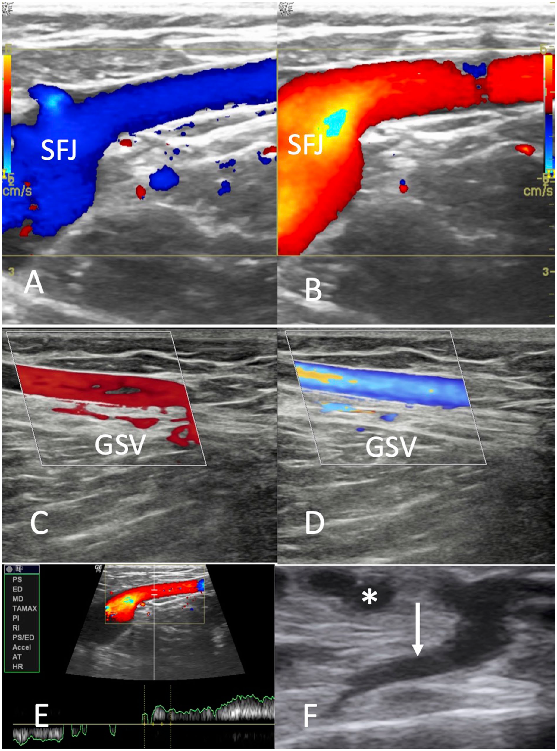
Duplex ultrasound images demonstrating saphenofemoral junction incompetence (A, B), axial reflux in the great saphenous vein (C, D), duration of axial reflux greater than 0.5 seconds (E), and presence of varicosities (depicted by * in F) and perforator (depicted by white arrow in F).
Procedure
The patients were treated in daycare setting in accordance with rigorous standardized protocol. In this technique, aliquots of glue are injected throughout the length of vein. The procedure was performed in supine position under local anesthesia and percutaneous access of saphenous was obtained under ultrasound guidance at the most distal point of reflux (below knee in most of the cases). In some cases, 2 simultaneous access were obtained above and below knee in cases of excessive tortuosity and difficult navigation of the intervening venous segment. A 7-French introducer was then inserted over guide wire. The dilator was replaced with the delivery catheter whose tip was 5 cm from the saphenofemoral junction and 3 cm from the saphenopopliteal junction. The dispenser gun releases 0.1 mL of glue with each trigger. The first administration releases 0.1 mL N-butyl cyanoacrylate (NBCA) with another aliquot of 0.1 mL at distance of 1 cm followed by pullback of 3 cm and compression for 3 minutes (Figure 2). Thereafter, aliquots of 0.1 mL are delivered every 3 cm along the length of vein followed by compression for 30 seconds. The last aliquot is delivered 3 cm from the catheter access site. Then the catheter is removed with duplex scanning to confirm venous occlusion and to rule out deep venous thrombosis (DVT). After the procedure, compression dressing was applied and the patients were advised to walk for 30 minutes. Patients were discharged on the day of procedure and can resume normal activities. Duplex scanning was done after 24 hours to rule out DVT. Adjunctive treatment such as sclerotherapy if needed for the large diameter tributaries was performed concomitantly. 3 The patients were prescribed grade 2 compression stockings for 1 month and compliance was ensured. A 6-, 12-, and 18-month follow-up was done for all patients. Patients were evaluated using clinical and duplex examination to assess for improvement in rVCSS and vein occlusion, respectively.

Photograph depicting various components of the VenaSeal system (A). Ultrasound image depicting 7F introducer sheath in the superficial vein (B), 5F delivery catheter with glue aliquot (C), and postprocedural venous occlusion (D).
Definitions and Outcome Measures
Clinical, Etiology, Anatomy, and Pathophysiology (CEAP) classification is widely adopted method to evaluate clinical signs and severity of varicose vein disease. The stage ranges from C0 to C6; C0: no varicose veins; C1: reticular veins or telangiectasia; C2: varicose veins; C3: venous edema; C4a: pigmentation; C4b: lipodermatosclerosis; C4c: corona phlebectasia; C5: healed venous ulcer; C6: active venous ulcer (Figure 3). The primary objective of this study was to determine treatment and clinical success. The treatment success will be determined based on venous occlusion/obliteration rate on duplex imaging. Closure was defined as complete, luminal occlusion along the entire length of vein. The clinical success was assessed by improvement in revised Venous Clinical Severity Score (rVCSS). Revised VCSS includes 10 hallmarks of venous disease and the severity is scored from 0 to 3. The parameters of this score are pain, varicose veins, venous edema, skin pigmentation, induration, inflammation, active ulcers (number, size, and duration), and compression. The secondary objective was to assess postprocedural complications. Major adverse events constitute those complications requiring admission or surgery. Minor events require only medical treatment and include pain, bruising, puncture site infection, or thrombophlebitis. The diameter of the truncal vein was measured at 3 levels: proximal, mid, and distal and mean of the 3 measurements was taken.
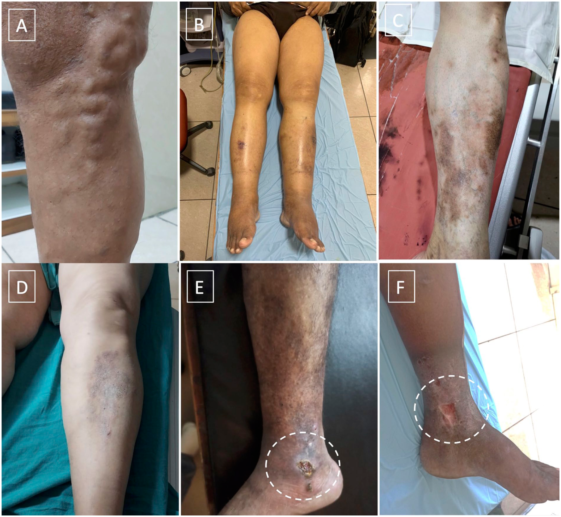
Clinical photographs depicting CEAP classification; (A) C2: varicose vein, (B) C3: edema, (C) C4a: pigmentation, (D) C4b: lipodermatosclerosis, (E) C5: healed venous ulcer; and (F) C6: active venous ulcer.
Statistical Analysis
SPSS software was used to analyze the data. The categorical variables were reported as counts and percentages, while the continuous variables were reported as mean and SD for variables with normal distribution. The difference in the distribution of categorical variables was analyzed using the Chi-square test and Students t test was used for continuous variables with normal distribution. A p-value less than 0.05 was considered statistically significant.
Results
A total of 175 patients (176 limbs) were evaluated over study period of 1 year 5 months with 6-, 12-, and 18-month follow-up.
Patient Demographics
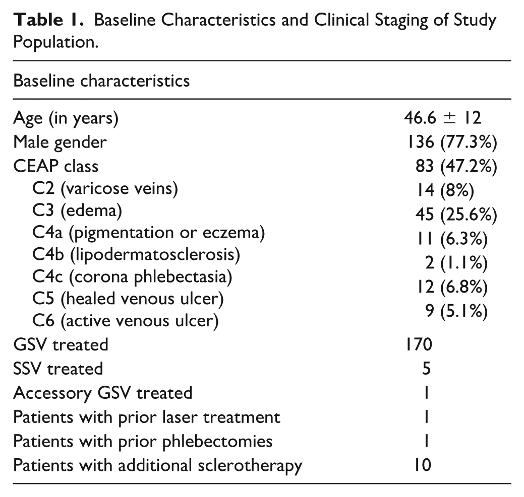
The mean age of the study group was 46.6 ± 12 years. Men constituted 77.3% of the study group. Previous phlebectomy was performed in 1 patient and 1 patient had previously undergone laser ablation. The most common CEAP classification was C2 seen in 83/176 (47.2%) limbs. The baseline characteristics and preoperative clinical staging of the study population are tabulated in Table 1.
Baseline Characteristics and Clinical Staging of Study Population.
Procedural Characteristics
Of the total 176 limbs, incompetent GSV were ablated in 170, incompetent SSV was ablated in 5 and incompetent accessory GSV in 1 patient. The preprocedure GSV diameter (supine position) was 5.7±1.8 mm and the length of ablated vein was 32.9±7 cm. The maximum diameter of GSV was 13 mm and minimum was 3 mm. Adjunctive treatment of target GSV in form of additional sclerotherapy was performed in 10 patients.
Anatomical and Clinical Success
The technical success was 100% with no device related complications. Duplex imaging done postprocedure and after 24 hours revealed complete occlusion of treated venous segments in all patients (100%). Closure rate was 100% at 6 months and 99.4% at 12 months and 18 months on follow-up Duplex scans (Figure 4). The preprocedural VCSS score was 5.8±2.2. There was statistically significant improvement in VCSS score at 6 months (2.1±1.2; p<0.001) with sustained improvement at 12 months (1.7±0.99; p<0.001) and 18 months (1.4±0.67; p<0.001) (Figure 5).
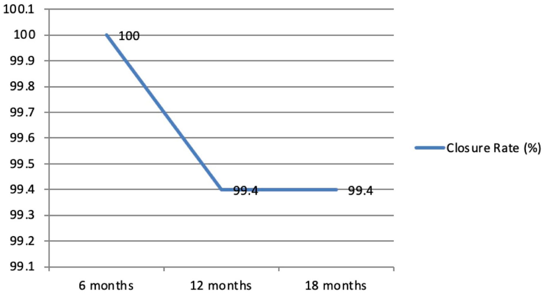
Graphical representation showing venous closure (in percentage) at 6-, 12-, and 18-month follow-up.
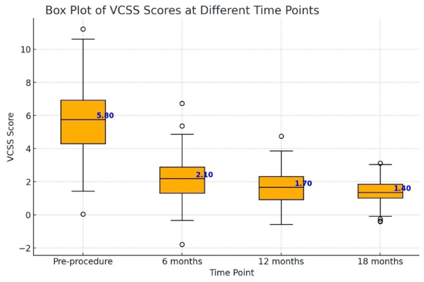
Box plot demonstrating Venous Clinical Severity Score (VCSS) at baseline with significant and sustained improvement at 6, 12, and 18 months.
Adverse Events
There was no major or life threatening adverse event. No patient experienced access site infection or significant hematoma formation. The most common side effect was thrombophlebitis seen in 13/176 (7.4%) limbs (Figure 6). In these patients, the saphenous vein was located in the suprafascial plane close to the skin with a depth less than 1 cm from the skin surface. The symptoms were mild and were managed conservatively with oral nonsteroidal anti-inflammatory drugs (NSAIDs). Hypersensitivity reaction with multiple superficial wounds along treated GSV was seen in 2/176 (1.1%) limbs 1 week after the procedure. Manual compression of these areas caused skin eruption with extrusion of solidified glue fragments (Figure 6). The patients were prescribed antibiotics and corticosteroids with resolution of symptoms and healing of wounds. Patch test confirmed hypersensitivity to glue. Five patients complained of dragging sensation during walking at 6-month follow-up due to fibrosed cord like GSV postembolization with no complaint at 12-month follow-up. One patient required reintervention for significant progression of below the knee GSV reflux. Asymptomatic partial DVT of the popliteal vein was seen in 1 patient. However, no patient demonstrated any signs or symptoms of pulmonary embolism or paraesthesia.
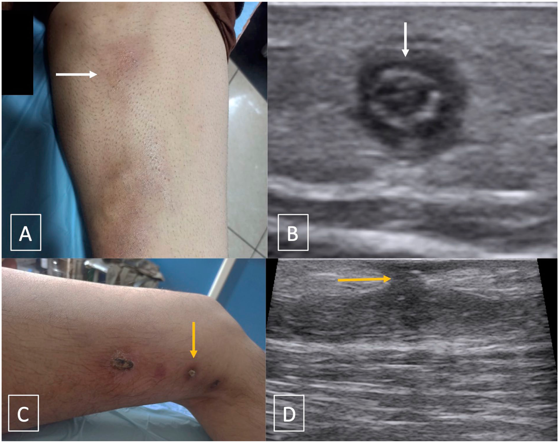
Clinical photograph and ultrasound image depicting postprocedural thrombophlebitis (shown by white arrow in A, B) and presence of hypersensitivity reaction with multiple superficial wounds along treated GSV in another patient (yellow arrow in C, D).
Discussion
This study is a single center retrospective analysis of the use of endovenous cyanoacrylate ablation for treatment of incompetent saphenous veins in Indian cohort. Our results have shown high vein closure rate of 99.4% at 12 months with sustained clinical outcomes through 18 months in Indian population. There was also significant improvement in venous disease-specific quality-of-life score (VCSS). The findings are concordant with the 3-year results of first-time use of cyanoacrylate glue for truncal reflux, European eScope registry and medium-term results of US VeClose randomized closed trial (RCT).5,6,9 A randomized controlled trial comparing surgical stripping versus glue embolization has demonstrated 100% vein occlusion in both the groups with less pain and bruising in the cyanoacrylate group. 13 Another randomized controlled trial comparing endovenous glue ablation with radiofrequency in 242 patients has demonstrated noninferiority of cyanoacrylate adhesive with less chances of bruising in the cyanoacrylate group.9,10 The 5-year results of this trial have shown occlusion rates of 91.4% with cyanoacrylate and 85.2% with radiofrequency ablation. There was sustained improvement in quality-of-life measures throughout the 5-year follow-up. 10 Similarly, a systematic review has demonstrated significantly higher 2-year closure rate with glue ablation (93.7%) as compared with radiofrequency (90.9%) and laser ablation (91.5%). 10 In addition, glue treated patients experienced the least complications. 14 Kolluri et al performed a network meta-analysis to compare VenaSeal with other endovenous ablative therapies for varicose vein disease and included 20 RCTs and 4570 patients. VenaSeal had the highest probability of being ranked first in terms of anatomic success at 6 months and reduction of postoperative pain. The occurrence of adverse events was also the least. 15 In this study, preprocedural VCSS score was 5.8±2.26 similar to study by Almeida et al 5 (6.1±2.7). The 6-month VCSS score was (2.1±1.2; p<0.001) in this study which ranged from 1 to 1.5 in the previous published studies.5,6,9,10
The results of our study are also consistent with 1 previous Indian study published by Shah et al.11,12 Shah et al studied 200 patients over a period of 3 years and the vein occlusion rates were 97% and 95% at 12 and 24 months, respectively. The reintervention rate was very low in our patients consistent with the study by Tang et al. 7 The reintervention in 1 patient was due to development of recent below the knee GSV reflux which was not present initially in the Duplex scan. Recanalization was seen in only 1 patient. This can be attributed to the fact that adjunctive procedures like sclerotherapy were performed concomitantly for large-diameter tributaries similar to study by Shah et al 11 and Tang et al. 7 In a study by Proebstle et al, 6 all 5 cases of recanalization were attributed to untreated tributaries. There is variability in literature regarding the use of adjunctive procedures along with cyanoacrylate closure. Only the truncal reflux was treated in feasibility study, eSCOPE, and VeClose study.5,6,9
Endo venous glue ablation has many advantages over thermal methods of ablation. In thermal techniques, tumescent anesthesia is administered to protect perivenous tissues from heat. This requires multiple subcutaneous injections which can potentially damage saphenous or sural nerves especially below knee. There can be injury to the vein wall, the fascial compartment and reticular veins causing pain and bruising. 16 However, cyanoacrylate is a nonthermal and nontumescent technique with no complications as paresthesia or bruising. In a meta-analysis by Amshar et al 17 cyanoacrylate group had lower skin pigmentation and nerve damage as compared with laser ablation. There was no case of paresthesia in this study.
The most common side effect was thrombophlebitis seen in 13 of the 176 (7.4%) limbs. Similarly, Shah et al 12 reported phlebitis in 6% patients. Type IV hypersensitivity reaction with multiple superficial wounds and extrusion of glue fragments was seen in 2 of the 176 (1.1%) limbs 1 week after the procedure. This is a T-cell mediated immune response that usually occurs 1 to 23 days after the procedure in response to foreign allergen. 18 Similar complication was also described by Louden et al. 19 It is usually difficult to control as the allergen is still present inside the vein and sometimes may necessitate surgical vein excision and removal of glue. Kim et al 20 reported hypersensitivity reaction in 3.7% patients after glue ablation.
Five patients complained of cord like sensation similar to study by Tang et al. 7 This can be attributed to the fact that endovenous glue ablation does not lead to thrombotic reaction and the venous walls are immediately coapted to the glue adhesive after external compression. This leads to delay in fibrosis with the patient feeling the “inflamed” GSV longer than as compared with thermal ablation. This complain was mainly seen when the truncal vein was superficial located just below the skin in thin individuals. The most dreaded complication of glue ablation is DVT with risk of pulmonary embolism. Deep venous thrombosis was seen in 1 patient with no case of pulmonary embolism. In a systematic literature review, the rate of DVT post cyanoacrylate glue embolization was 0.5% slightly lower than radiofrequency (0.9%) and laser ablation (0.6%). This complication can be prevented by manual compression under ultrasound guidance across saphenofemoral and saphenopopliteal junctions by ultrasound probe.
Limitations
The current study has few limitations. First, our study was a single center retrospective study with a small sample size. The recruitment at a single institution may have resulted in selection bias. Second, we did not compare endovenous glue ablation with other treatment methods. Thirdly, standardized assessment tool was not used to measure intensity of pain or bruising. Finally, as recanalization was only observed in 1 patient, we could not study the predictors of recanalization like increased GSV diameter. However, in future, a randomized control trial with larger sample size and longer follow-up is needed in Indian population to compare the efficacy and clinical outcomes of glue with thermal ablation techniques.
Conclusion
This study has demonstrated high efficacy and safety of endovenous glue ablation in terms of truncal closure, technical and clinical success in symptomatic patients with chronic venous insufficiency in Indian population. However, complications like thrombophlebitis and hypersensitivity reactions are reported with cyanoacrylate ablation.
Footnotes
Ethical Considerations
Obtained.
Funding
The authors received no financial support for the research, authorship, and/or publication of this article.
Declaration of Conflicting Interests
The authors declared no potential conflicts of interest with respect to the research, authorship, and/or publication of this article.