





Abstract
We present an unusual case of a 23-year-old man who had symptomatic lower extremity varicosities that have been present since birth. He was complaining of pain and swelling of several years duration. Evaluation revealed Klippel–Trenaunay syndrome with associated compression of the left common iliac vein by the overriding left common iliac artery (May–Thurner syndrome). The patient was treated with left common iliac vein stenting followed by high ligation and inversion stripping of his grossly incompetent left small saphenous vein, stripping of his aberrant left lateral embryonic veins, and also by multiple stab phlebectomies, with excellent early result.
Introduction
Klippel–Trenaunay syndrome (KTS) is a rare malformation characterized by a constellation of signs and symptoms, most notably capillary malformation (port-wine stain), bony or soft tissue hypertrophy, and varicose veins or venous malformations. 1 Lymphatic malformation with lymphedema is frequent and many patients have abnormal development of deep veins, including hypoplasia, agenesis, or congenital aneurysms of the deep venous system. Although many different etiologies have been proposed, the persistence of an embryonic venous system and mutations in angiogenic factor VG5Q are among the most widely accepted explanations for the vascular component of the phenomenon. The mutation predisposes patients to unregulated endothelial cell secretion, proliferation, and survival giving rise to the KTS’s characteristic clinical findings. 2 Thrombophlebitis, deep vein thrombosis, and pulmonary embolism are somewhat more frequent in patients with KTS than in the normal population. 3
May–Thurner syndrome (MTS) refers to compression of the left common iliac vein by the overlying right common iliac artery. This was first described in 1957 by May and Thurner 4 who observed the phenomenon in autopsies of 430 patients and later in 1965 by Cockett and Thomas. 5 May and Thurner suggested that the predominance of left-sided deep vein thrombosis and symptoms were the result of the left common iliac vein outflow obstruction.
An association of MTS with KTS is rare and combined treatment of this condition, according to our knowledge, has not been previously reported. This article illustrates an unusual case of MTS in a patient with KTS, his clinical workup and subsequent medical, endovascular, and open surgical treatment.
Case Report
A 23-year-old male patient with KTS presented at Mayo Clinic, Rochester in April 2012 with complaints of left leg edema, pain with exercise, heaviness, and painful, unsightly varicose veins. He also had a recent history of frequent rectal bleeding. His varicosities were noted at birth; however, medical treatment and clinical course were not documented until 1999, after which he had treatment with compression stockings, endovascular laser ablation of his varicose veins, and multiple ambulatory phlebectomies. Gluteal and perforator vein coil embolization was performed at an outside facility but it was complicated with pulmonary embolism of the coils. Family history was negative for any vascular malformations.
Physical examination revealed a well-developed young man with normal cardiac, pulmonary, and gastrointestinal systems. He had a slightly enlarged left lower extremity (LLE) with large lateral varicosities and port-wine stains. He also had prominent changes over the plantar surface of his toes (Figure 1A and B). Preoperative plethysmography revealed a patent deep venous system with hemodynamically insignificant venous incompetence in the right common femoral and left popliteal veins. Ultrasonography confirmed a mildly incompetent left common femoral vein and a grossly incompetent left popliteal vein. The left great saphenous vein was incompetent below the knee, and the left small saphenous vein was severely incompetent throughout with several varicosities. Contrast ascending venography (Figure 2A-D) showed large dilated lateral veins vein draining directly into the pelvic vessels, extending from the left lateral thigh and calf. This dilated lateral vein was draining into 4 incompetent perforating veins at the level of the gluteus. Magnetic resonance venography of the pelvis and bilateral lower extremities showed extensive superficial varicosities consistent with KTS. The deep venous system was intact and there was no arteriovenous shunting. High-grade stenosis of the left common iliac vein from right iliac artery compression revealed previously undiagnosed MTS (Figure 3). The arterial anatomy was otherwise normal bilaterally without evidence of disease.
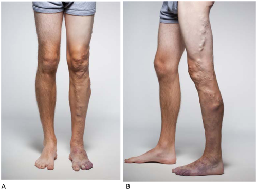
(A, B) Photographs of a 23-year-old man with left lower limb Klippel–Trenaunay syndrome affecting the left lower limb and pelvis. Note large lateral persistent embryonic veins and capillary malformations (port-wine stains).
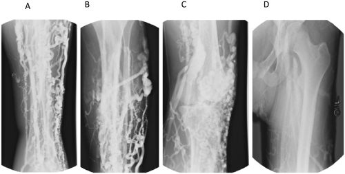
(A-D) Contrast ascending venogram of the left leg showed extensive varicosities, particularly over the lateral aspect, from the mid-thigh to foot. Note the patent but dilated popliteal vein.
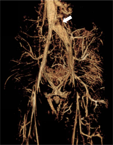
Magnetic resonance venogram showed marked narrowing of the left common iliac vein close to the inferior vena cava, compressed by the overriding right common iliac artery. Note the large amount of venous collaterals.
Preoperative laboratory values were within normal limits, except for a clinically insignificant microcytic anemia (hemoglobin 10.6 g/dL; mean corpuscular volume 74.1 fL) attributable to his hemorrhoidal bleeding, and he was started on oral ferrous sulfate (325 mg) for 2 months.
Since the patient’s symptoms in part were compatible with MTS, we recommended endovascular treatment of the high-grade left common iliac vein stenosis with stenting prior to further intervention for his KTS. An intraoperative venogram, performed with percutaneous access of the left common femoral vein, confirmed the high-grade left common iliac vein stenosis with a widely patent inferior vena cava and right iliac system (Figure 4A). There was a significant amount of collateral venous circulation. Intravascular ultrasound supported these findings. Two self-expanding 16 mm Wallstents (6 cm × 16 mm and 4 cm × 16 mm) were deployed at the area of stenosis and the stents were dilated with a 4 cm × 16 mm balloon under 10 atmospheres of pressure. A completion venogram and intravascular ultrasound showed a patent lumen without residual stenosis (Figure 4B and C). The patient did well from the procedure despite a small amount of bleeding at the groin puncture site and a brief episode of transient orthostatic hypotension that spontaneously resolved. No transfusion was required and no groin hematoma was present. He was discharged from the hospital after 48-hour observation on 162 mg of aspirin for antiplatelet therapy.
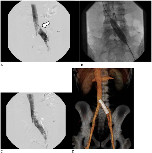
(A) Pre-stent venogram confirms high grade left common iliac vein stenosis. (B) Balloon angioplasty after placement of two 16-mm Wallstents. (C) Completion venogram after stenting. (D) Computed tomography venogram one week after stenting confirms a widely patent stent in the left common iliac vein.
The patient returned to clinic 1 week later for follow-up ultrasound and computed tomography venogram, which showed a widely patent stent in the left common iliac vein (Figure 4D) without hematoma or thrombotic complications. The next day he underwent treatment of his lateral varicosities under general anesthesia. An Esmarch bandage was used to exsanguinate the extremity and a tourniquet was placed on the proximal thigh and inflated to 300 mmHg to minimize blood loss. The left small saphenous vein was cannulated with a stripper via a small incision on the lateral aspect of the foot. The stripper advanced to the knee and the vessel removed using the inversion technique without complications, after high ligation of the proximal stump of the small saphenous vein (Figure 5). A portion of the lateral embryonic vein in the thigh was similarly removed. Additional varicose veins and venous malformations on the extremity were removed using stab phlebectomy. Total tourniquet time was 72 minutes and the patient recovered overnight in the hospital without complications. He was discharged the following day with 75 mg of Plavix and oral pain medications.
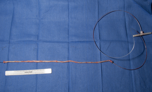
Small saphenous vein removed after high ligation and inversion stripping.
Follow-up at 5 months reveals significant symptomatic improvement with very little to no discomfort. The patient continues to wear compression stockings on a regular basis and has not had any recurrent varicosities. He is able to work and exercise freely without limitations.
Discussion
Klippel–Trenaunay syndrome is a mixed mesenchymal malformation that affects the development of blood vessel, soft tissue, and bones. The classic clinical triad includes varicose veins, bone and soft tissue hypertrophy, and capillary malformations. In the Mayo Clinic registry of 252 patients, Jacob et al 3 in 1998 observed that the most prevalent features were capillary malformations (98%) followed by venous malformations (72%) and limb hypertrophy (67%). Although they predominantly involve one lower extremity (69%), no significant laterality has been shown (right 36%; left 33%). 3 Patients typically present with pain and heaviness of the limb (37%) but may also have lymphedema (10%), cellulitis (13%), lymphorrhea from lymph vesicles, superficial phlebitis, deep venous thrombosis (4%), pulmonary embolism (PE, 4%), and hematuria or rectal bleeding 3 . External compression helps to diminish symptoms of venous insufficiency and lymphedema, and bouts of recurrent cellulitis can be managed with prophylactic antibiotics in selected cases. Cosmetic surgery generally yields limited results. Multiple laser therapy treatments are frequently required to decrease discoloration of capillary malformations. Direct injection of absolute alcohol into cavernous venous malformations is quite effective but it may frequently lead to skin breakdown and scarring. Removal or ablation, if possible with radiofrequency or laser, of symptomatic varicose veins in patients with a functional deep venous system improves cosmesis and significantly improves symptoms, although recurrence is common. Epiphysiodesis is effective in children with a leg length discrepancy greater than 2.5 cm; timing is crucial to preserve adult height at skeletal maturity.
The risk of deep vein thromboses and pulmonary embolisms is increased due to aneurysmal dilations, phlebectasias, and likely thrombophilia in many patients. Venous duplex ultrasonography is an excellent test to assess patency of the venous system, anatomic abnormalities, as well as deep venous thrombosis and valvular incompetence in these patients. However, a dedicated vascular laboratory with expert sonographers is necessary due to the complexity and range of possible findings. Magnetic resonance venography allows for better visualization of the deep venous system and is a useful prerequisite study to guide duplex ultrasonography. 6 Its accuracy, consistency, and availability make magnetic resonance venography an increasingly popular diagnostic tool for evaluation of venous anatomy.
May–Thurner syndrome is caused by stenosis of the left comon iliac vein due to compression by the overriding right common iliac artery. In our patient this was coupled with the increased venous outflow from his LLE because of the persistence of large embryonic veins. The large varicosities draining into the pelvic vessels contributed to pelvic congestion and dilation of his left common iliac vein and collecting system due to the proximal venous stenosis. MTS is frequently asymptomatic for decades but it can also present with unilateral LLE pain, swelling, and color changes; intra-abdominal and pelvic varices have also been reported. 7 Pulmonary embolism secondary to lower extremity deep venous thrombosis remains a feared presentation of MTS but is relatively rare. MTS-related deep venous thromboses were classically thought to make up only 2% to 3% of all deep venous thromboses, but this number has greatly increased in recent years because of high index of suspicion and improvement in imaging studies.8,9 Our patient’s LLE symptoms likely arouse from a combination of KTS- and MTS-related factors. The true incidence of this phenomenon is unknown, as according to our knowledge this is the first documented case report of these syndromes treated concomitantly
May–Thurner syndrome can cause acute left iliofemoral deep venous thrombosis; in these patients, catheter-directed thrombolytic therapy is followed by angioplasty and stenting. The minimally invasive nature of endovascular techniques and high rates of technical success has made angioplasty and stenting the treatment of choice also for uncomplicated chronic MTS.8,10,11 Accordingly, we opted to stent our patient’s left common iliac vein to increase venous outflow and alleviate congestion in the venous circulation of the leg. In a retrospective study of 189 consecutive patients, Kurklinsky et al 12 demonstrated 100% technical success using venous self-expanding stents in the setting of chronic iliac and femoral vein thrombosis. Primary patency at 30 days, 1 year, and 3 years were 99%, 81%, and 71%, respectively. 12 Complications are rare but can include acute iliac vein rethrombosis, access site hematomas, intimal hyperplasia, and bleeding. In one study, 85% of patients reported complete or partial symptomatic improvement compared to before the procedure. 8
By combining venous stenting with high ligation and stripping of the saphenous vein, Meng et al 13 reported on a patency rate of 91.7% in 296 patients over a median follow-up of 46 months. Symptomatic improvement was similarly very high in this report that included patients with varicose veins, lower extremity edema, and venous ulcers. 13 The age range was from 15 to 63 years and younger patients required reintervention for restenosis more frequently. Stenting in pediatric patients or young adults, however, is still quite rare and long-term results are not well known. Agnoletti et al 14 reported on 32 patients who underwent 39 interventional catheterizations aimed to dilate or recanalize occluded iliofemoral veins, inferior or superior venae cavae. Median age of the patients was 5 years (range 0.1-18.0 years). Fifty-two stents were implanted in 29 patients (32 vessels). There were no major complications but there were 2 procedural complications (5.1%): one acute stent occlusion and one local dissection. At a median follow-up of 2.5 years (range 1-10 years) there were 6 complications of stenting (11.5%): 2 fractures, 2 occlusions, and 2 restenoses.
The management of KTS is largely conservative with compression therapy making up the mainstay of treatment. 1 If conservative methods fail, less invasive techniques, including ablation and sclerotherapy can be attempted but are generally less effective compared with traditional surgical procedures. Noel et al 15 reported success by stripping the superficial embryonic veins and varicosities in a series of 20 KTS patients. All patients initially improved with treatment and mean follow-up was 63.6 months. At last follow-up, 90% of patients were pain free without swelling with compression garments; the remaining 10% had persistence of presenting symptoms. Some varicosities reoccurred in 50% of patients. 15 Burrows and Mason 16 reported similar positive results using serial sclerotherapy in 75% to 90% of patients with low flow vascular malformations. Our patient had recurrence of lower extremity varicosities following endovenous laser ablation and attempts at coil embolization of the perforating veins. Increased venous pressure from concurrent MTS likely contributed to recalanization of the previously occluded small saphenous vein. High ligation, division, small saphenous stripping using the inversion technique, stripping of the lateral embryonic veins, and multiple stab phlebectomy were performed with good early results. Left untreated, patients are at increased risk of pain, bleeding, superficial thrombophlebitis, and deep venous thromboses. Surgical management even in pediatric patients have shown promising reductions in venous hypertension and improvement of symptoms. 17
Conclusion
The concurrent treatment of KTS and MTS in a single patient according to our knowledge has not yet been reported. Their overlap of clinical features makes diagnosis a challenge and requires a high index of suspicion to rule out proximal venous occlusion. Increased ambulatory venous hypertension and chronic venous insufficiency can be caused by both KTS and MTS. Failure to treat one of the syndromes may delay benefit from treatment of the other. Management of this concurrent condition requires careful consideration to minimize patient morbidity and mortality. Iliac venous tenting of symptomatic MTS even in the pediatric age-group and in young adults appears warranted.
Footnotes
Declaration of Conflicting Interests
The author(s) declared no potential conflicts of interest with respect to the research, authorship, and/or publication of this article.
Funding
The author(s) received no financial support for the research, authorship, and/or publication of this article.