






Abstract
Chronic wounds unresponsive to existing treatments constitute a serious disease burden. Factors that contribute to the pathogenesis of chronic ulcers include oxidative stress, comorbid microbial infections, and the type of immune system response. Preclinically, and in a case study, a formulation containing a Ceratothoa oestroides olive oil extract promoted wound healing. Patients with chronic venous and pressure ulcers, clinically assessed as being unresponsive to healing agents, were treated for 3 months with an ointment containing the C oestroides extract combined with antibiotic and/or antiseptic agents chosen according to the type of bacterial infection. Treatment evaluation was performed using the Bates-Jensen criteria with +WoundDesk and MOWA cell phone applications. After 3 months of treatment, C oestroides resulted in an average decrease of 36% in the Bates-Jensen score of ulcers (P < .000), with the decrease being significant from the first month (P < .007). The combined use of topically applied antibiotics and antiseptics efficiently controlled microbial ulcer infection and facilitated wound healing. In relation to other factors such as initial wound size, chronicity appeared to be an important prognostic factor regarding the extent of wound healing. Future clinical investigations assessing the wound healing efficacy of the C oestroides olive oil extract are warranted.
Skin wounds heal through an orderly and timely repair process that results in anatomical and functional integrity.1-3 The physiological wound healing process is dynamic, and if interrupted, an ulcer may become chronic. 4 An estimated 2% of the population in Western countries will suffer from a chronic wound during their lifetime.5,6 Venous, arterial, diabetic, pressure, and traumatic ulcers may become nonhealing lesions.7-10 Moreover, aging and comorbidities such as diabetes, venous insufficiency, obesity, and the presence of a microbial biofilm may contribute to healing failure.8,9,11-14 Parameters such as oxidative stress, the state of the innate immune system, and cutaneous neuromediators also significantly influence the wound healing process.15-17
Treatments for chronic, difficult-to-heal ulcers include debridement of the necrotic tissue, provision of a moist environment, pressure relief in the wound area, infection control, ischemia reversal, and comorbidity management.18-22 Various other therapies have been implemented, such as vacuum-assisted closure, warming, oxygenation, transplantation of cells or skin grafts, administration of protein-rich plasma, and several types of impregnated dressings and natural products.23,24 Nonetheless, many chronic ulcers fail to heal and persist for months or even years. 25 Moreover, the ulcer may recur after healing, requiring additional wound care therapies. 22 Currently, there is no fully satisfactory treatment, and new, efficient healing agents must be developed and introduced in clinical practice. 1
During the past decade, a novel healing agent was investigated in our laboratory: an extract of the isopod Ceratothoa oestroides (Isopoda: Cymothoida). In preclinical trials in mice, an olive oil extract of C oestroides exhibited substantial wound healing efficacy for both wounds and burns, without inducing toxicity. 26 Clinically, when the extract was combined with sodium eosin, it showed a remarkable capacity to heal very difficult-to-treat diabetic foot ulcers. 27
Ceratothoa oestroides is a ubiquitous protandric hermaphrodite fish parasite that settles in the buccal of wild fish species and is incapable of active migration to another host. The parasite initiates hematophagic nourishment, which comprises alternating cyclic periods of bloodsucking and blood absorption by its intestine. 28
The present study investigated whether the administration of a C oestroides olive oil extract in combination with antiseptics and/or antibiotics promotes the healing process of chronic venous and pressure ulcers in patients who previously received the standard-of-care treatment, which failed to induce wound healing.
Materials and Methods
Study Design
Outpatients were recruited from January to June 2017. All patients were inhabitants of Zakynthos Island in Southwestern Greece and were thus also monitored by the local primary hospital, Zakynthos General Hospital “Agios Dionysios.” Protocols were performed in compliance with the International Conference on Harmonization guidelines for Good Clinical Practice and the principles of the Declaration of Helsinki. All participants were thoroughly informed about the purposes of the study and provided written consent.
Eligible participants were adult patients who were diagnosed with one or more chronic skin ulcers that did not improve with prior conventional treatment. Exclusion criteria included a known allergy to seafood and/or the antibiotics or antiseptics used in the study, serious life-threatening complications, poorly controlled diabetes (glycated hemoglobin A1c [HbA1c]) >8.5% (>68 mmol/mol), pregnancy, breastfeeding, or an inability to read or write in Greek.
Patients
Fourteen volunteers aged 40 to 97 years (mean = 72 years and median = 75 years) were enrolled in the study. All patients were of Caucasian origin; 11 were female and 3 male. The patients suffered from one or more chronic, nonhealing wounds (mean = 1.357 and median = 1) that either failed completely to close or exhibited a ≤20% reduction in the ulcer area after proper standard-of-care treatment, including cleansing, debridement, infection control, and off-loading of the ulcer area, over 12 weeks. 29 The total number of wounds examined was n = 19. Chronic ulcers were identified as pressure (n = 12), venous (n = 6), and a mixture of venous and arterial (n = 1) ulcers. All patients were incompetent, and the initial mean Bates-Jensen Wound Assessment Tool (BWAT) score of the wounds was 39.05 ± 4.23 (median = 39); accordingly, these wounds were considered very challenging from a therapeutic perspective.
Interventions
The extract was prepared after the addition of homogenized C oestroides to olive oil at a concentration of 10% w/w with stirring for 24 hours. The extract was then formulated as a fatty ointment. Wounds were cleansed with Ringers lactate or normal saline. Wounds infected with Pseudomonas aeruginosa were cleansed with 2% w/w acetic acid in normal saline.
Microbiological analysis of the wounds and related antimicrobial susceptibility tests were performed. Based on the findings from these assays, the antibiotics of choice were 0.5% gentamicin (Fagron Hellas SA, Trikala, Greece), 0.3% tobramycin (Fagron Hellas SA), and 10 000 IU/g polymyxin B (Fagron Hellas SA) or 500 IU/g bacitracin (Fagron Hellas SA). A treatment for gram-positive bacteria, 2% w/w sodium eosin (Fagron Hellas SA), was incorporated into the ointment as needed.
Wound care treatment was performed once daily. After cleansing the wound, a thin layer of the ointment was applied, covering the entire wound area. The wounds were then bandaged with suitable sterile dressings.
Assessments
Patients were visited at home at least once every 2 weeks (0.5 month) for 12 weeks (3 months). Images of the wounds were captured and assessed using the BWAT score, 30 either by conventional methods or the innovative cell phone applications +WoundDesk (digitalMedLab, Zürich, Switzerland) 31 and MOWA, 32 thus minimizing the subjectivity of the results.
Briefly, photographs were obtained at every meeting. Wound dimensions were measured automatically by the mobile applications (+WoundDesk and MOWA). In particular, a single-use paper reference area marker was placed beside the wound to enable automatic calculation of the wound surface (Figure 1). The following parameters were also scored: depth, edges, undermining, necrotic tissue type and amount, exudate type and amount, skin color surrounding the wound, peripheral tissue edema and induration, granulation tissue amount, and epithelialization. These data were stored in the +WoundDesk application (Figure 1).
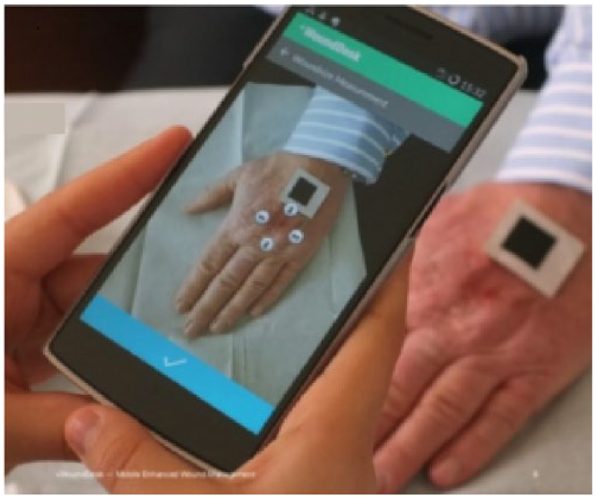
Wound evaluation by the +WoundDesk application.
Microbiological Tests
Microbiological tests were conducted every 15 days. Wound sampling was performed by rotating a sterile swab across the entire wound area. Cultures were performed on media suitable for microorganisms reported to contaminate and cause infections in chronic wounds (Blood Agar, MacConkey No. 2 Agar, and Sabouraud Dextrose Agar, Bioprepare, Athens, Greece). Microbes were identified by microscopic observations (Gram staining) combined with biochemical testing (Kits and Reagents: Liofilochem Srl, Italy). Antimicrobial susceptibility tests were performed according to Clinical and Laboratory Standards Institute performance standards for antimicrobial susceptibility testing.
Data Analysis
Patients were divided into 2 groups: those suffering from pressure ulcers and those suffering venous ulcers. Data are presented as means ± standard error of mean. The normality of BWAT scores was examined using Kolmogorov-Smirnov and Shapiro-Wilk tests with SPSS software (IBM SPSS software version 24.0 for Windows, Armonk, NY). As all data exhibited a normal distribution, repeated-measures analysis of variance followed by the least significant difference post hoc test were performed in order to examine statistically significant differences between the mean BWAT scores recorded at different time points. T tests were employed to assess statistically significant differences between mean BWAT scores at 2 time points. Pearson’s correlation testing was conducted to evaluate correlations between wound healing and wound age or the wound surface.
Results
Microbiological Evaluation
The majority of wounds had a polymicrobial profile (Table 1). Necrotic tissue related to microbes, which was considered a microbial biofilm, was evident in most wounds on initiation of treatment (Figure 2). The most common infections were caused by P aeruginosa, Staphylococcus spp, and Staphylococcus aureus (Table 1). Six wounds were infected with both P aeruginosa and S aureus (a mean 31.57% of the total wounds). Additionally, multidrug-resistant microbes were identified in 3 cases: extended-spectrum β-lactamase–positive Escherichia coli, methicillin-resistant S aureus, and multidrug-resistant (MDR) P aeruginosa. Notably, the strain of MDR P aeruginosa was resistant to all antibiotics tested, with the exception of colistimethate sodium, which was used in combination with a 2% w/w acetic acid cleansing solution and eradicated the infection after 20 days of treatment.
Microbial Species That Colonized the Wounds Before Treatment.
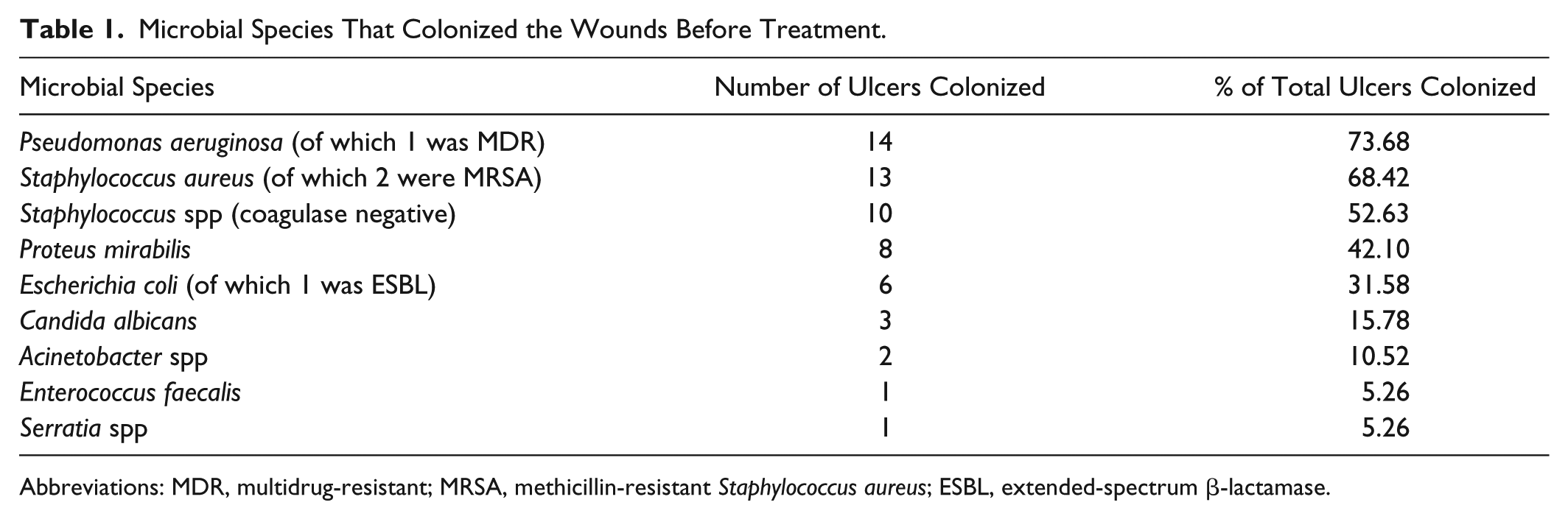
Abbreviations: MDR, multidrug-resistant; MRSA, methicillin-resistant Staphylococcus aureus; ESBL, extended-spectrum β-lactamase.

Three months after treatment. (A) Pressure and (B) venous wounds.
Clinical Evaluation
The main characteristics of the patients and wounds are summarized in Table 2. The patients enrolled in the present study included 3 males and 9 females with 1 ulcer each, 1 female with 2 ulcers, and another female with 5 ulcers. The wound durations ranged from 3 months to 21 years (mean = 32.71 months and median = 8.5 months). All the wounds were assessed prior to the initiation of treatment. The average BWAT score was 39.05, and the median score was 39 (Table 3). As shown in Table 3, BWAT scores decreased after 3 months of treatment in all cases, ranging from 14.7% to 67.5% (mean = 36% and median = 38%).
Patient Characteristics.

Changes in the BWAT Score During 3-Month Treatment With the Ceratothoa oestroides Olive Oil Extract.
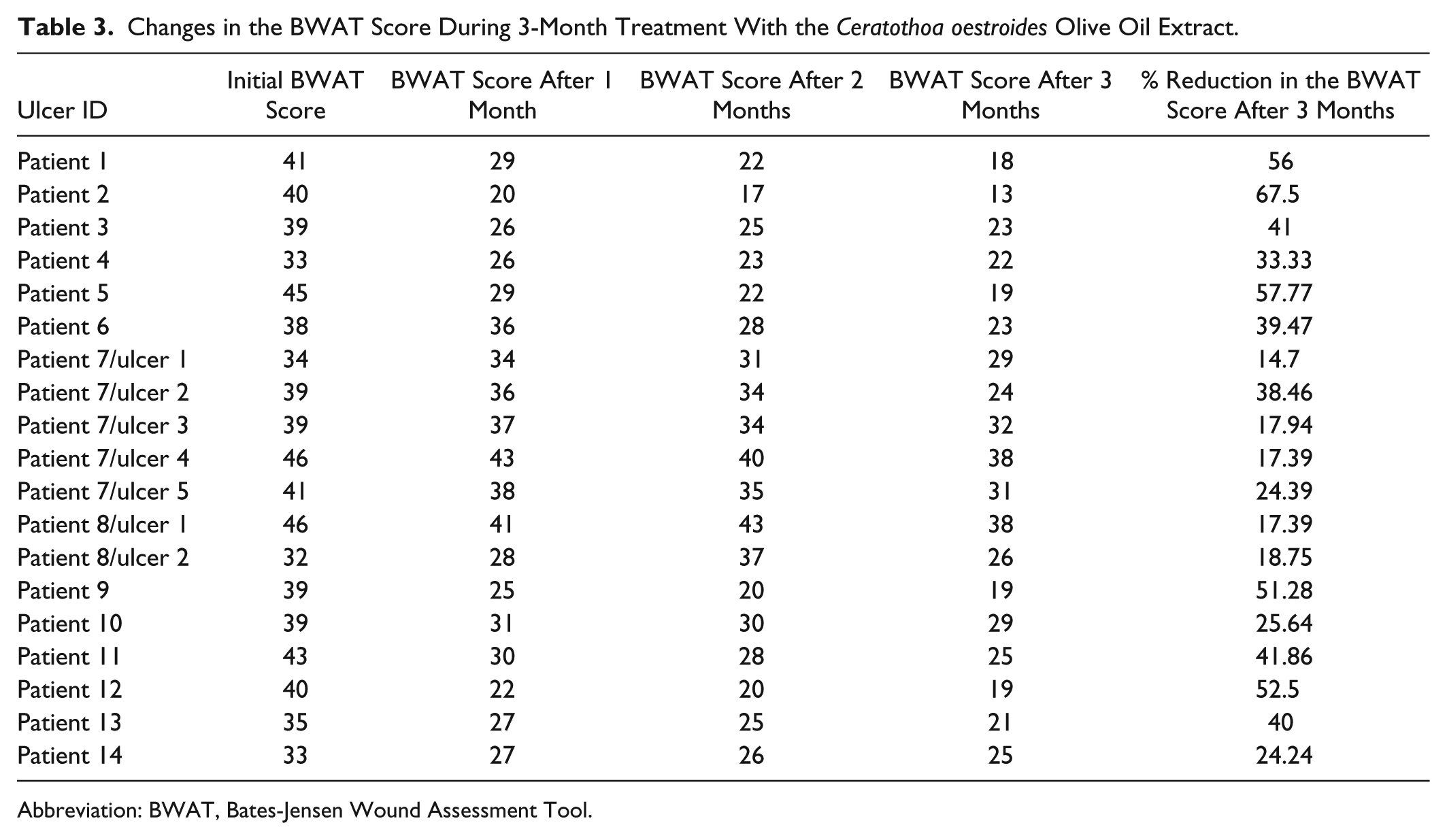
Abbreviation: BWAT, Bates-Jensen Wound Assessment Tool.
Complete healing was achieved in 5 patients (1 venous and 4 pressure ulcers) during the study. In all cases, complete healing was observed between the 10th and 12th weeks. For pressure and venous ulcers, primary healing end points were calculated to be 33.33% and 14.28%, respectively.
Representative images of the progressive healing of pressure and venous ulcers after 3 months of treatment with the C oestroides olive oil extract are presented in Figure 2. Both types of ulcers exhibited substantial improvement during the first month of treatment (Figure 3A). C oestroides exerted similar effects on both venous and pressure ulcers, and repeated-measures BWAT analysis of variance showed P < .000 in both cases. For both venous and pressure ulcers, the least significant difference post hoc test revealed a statistical significant decrease between the initial BWAT score and that of first month and all 0.5-month periods up to the third month (P values ranging from .007 to .000). However, no statistical significant decrease was obtained only for the first 0.5-month period (P > .09).

Bates-Jensen Wound Assessment Tool (BWAT) score: (A) Change in relation to treatment time. (B) Correlation with wound age. (C) Correlation with the initial wound size.
Based on the above observations, the reduction in the total BWAT scores for the venous and pressure ulcers following extract administration for 3 months were combined and plotted against the chronicity of the wound or the initial wound size. The degree of wound healing was negatively correlated with the chronicity of the wound (Figure 3B), whereas the initial size of the wound surprisingly did not influence wound healing (Figure 3C).
Discussion
The C oestroides olive oil extract significantly regenerated chronic ulcers that were unresolved by other agents possessing antibacterial activity. Topical administration of appropriate antibiotics and antimicrobial agents was effective against wound infections, inhibiting biofilm formation. Moreover, cleansing wounds with 2% w/w acetic acid solution helped significantly eradicate P aeruginosa. Wound chronicity should be considered as a major factor when treating such wounds, whereas the wound size or patient age may be only a secondary concern (Figure 3B and C).
In this study, all wounds were infected with bacteria, and the majority of the wounds were colonized by different types of microbes (Table 1). Many of those microbes were found to be resistant to antibiotics due to the formation of a biofilm with a polymicrobial profile. Gram-positive, pathogenic S aureus and nonpathogenic Streptococcus spp, as well as gram-negative P aeruginosa (Table 1), were the most commonly identified bacteria. All 3 bacteria have been reported to create biofilms at high rates, significantly impairing wound healing.33-35 In addition, 32% of the patients presented a coinfection with P aeruginosa and S aureus, and coinfection with these 2 pathogens has been reported to exacerbate wound healing. 36 According to Lindsay et al, 37 P aeruginosa, S aureus, and Streptococcus spp produce proteases that destroy the host tissue, leading to the development of inflammatory responses and impaired wound healing. As shown in our study, chronic wounds are related to the existence of polymicrobial infections and biofilms, which is consistent with the literature. 38 In 20% of the cases in our study, the wounds were infected with MDR microbes. Remarkably, all patients infected with MDR microbes had a chronic wound that lasted for 2 to 3 years or more and were repeatedly treated with antibiotics per os (by mouth) for long periods of time. This high incidence confirms that resistant microbes preferentially develop in chronic wounds. 39
A 2% w/w acetic acid solution was used as a cleansing agent and significantly contributed to P aeruginosa eradication, which has been confirmed by several studies.40,41 Based on microbiological susceptibility tests, the appropriate antibiotics were incorporated into the ointment and administered by topical application. In all cases, these antibiotics effectively controlled the infection during the first month of treatment. Therefore, microbial control via combinations of topically applied antibiotics and antiseptics was very efficient against all types of bacteria, even MDR bacteria. Accordingly, infection control is a predominant parameter contributing to effective wound healing. However, in preliminary studies performed in our laboratory in mice with noninfected wounds treated with different antibiotics and sodium eosin, we observed a significant delay in wound healing (unpublished data). Thus, infection control is a prevalent factor contributing to effective wound healing, but antimicrobial agents lack wound healing efficacy. The C oestroides olive oil extract also shows significant antibacterial activity against gram-negative bacteria (unpublished data), possibly contributing to the eradication of bacteria in infected wounds and facilitating wound healing.
Ceratothoa oestroides notably promoted skin regeneration, as it was an effective medication for chronic pressure or venous wounds. Considering the short study period and the severity of the wounds treated, the primary end point rate, especially for pressure ulcers, was remarkable. As shown in Table 3 and Figure 3A, the extract was most effective during the first month of treatment, and in the second and third months, the extract continued to promote wound healing but at a lower rate. This decrease in the wound healing rate has also been observed for chronic treatments with other commercial wound healing agents. 42 Interestingly, patients with multiple wounds exhibited less of an improvement than did patients with a single wound. The reduction in BWAT scores was not correlated with the initial wound surface or the age of the patient, though wound chronicity was apparently negatively correlated with reduced BWAT scores (Figure 3B and C). Therefore, relatively recent wounds heal faster than do older ones.
The C oestroides olive oil extract significantly promoted wound healing but did not completely heal them in 3 months; it should be noted that the study was approved to be conducted for only 3 months. Therefore, studies investigating the healing of chronic, untreated wounds must be performed for a longer period of time. For example, when using allogeneic human keratinocyte cultures on chronic venous leg ulcers, complete wound closure was achieved after 6 months at a rate of 41%, and this period was much longer than that of our study. 43 In another study testing low-level laser therapy for chronic venous leg ulcers, no effect was observed for the same time period of 3 months, leaving in question possible delayed effects. 44
In addition to the above-mentioned study time limitation, another main restriction was the decreased wound healing rate for such long-lasting chronic wounds, despite an initial relatively rapid healing period, achieving only 27.8% complete healing. Future studies are required to investigate the long-term efficacy of this extract. Another factor somewhat limiting the study was the lack of consideration of the nutrition status of the patients, a parameter that is strongly associated with wound healing. 45
The C oestroides extract has been consistently shown to promote skin regeneration in recent preclinical and clinical studies. The extract improved the healing of excision wounds, second-degree burns, and wounds induced by UV (ultraviolet) irradiation in preclinical studies, and healing in these cases was associated with a significant decrease in oxidative stress and inflammation of the wound area. The C oestroides extract has also been shown in clinical studies to heal diabetic and venous ulcers.27,42
The mechanism of action of the C oestroides extract may be similar to that of other parasites such as helminths and fly larvae, but further studies are needed to elucidate the mechanism.46,47 In particular, some parasites activate Th2-mediated immunity, which appears to improve inflammation and wound healing. 48
New technologies such as cell phone applications may substantially improve patient monitoring and contribute to a more effective evaluation of chronic wounds. Overall, immediate and direct communication between care team members may be beneficial for patients. 32
In conclusion, the C oestroides olive oil extract showed substantial healing properties when applied to chronic, untreated wounds inducing significant decrease in wound size. Furthermore, the efficient microbial control by topically applied agents facilitated wound healing, inhibiting recurrence of wound infection. In relation to other factors such as the initial wound size, chronicity appeared to be an important prognostic factor for the extent of wound healing. Future clinical investigations assessing the wound healing efficacy of the C oestroides olive oil extract are warranted.
Footnotes
Acknowledgements
We are grateful to digitalMedLab for generously providing the mobile phone application +WoundDesk.
Authors’ Note
Permission of the Scientific Committee of Laikon General Hospital, Athens, Greece (Protocol No. 1462/2016) and the Hellenic Data Protection Authority (License No. 1834, Athens, Greece).
Declaration of Conflicting Interests
The author(s) declared no potential conflicts of interest with respect to the research, authorship, and/or publication of this article.
Funding
The author(s) received no financial support for the research, authorship, and/or publication of this article.
