






Abstract
Nonhealing leg ulcers are a major health problem worldwide with a high economic burden since they require human and material resources. Moreover, nonhealing ulcers are a major nontraumatic cause of lower limb amputations. Dermal substitutes have emerged as an effective therapeutic option for treatment of skin lesions, but data on leg ulcers are scarce. We evaluated safety and efficacy of a porcine-derived dermal substitute in the treatment of chronic vascular leg ulcers. Records of patients with nonhealing ulcers seen at our unit from 2018 to 2019 were retrospectively reviewed. Wound etiology, wound area, and complications were evaluated. Each patient received one application of porcine-derived dermal substitute and was followed-up. Six patients (5 females and 1 male) with a mean age of 61.3 (52-81) years presented with nonhealing leg ulcers. After surgical debridement and wound bed preparation, porcine-derived dermal substitute was applied onto the ulcer. Granulation was satisfactory within 10 days. All wounds healed after an average time of 14 weeks. Graft take was good, and no graft loss, rejection, or associated infection were observed. In conclusion, the data presented indicate that dermal substitutes are safe and effective for treatment of chronic nonhealing vascular leg ulcers.
Introduction
Chronic or nonhealing ulcers can be defined as lesions, typically in lower extremities, that do not respond to initial therapy or that persist despite adequate wound care. Many chronic leg ulcers (CLUs) last approximately for 12 months and have a high recurrence rate. 1
Dermal substitutes (DS) represent a valid therapeutic approach and are used for providing early wound coverage and neodermis formation.
The introduction of DS could be considered a revolution in the field of the management of nonhealing ulcers. Indeed, DS can provide long-term functional and cosmetic outcomes with improved skin texture and pliability and results are comparable with that of autologous skin graft.2,3 Other advantages of this approach include simplicity and reliability of technique.
The role of DS in reconstruction of dermal defects other than burns has not been fully investigated so far, and data are not available in the literature. The purpose of the present study was to evaluate safety and efficacy of porcine-derived DS in treating nonhealing vascular ulcers of lower extremities.
Materials and Methods
Patients
We performed a retrospective review that included 6 patients seen from January 2018 to December 2019 treated with porcine-derived DS for complex wound defects. Patient medical records were reviewed, and the following variables were evaluated: wound area, wound etiology, wound location, acute complications, and patient satisfaction. We used Visual Analogic Scale (VAS) to evaluate pain, that severely decreases quality of life and functional outcomes in these patients. We also included pictures taken before and after treatment, after the informed consent was obtained from all participants in the study, thus respecting their privacy.
Surgical Technique
We used porcine-derived DS that is an acellular matrix made from atelocollagen derived from porcine tendon. All surgical procedures were performed under aseptic conditions in patients with a previous negative culture for bacteria. Exclusion criteria were silicone allergy, hypersensitivity to proteins of animal origin, infected or bleeding wound, and excessive exudate. After debridement and hemostasis, wound was rinsed with super-oxidized solution. Porcine-derived dermal matrix was modelled according to the shape and the size of the wound, then put in sterile saline for 15 seconds before coverage. After that it was applied on the ulcer without staples in order to minimize perilesional trauma and covered with a nonadherent dressing that is gauze and crepe bandage that were changed every 5 days. The fenestrated DS allowed drainage in case of exudating wounds or hematomas. The silicone film naturally peeled off when the collagen layer was replaced by new tissue, typically after 7 to 14 days.
Results
For the present study, a total of 6 patients’ medical records were examined. The average age of patients was 61.3 years (range 52-81 years). There were 5 females and 1 male. Additional demographic and wound details are presented in Table 1.
Patient Demographic and Ulcer Characteristics.

Abbreviation: NPWT, negative-pressure wound therapy.
Porcine-derived dermal matrix was used in nonhealing ulcers from different etiologies, including venous, arterial, diabetic, and traumatic leg ulcers. We made the first control after 5 days and silicon peeled off spontaneously typically at day 10. There were no major infections, failures, or rejections in all the patients. Indeed, after 14 days, there was a well-vascularized neodermis due to autografting. All patients received meticulous wound care that included aseptic dressing changes with an appropriate choice of dressing as wound evolved. All wounds healed after an average time of 14 weeks without complications. The restored skin area displayed appearance and morphological features like those of the neighboring skin tissue. During the follow-up, all patients had good functional outcomes and pain improved. In fact at T0 83% of patients had severe pain; after two weeks 60% of them showed significant pain reduction; after one month 100% of them had non pain. After two weeks from PELNAC application patients were able to climb several flights of stairs and to do moderate activities without limitations. Their emotional status improved because they felt to be healthier.
Case Presentations
Patient 1, an 81-year-old female, had a history of hypertension, T2 diabetes mellitus, and hepatitis. She developed bullous lesions after oral administration of ibuprofen. This reaction was suspected for Steven-Johnsons syndrome since the patient had flu-like symptoms the week before. The largest bullous injury was located on the medial region of the leg, and it had showed delayed wound healing probably due to the concomitant chronic venous insufficiency (CEAP2) and diabetic arteriopathy with tibial critical stenosis. The patient presented to our unit with severe uncontrolled pain despite the use of tramadol. After sterile serum aspiration, adequate debridement and wound bed preparation, porcine-derived DS was placed onto the ulcer. Follow-up showed complete pain control at day 5. At month 6 satisfactory healing with a small skin defect was observed but advanced medications were not required.
Patient 2, a 54-year-old female, developed chronic venous ulcer due to postthrombotic syndrome treated conservatively with 20 to 30 mm Hg knee-length elastic compression stockings worn daily and anticoagulant therapy. Moreover, she was affected by hypertension, dyslipidemia, hypothyroidism, and antithrombin III deficiency with a history of recurrent deep venous thrombosis. The chronic ulcer, located on the medial malleolus, presented a fibrous wound bed with severely inflamed perilesional skin (Figure 1a). After debridement and wound bed preparation we put porcine-derived DS onto the lesion. After a 2-month follow-up (Figure 1b) complete wound healing was obtained.
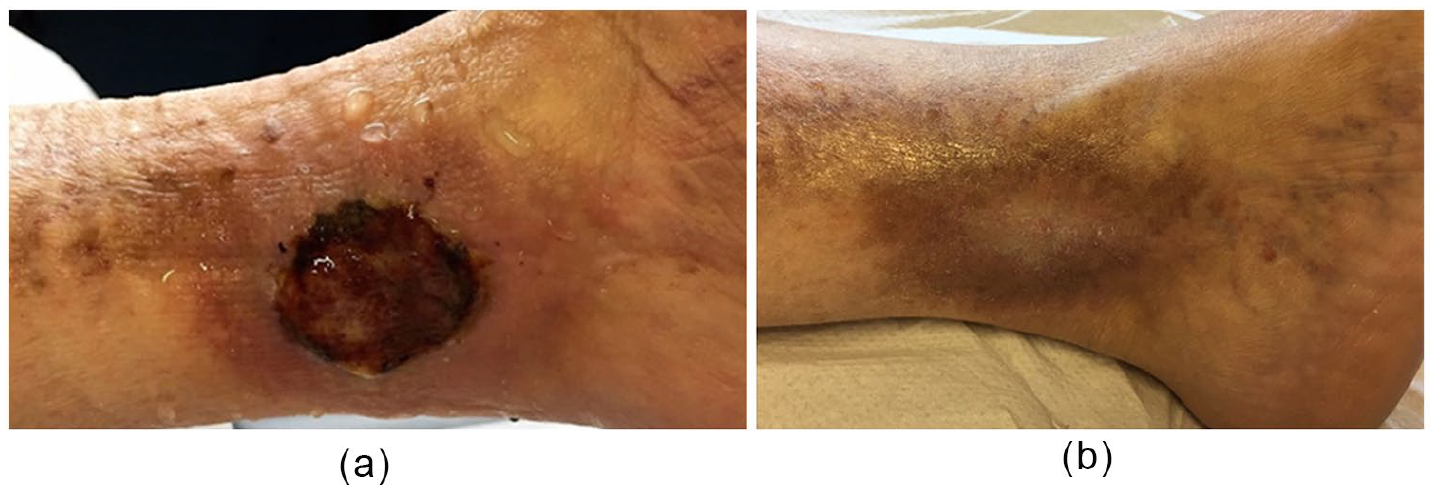
Patient 2: (a) A 54-year-old female developed chronic venous ulcer due to postthrombotic syndrome. (b) Two-month follow-up showed complete wound healing.
Patient 3, a 55-year-old female, had a history of hypertension, hyperthyroidism, dyslipidemia, and diabetes mellitus complicated with diabetic neuropathy and macroangiopathy causing stenosis of femorotibial axis without hemodynamic effect. She developed a circular mid-calf mixed ulcer without pain due to regional neuropathic anesthesia. After wound bed preparation, porcine-derived DS was placed on the ulcer. After 4-months there was a complete wound healing.
Patient 4, a 52-year-old female, was affected by chronic venous insufficiency, postthrombotic syndrome, and obesity. She presented to our unit with a nonhealing traumatic ulcer. The leg showed edema and lymphangitis. Antibiotic treatment was administrated with subsequent inflammation reduction. Grade class 2 compression tights were prescribed in order to improve lymphedema. After surgical debridement and wound bed preparation, porcine-derived DS was placed onto the ulcer. The end of 2-month follow-up showed complete wound healing.
Patient 5, a 70-year-old female, had developed a traumatic ulcer. She was affected by chronic venous insufficiency (CEAP4) and recent microcytic hypochromic anemia. The lesion was located on the pretibial region and showed a bleeding fundus with severe perilesional inflammation (Figure 2a). We performed a culture that was positive for Pseudomonas aeruginosa. Local hemostatics were used to control the bleeding. Oral therapy was supplemented with vitamin K, iron, B-complex vitamins, and adequate antibiotics according to the in vitro sensitivity tests until culture showed no bacterial growth. Then fenestrated porcine-derived DS was put on the ulcer (Figure 2b). After a 6-month follow-up (Figure 2c) complete wound healing was observed.
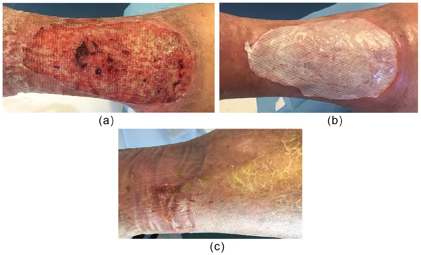
Patient 5: (a) A 70-year-old female, developed a traumatic ulcer with a bleeding fundus and severe perilesional inflammation. (b) Cultural examination was positive for Pseudomonas aeruginosa. (c) Follow-up at 6 months showed complete wound healing.
Patient 6, a 56-year-old man, presented to our unit with a nonhealing venous ulcer. He was affected by chronic venous insufficiency and postthrombotic syndrome. Orthopedic surgery for traumatic malleolar fracture had been performed some months before. After surgical debridement negative-pressure wound therapy was used to reduce exudate and prolonged for 3 weeks. Then, we placed a fenestrated double-layer porcine-derived dermal matrix that obtained a complete wound closure after 1 month.
Discussion
Chronic leg ulcers may have different underlying etiologies that may be related to systemic disease or local disorders 4 and are challenging clinical problems. There are many types of CLU classified as vascular (either arterial, venous, or mixed), diabetic, traumatic, or pressure ulcers.
The incidence of CLU is rising due to an increase in both population’s age and risk factors for atherosclerotic disease such as smoking, obesity, and diabetes.
These types of ulcers impair the quality of life and the patient productivity posing a substantial economic problem for the patient and the health care system. 5 Despite standard and advanced treatment, many CLU may remain open and do not heal for long time periods. These lesions fail to proceed through the normal phases of wound healing due to factors including an excessive level of pro-inflammatory cytokines, proteases, reactive oxygen species, and senescent cells. Persistent infection, deficiency of stem cells, or the possible presence of a biofilm also account for resistance to many forms of treatment. 1
During past years, DS have emerged as a relevant therapeutic approach to dermal wounds. They are biomatrices that fulfil the functions of the cutaneous dermal layer that are control of pain and scarring. They act as scaffolds promoting dermal regeneration, vascularization, and wound healing6,7 with enhanced pliability and a greater scar quality. 8 Moreover, DS replace the absent or dysfunctional extracellular matrix and reduce proteases, thus preventing inflammatory or immunogenic response to occur.9,10 The new collagen that was made is indistinguishable from normal dermal collagen.6,7 The silicone layer acts as a physical barrier to bacterial contamination preserving wound environment and contributing to immediate wound closure. 11
This case series indicate that DS are safe and effective in the treatment of nonhealing vascular ulcers. The porcine-derived DS used is PELNAC (Gunze Medical Materials Center) that is an acellular matrix, made from atelocollagen, which is a highly purified type 1 collagen obtained from porcine tendon. The amino acidic composition of atelocollagen is almost identical to endogenous collagen also because the highly antigenic telopeptides are removed by pepsin during the production process. This lowers the immune response to the matrix, its rejection, and failure. Also, inflammatory response is decreased and this is an additional benefit when dermal loss to be treated is contaminated by bacteria. 12 This provides a result that mimics endogenous dermis reducing disabling scar contractures and therefore improves cosmetic outcome. 13 The use of porcine-derived DS is practical for several reasons. It is biocompatible, porous, and elastic and ensures high take rates of the graft. The soft collagen sponge structure ensures good adhesion to the irregular wound surface and prevents the development of surface irregularities. PELNAC does not require prewashing; it can be stored at room temperature and it can be used directly from the package or after presoaking in a small amount of sterile saline. Several types of PELNAC are available (fenestrated type, fortified type, and single layer) and this allows to select the one more appropriate for treatment based on wound features. There are several other porcine-derived products that differ from PELNAC for structural characteristics, which include pore diameter and pore shape. The pore size of PELNAC ranging from 70 to 110 mm prevents the formation of a tissue capsule and facilitates cell migration into the matrix, allowing the formation of a consistent and elastic neodermis. 14 This pore diameter promotes cell penetration into the scaffold and their binding to the ligands present on scaffold surface. Moreover, pores have an elliptical shape that is easier to collapse than a circular shape.
Anyway further in vivo experiments are required to build a consensus of the different uses of DS that is not based on preference but on their adequacy.
However, in this study, DS show safety and efficacy along with improvement of quality of life. Porcine-derived DS was designed to provide coverage on avascular beds promoting dermal regeneration and reepithelialization11,15 and therefore it could play an important role in the treatment of chronic, hard-to-heal vascular ulcers.
Conclusions
Dermal substitutes have emerged as a relevant therapeutic approach to dermal wounds in acute and chronic settings. Functional and cosmetic results are satisfactory. Quality of life is improved.
Our data indicate that DS may be beneficial in the treatment of vascular nonhealing ulcers irrespective of their etiology. Despite the progress of bioengineered skin substitutes, we still need a DS that replaces morphology and function of the entire skin. The development of a combined dermal-epidermal skin substitutes will be entirely welcome.
Footnotes
Declaration of Conflicting Interests
The author(s) declared no potential conflicts of interest with respect to the research, authorship, and/or publication of this article.
Funding
The author(s) received no financial support for the research, authorship, and/or publication of this article.
