
Abstract
Wound healing is interaction of a complex cascade of cellular/biochemical actions leading to restoration of structural and functional integrity with regain of injured tissues strength. This study was aimed at evaluation of application of ethanolic extract of propolis-loaded poly(-lactic-co-glycolic acid) nanoparticles (EEP-PLGA NPs) on wound healing in diabetic rats. Sixty rats were randomized into four groups of 15 rats each: In control group (Control) diabetic wound was treated with normal saline. In Carrier 1 group diabetic wound was treated with PLGA nanoparticles based solution. In Carrier 2 group the diabetic wound was treated with EEP. In Treatment group animals received EEP-PLGA NPs on the wound. Wound size was measured on 7, 14 and 21 days after surgery. The expression of p53, bcl-2, Caspase III, were evaluated using reverse-transcription PCR and Immunohistochemical staining. The Treatment group had significantly reduced the wound size compared to other groups (P = 0.001). histological and morphometric studies, and mean rank of the qualitative studies demonstrated that there was significant difference between Treatment group and other groups (P < .05). Observations demonstrated that ethanolic extract of propolis-loaded PLGA nanoparticles significantly shortened the inflammatory phase and accelerated the cellular proliferation. Accordingly, the animals in Treatment group revealed significantly (P < .05) higher fibroblast distribution/one mm2 of wound area and rapid re epithelialization. The mRNA levels of bcl-2, p53 and caspase III were remarkably (P < .05) higher in Treatment group compared to control and animals. The immunohistochemical analyzes confirmed the RT-PCR findings. EEP-PLGA NPs offered potential advantages in wound healing acceleration and improvement through angiogenesis stimulation, fibroblast proliferation and granulation tissue formation in early days of healing phases, acceleration in diabetic wound repair associated with earlier wound contraction and stability of damaged area by rearrangement of granulation tissue and collagen fibers.
Introduction
Diabetes mellitus, commonly known as diabetes, is one of the most serious and common metabolic chronic diseases characterized by a hyperglycemic state related to insulin deficiency. The main classes of diabetes include type 1 and type 2. Type 1 diabetes occurs due to the immune-mediated destruction of the pancreatic β-cells leading to insulin deficiency. Type 2 diabetes is polygenic disorder that involves an impairment of insulin secretion related to the insulin resistance. 1 Even though most of diabetes complications are linked to its chronicity, one of the most severe complications of diabetes is impaired wound healing even in the early stage of the disease. 2
Cutaneous wound healing, which is triggered by tissue injury, is an intricate and highly coordinated process that includes four phases: (i) Hemostasis (ii) inflammation which is mediated by inflammatory cell recruitment, and growth factor and cytokine secretion (iii) proliferation characterized by the formation of the granulation tissue and re-epithelialization; and (iv) remodeling, in which granulation tissue is reorganized and the mature scar is formed.3,4 However, under hyperglycemic conditions the natural healing process is ineffective to recover the damaged skin as all the stages of the wound healing process are disrupted and the wound repair is delayed. 5 Indeed, the cutaneous wound healing in acute diabetic patients is associated with abnormal and delayed inflammatory response characterized by the deficiencies in the number of neutrophils in the wound site. 2 This may increase the risk of bacterial infection that leads to chronic non-healing wound state posing a significant healthcare burden with consequences that include hospitalization and lower limb amputation. Topical antimicrobial therapies have been used on diabetic foot ulcers, either as a treatment for clinically infected wounds, or to prevent infection in clinically uninfected wounds. 6 However, The incidence of impaired healing process in diabetic patients is constantly increasing. 6 Therefore, it is important to develop novel therapies to avoid any serious complication and accelerate wound healing process in the early diabetic stage.
Propolis is a natural product collected by bees from the poplar and conifer trees. Bees use propolis as an antibiotic against foreign organisms and also to repair the cracks of their hives. 7 It has vast majority of biological activities such as anti-inflammatory, anti-fungal, antioxidant, and immune-stimulating activity. 8 Most of these effects have been related to the remarkable in vitro antioxidant activity and free radical scavenging ability of propolis. 9 The major components of propolis are polyphenolics including aldehydes, caffeic acid, and caffeic acid phenethyl ester which plays a critical role in neurological disorders, cardiovascular diseases, pathophysiology of cancer and diabetes.10–13 Although propolis is widely used in many applications as described above, the solubility of poorly soluble active compounds has been a limitation. 14 Currently, nanotechnology is applied in life sciences, especially the nanoparticles as a drug delivery system. 15 Advances in this system have led to the development of several aspects such as improved drug efficacy for infection diseases, targeted delivery for cancer therapy, and in cosmetics.16–18 In particular, nanotechnology may be likely to accomplish enhanced delivery of poorly wate rsoluble phytomedicine. 19 Polymeric nanoparticles (PNPs) are one of the smart drug delivery platforms. 20 Many materials such as natural or synthetic polymers are needed to formulate PNPs. 21 PNP preparation has been reviewed elsewhere. 22 Poly(-lactic-co-glycolic acid) (PLGA) is one of the most synthetic polymers for elaborating PNPs because it has a biodegradable property and has been approved by the Food and Drug Administration for drug delivery. 23 Moreover, PLGA-based nanoparticles have been reviewed for various biomedical applications. 24 Therefore, propolis loaded into PLGA nanoparticles may overcome the limitation of water solubility and easily dispersed in aqueous media.
The wound healing effect of biologically synthesized ethanolic extract of propolis-loaded PLGA nanoparticles on diabetics is yet to be determined. Therefore, this study was aimed at evaluation of application of ethanolic extract of propolis-loaded PLGA nanoparticles on wound healing in diabetic rats.
Materials and Methods
Preparation of Ethanolic Extract of Propolis-Loaded PLGA Nanoparticles (EEP-NPs)
EEPNPs was prepared using PLGA matrix and EEP nanoparticles. The suspensions of nanoparticles (NPs) were prepared by the modified oil-in-water (o/w) single-emulsion solvent evaporation method. 25 In brief, the organic solution consisting of EEP and PLGA were dissolved in absolute ethanol and dichloromethane (DCM), respectively. EEP and PLGA were mixed at room temperature until all materials were completely dissolved. The organic solution was emulsified into 4 ml polyvinyl alcohol (2% w/v, polyvinyl alcohol (PVA)) in a dropwise manner. The resulting solution was stirred and then sonicated (output power 70 W, 2 min) using an ultrasonic processor UP100H (Hielscher Ultrasonics, Germany) within an ice bath. The mixed solution was incubated overnight at room temperature in dark condition for complete polymerization. The resulting nanoparticles were centrifuged at 8800 g for 40 min at 4 °C (Beckman Coulter, CA), washed once with deionized water and then lyophilized until use.
Analytical and Characterization Methods of EEP-NPs
The characterization methods were performed based on the others. 15 Transmission electron microscopy (TEM), field emission scanning electron microscopy were adopted to assess the morphology and topography of the samples as well as elemental chemical surface analysis. The size distribution of the nanoparticles was detected through Zetasizer Nano ZS (Malvern Instruments Limited) particle analyzer. 26
Ethical Considerations
The Guide of the National Academy of Sciences published by the National Institutes of Health (NIH Publication No. 85-23, revised 1985) were followed in the present study. The procedures adopted to clear any potential pain in the animals were approved by Institutional Ethical Committee of the University.
Induction of Diabetes and Monitoring the Blood Glucose Levels During the Study Period
Induction of diabetes was based on methods of others. 27 For insulin-deficient diabetes, the animals were fasted overnight prior receiving a single intraperitoneal injection (60 mg/kg in 0.9% sterile saline) of streptozotocin. Hyperglycemia (15 mmol/L or greater) was confirmed 2 days later by measurement of tail vein blood glucose concentration (Ames Glucostix; Miles Laboratories). The rats underwent the procedures 3 days after induction of diabetes. Levels of blood glucose in the animals within the study period was performed to ensure that the animals were diabetic throughout the study period.
The Procedures for Wound Creation
The processes for creation of wound was done based on methods of others. 16 “Intraperitoneal injection of ketamine (70 mg/kg of body weight) and xylazine (5 mg/kg of body weight), were used to anesthetize the animals. The area on their back was shaved, cleansed with 70% alcohol solution and after aseptic preparation, a circular wound, approximately 20 mm in diameter, in full thickness was excised from predetermined area on the anterior-dorsal side of each rat.
Randomization and Grouping of Animals
Randomization and grouping of animals were according to the methods of others. 27
A random number generator was used to create a list of random numbers. Therefore, sixty diabetic male rats were divided into 4 groups of 15 animals each, randomly.
Sixty rats were randomized into four groups of 15 rats each: In control group (Control) the diabetic wound was treated with normal saline. In Carrier 1 group the diabetic wound was treated with PLGA nanoparticles based solution. In Carrier 2 group the diabetic wound was treated with ethanolic extract of propolis. In Treatment group animals received ethanolic extract of propolis-loaded PLGA nanoparticles on the wound.
Application of test formulations was lasted for 5 days and twice a day. Animal houses were in standard environmental conditions of temperature (22 ± 3 °C), humidity (60 ± 5%), and a 12-h light/dark cycle. The animals were maintained on standard pellet diet and tap water. All rats were closely observed for any infection; and if they showed signs of infection, they were separated, excluded from the study, and replaced.
Planimetric Studies in Excisional Wound Model
Percentage of wound contraction = (At-A1) / At × 100
Where A0 is the original wound area and At is the wound area at the time of imaging. 27
Biochemical Investigations
The tissue samples of wounds were kept at −80 °C for 3 days, and then enzyme activities were determined in the rat tissues. The tissues were ground with liquid nitrogen in a mortar. One half gram was weighed for each group and then treated with 4.5 mL of an appropriate buffer. This mixture was homogenized on ice with use of an Ultra-Turrax homogenizer (IKA® Werke) for 15 min. Homogenates were filtered and centrifuged by using a refrigerator centrifuge at 4 °C. Then the supernatants were used to determine the enzymatic activities. All assays were carried out at room temperature. Antioxidant activities including superoxide dismutase (SOD), total nitric oxide synthase (tNOS), malondialdehyde (MDA), myeloperoxidase (MPO), total glutathione (tGSH), glutathione peroxidase (GPO), glutathione reductase (GSHRd), glutathione S-transferase (GST) analyzes, isolation of DNA from tissue, and cDNA hydrolysis with formic acid measurement of 8-hydroxy-2′-deoxyguanosine (8-OH Gua) were performed. 27
Histological Preparation and Quantitative Morphometric Studies
The tissue samples were taken on 7, 14, and 21 days after surgery from periphery of the wound along with normal skin and fixed in 10% buffered formalin, dehydrated and embedded in paraffin wax, sectioned at 5 µm, and stained with hematoxylin and eosin (H&E) and Masson's trichrome stains. To investigate the predominant stage of wound healing, photomicrographs were taken under light microscopy. Cellular infiltration including the number of mononuclear cells, polymorphonuclear cells, and fibroblastic aggregation were quantitatively investigated. Acute hemorrhage, congestion, vascularization, epithelialization, collagen production, and density were also investigated qualitatively. 27 Morphological findings were scored using image analyzing software (Image-Pro Express, version 6.0.0.319, Media Cybernetics). 16 The histological parameters were classified according to the intensity of occurrence in 5 levels (−, absence; +, discrete; ++, moderate; +++, intense; and ++++, very intense).
Immunohistochemical Staining for Angiogenesis
Tissue section slides were heated at 60 °C for approximately 25 min in a hot air oven (Venticell®, MMM Einrichtungen). The sections were dewaxed in xylene and rehydrated using an alcohol gradient. The antigen retrieval process was performed in 10 mM sodium citrate buffer. Immunohistochemical staining was conducted according to the manufacturer's protocol (Biocare). Briefly, endogenous peroxidase was blocked in a peroxidase blocking solution (0.03% hydrogen peroxide containing sodium azide) for 5 min. Tissue sections were washed gently with washing buffer and subsequently incubated with CD31 (Biocare) biotinylated primary antibodies (Rabbit/anti-mouse, 1:500) for 15 min. The sections were rinsed gently with washing buffer and placed in a buffer bath. The slides were then placed in a humidified chamber with a sufficient amount of streptavidin-horseradish peroxidase (HRP) (streptavidin conjugated to horseradish peroxidase in phosphate-buffered saline (PBS) containing an anti-microbial agent). The slides were incubated for 15 min. Subsequently, the sections were rinsed gently in washing buffer and placed in a buffer bath. A DAB (3,3′-diaminobenzidine) chromogen was added to the tissue sections and incubated for 5 min followed by washing and counterstaining with hematoxylin for 5 s. The sections were then dipped in weak ammonia (0.037 M) 10 times, rinsed with distilled water, and cover slipped. Positive immunohistochemical staining was observed as brown stains under a light microscope. 27
RT-PCR for Caspase-3, Bcl-2, and p53
Reverse transcriptase-polymerase chain reaction (RT-PCR) for Caspase-3, Bcl-2, and p53 were according to the method of others. 16 As previously described. 27 The polymerase chain reaction (PCR) was carried out in a total volume of 25 µL containing PCR master mix (12.5 µL), forward (FWD) and reverse (REV) specific primers (each 0.75 µL), and cDNA as a template (1 µL) and nuclease-free water (10 µL). PCR conditions were run as follows: General denaturation at 95 °C for 3 min, 1 cycle, followed by 40 cycles of 95 °C for 20 s; annealing temperature (62 °C for Bcl-2, 52 °C for p53, and 50 °C for caspase-3) for 60 s; elongation: 72 °C for 1 min and 72 °C for 5 min. The reaction products were separated on 1.5% agarose gel and visualized by ethidium bromide staining using Gel Doc 2000 (Bio-Rad). Table 1 shows the forward and reverse primers caspase-3, Bcl-2, and p53.
The Forward and Reverse Primers Caspase-3, Bcl-2, and p53.
Statistical Analysis
Kruskal-Wallis variance analysis was adopted for differences among groups. Where the P value (from the Kruskal-Wallis test statistics) was statistically significant, multiple comparison tests were utilized to get the differences. Mann-Whitney U test was used for comparison among days. For retrieving possible multiple comparisons, the Bonferroni correction was utilized. We utilized SPSS 11.5 (SPSS Inc) for the analyzes and considered P value <0.05 as significant level.
Results
Analytical Assessments
Microstructural and Physicochemical Characterization
The TEM image of ethanolic extract of nanopropolis (EENPs) (Figure 1A) evidently illustrates that the particles were with a size range of less than 100 nm. The mean value for sizes of the NPs was about 57 ± 7 nm based on the dynamic light scattering technique (Figure 1B). The results of fourier-transform infrared spectroscopy (FTIR) indicated there was no significant difference between nanoparticles of (Figure 1C).
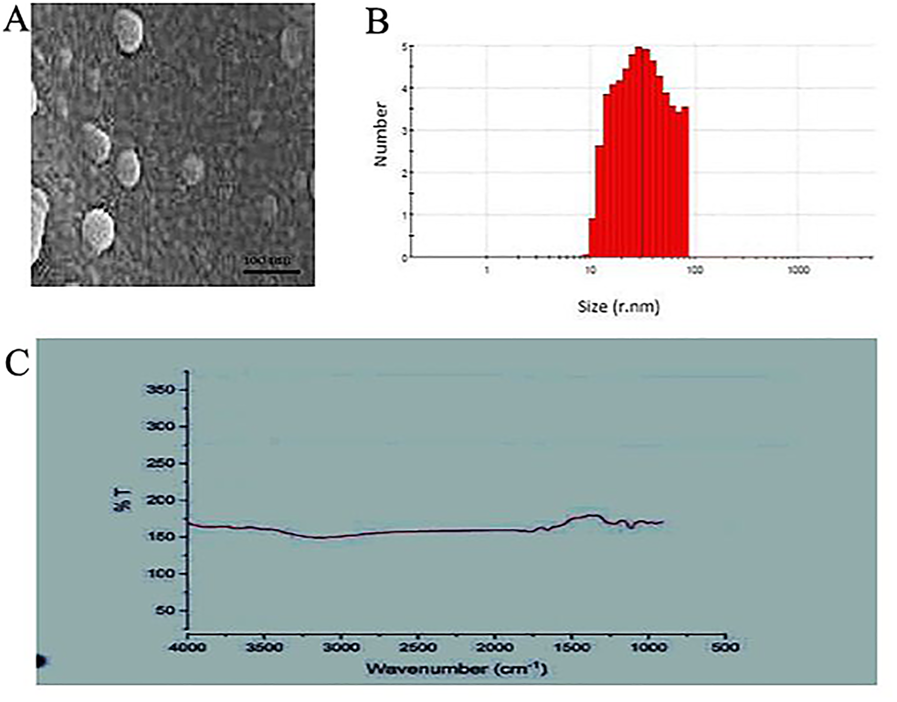
(A) FESEM image of the synthesized EENPs. (B) Size distribution of the nanoparticles by dynamic light scattering. (C) Spectrum of Fourier-transform infrared spectrophotometry EENPs.
Planimetric Studies
Table 2 shows wound contraction percentage in different groups within the study period. The healing rate of wounds in Treatment group was significantly different compared to other groups (P < .05).
Impact of Application of Biologically Synthesized Ethanolic Extract of Propolis-Loaded PLGA Nanoparticles on Circular Excision Wound Contraction Area (mm2). Values are Given as Mean ± standard error of mean (SEM).
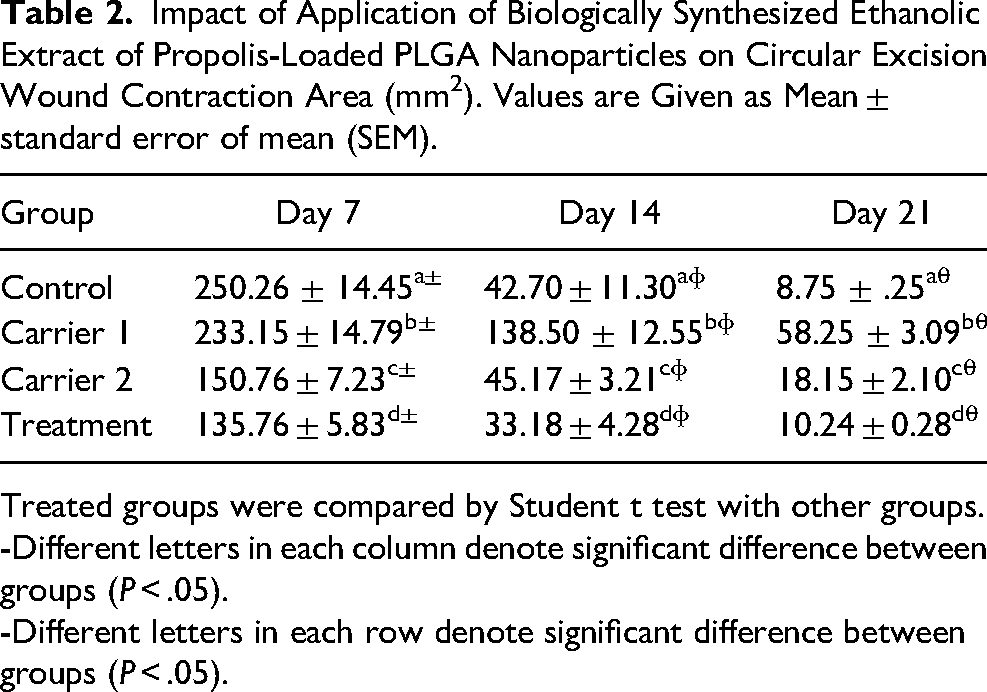
Treated groups were compared by Student t test with other groups.
-Different letters in each column denote significant difference between groups (P < .05).
-Different letters in each row denote significant difference between groups (P < .05).
Biochemical Findings
Topical usage of cinnamon nanoparticles resulted in significant augmentation in the activity of SOD in Treatment group in comparison with activity of SOD in other groups (P < .05). The activity of tNOS was declined in Treatment animals with a significant decrease compared to other groups (P < .05). Cinnamon nanoparticles significantly diminished MDA level in Treatment group in comparison with other experimental groups (P < .05). The cinnamon nanoparticles resulted in significant diminution level of MPO in tissues of Treatment animals (P < .05). Levels of GSH, GPO, GSHRd, and GST in Treatment animals were significantly increased in comparison with other experimental groups (P < .05). Significant decrease was found in the levels of 8-OHGual/Gua, a DNA damage product, in Treatment group in comparison with other experimental groups (P < .05, Table 3).
Comparison of the Activities of TAC, SOD, MDA and GPO in the Animals of the all Experimental Groups. Data are Expressed as Mean ± standard deviation (SD).
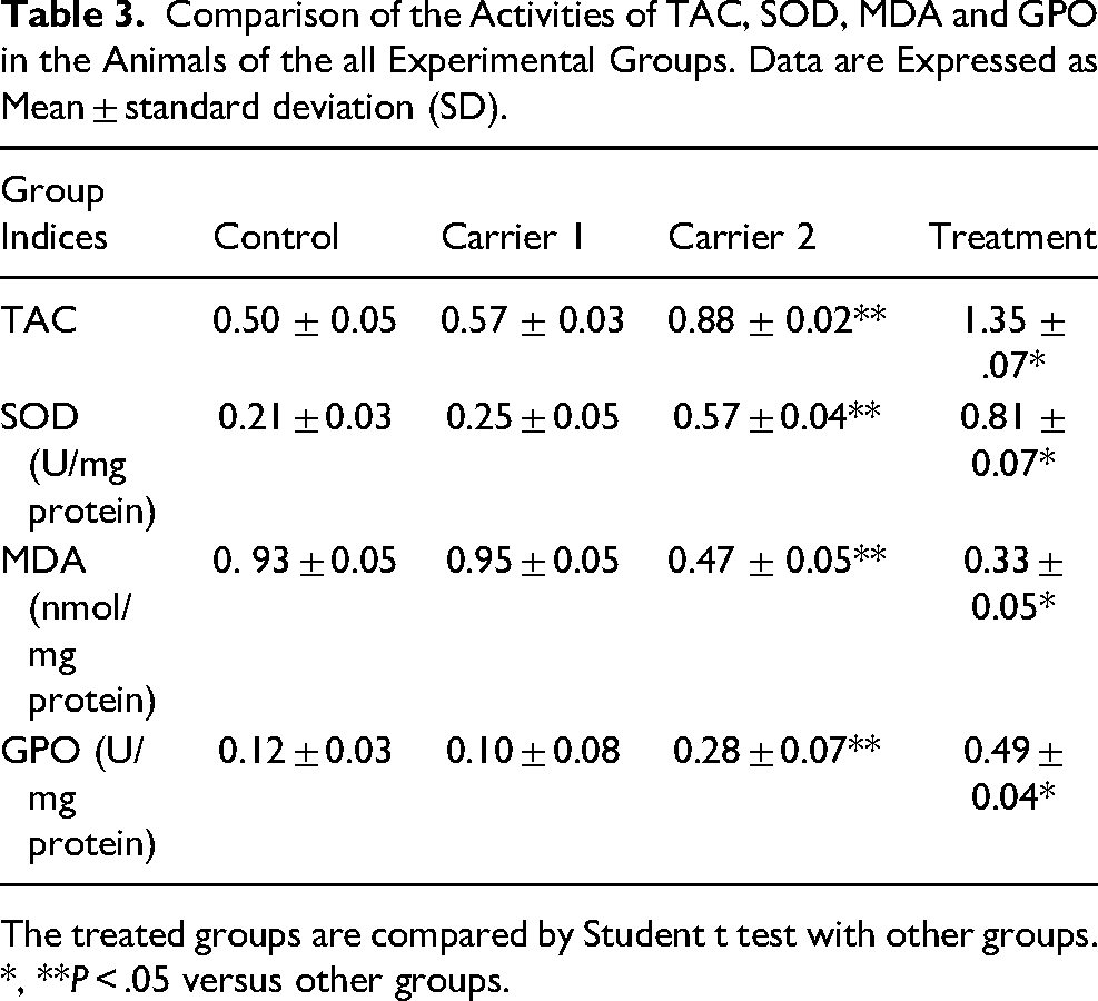
The treated groups are compared by Student t test with other groups. *, **P < .05 versus other groups.
Histological and Morphometric Findings
Significant differences between Treatment and other experimental groups were found regarding re-epithelialization, new vessel formation, acute hemorrhage, congestion, cellular infiltration, collagen production and edema. In the present study, significantly higher scores were observed in re-epithelialization and new vessel formation in Treatment group rats compared to other groups (P < .05). Mean values for cell count and also mean rank of the qualitative study of wound score in Treatment animals were significantly higher than those of othes animals (P < .05; Figures 2–4).
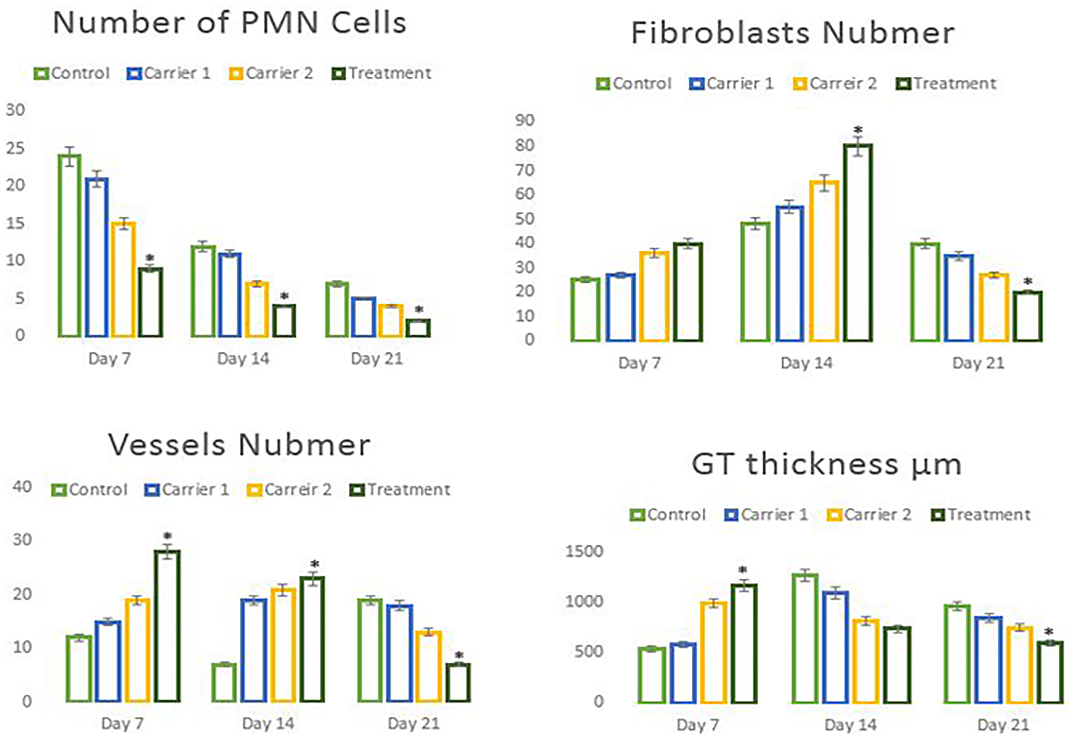
Line graphs show the quantitative results of number of PMN, fibroblasts, angiogenesis and granulation tissue (GT) thickness in experimental groups. Data are presented as mean ± SD. * P < .05 among experimental groups.
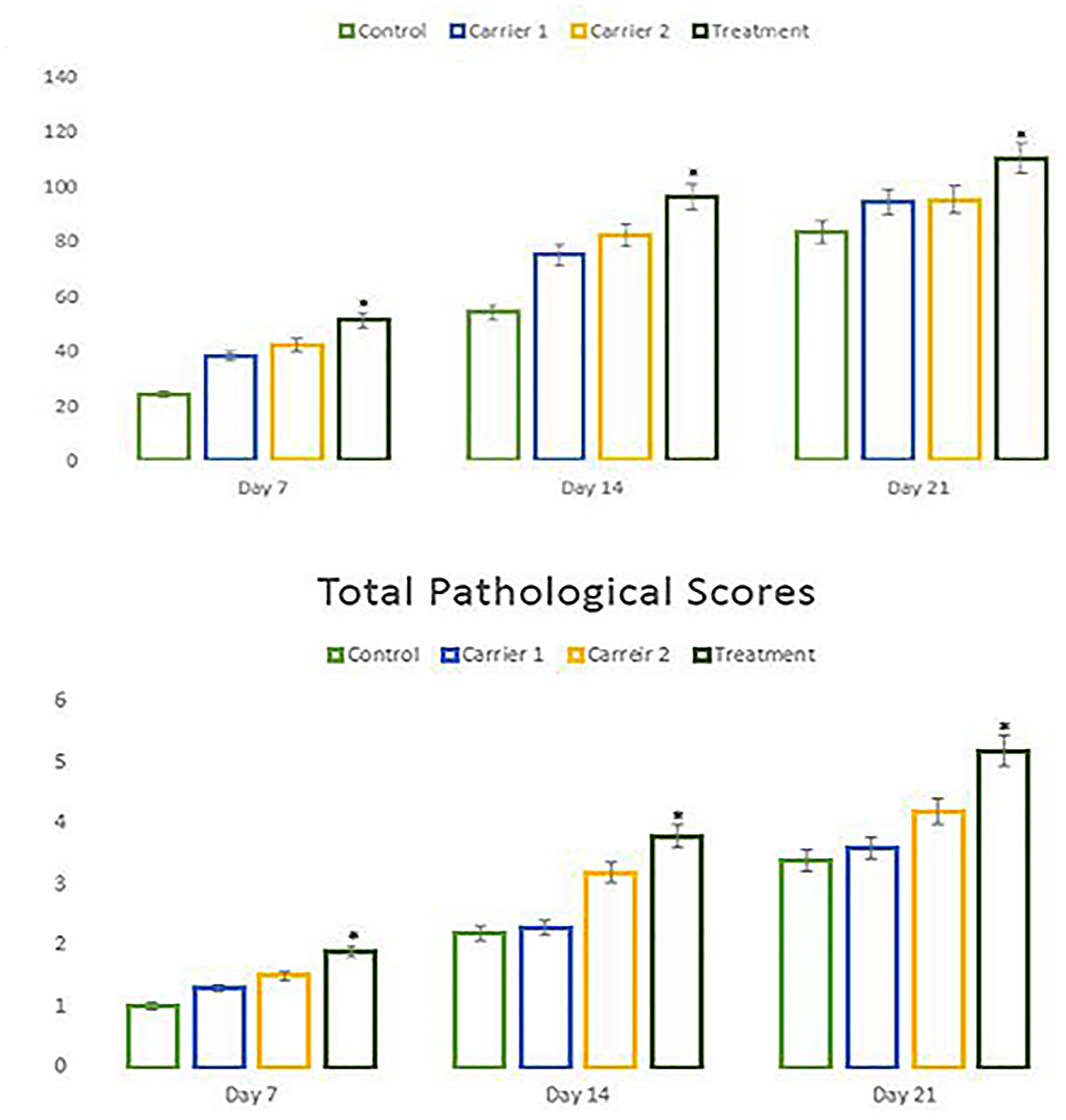
Line graphs show the quantitative results of epithelialization and total pathological scores in experimental groups. Data are presented as mean ± SD. * P < .05 among experimental groups.

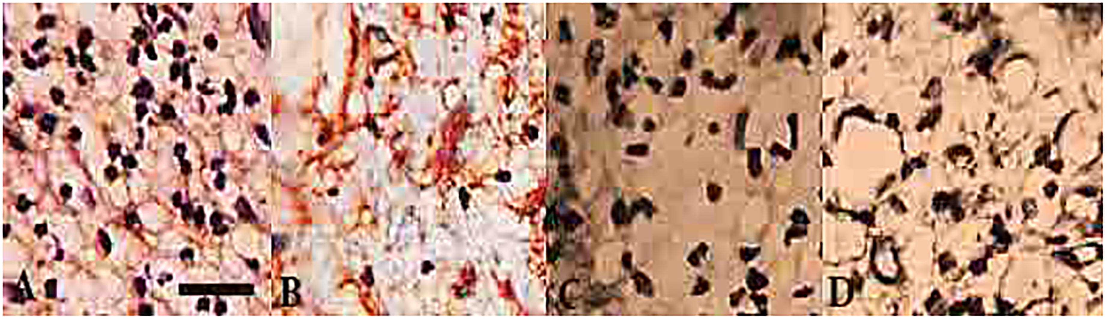
Histological characteristics of the skin on days 7 (A-D), 14 (E-H) and 21 (I-L) after wound creation in experimental groups. Wounds with surrounding skin were prepared for histological microscopic evaluation by H&E staining. Tretament (A,E,L), Carrier 2 (B,F,J), Carreir 1 (C,G,K) and Control (D,H,L)groups.
Results of Immunohistochemistry for new Vessel Formation
Analyzes of immunohistochemistry demonstrated that the angiogenesis was significantly augmented in treatment group (P < .05; Figure 5).
Immunohistochemical staining for CD31 on day 14 post wound creation: (A) control, (B) carreir 1, (C) carreir 2 and (D) treatment groups. Scale bar: 40 μm.
RT-PCR Results for Caspase-3, Bcl-2, and p53
The levels of mRNA of caspase-3, Bcl-2, and p53 genes were analyzed to assess the ratio of proliferation of cells on day 8 post-wounding. The findings indicated that application of the EEPNPs ended up a significant rise at mRNA level of caspase-3 in comparison with other animals (P < .05) (Figure 6).
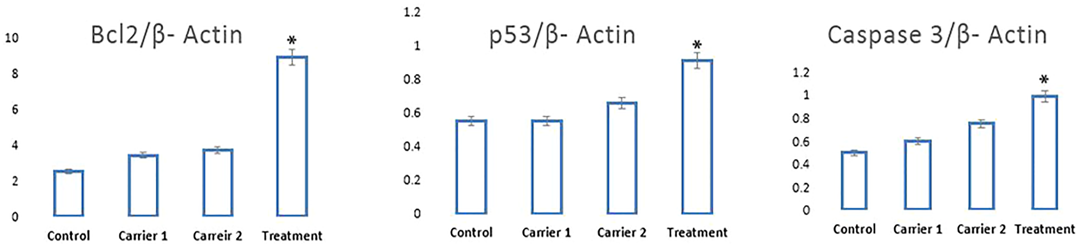
Reverse transcription–polymerase chain reaction results for mRNA levels of Bcl-2, caspase 3, and p53 based on β-actin intensity. EEPNPs increased the mRNA level of caspase 3, Bcl-2, and p53 on day 7 post operation. All data are presented in mean ± SEM. *P < .05 versus other groups.
Discussion
Inflammation, proliferation and tissue remodeling are three phases of healing process which occur following tissue damages as closely as possible to its natural state. The healing process is activated when platelets come into contact with exposed collagen leading to platelet aggregation and the release of clotting factors resulting in the deposition of a fibrin clot at the site of injury. The fibrin clot serves as a provisional matrix and sets the stage for the subsequent events of healing. Inflammatory cells also arrive along with the platelets at the injury site providing key signals known as growth factors. The fibroblast is the connective tissue cell responsible for collagen deposition required to repair the tissue injury. The collagen is the main constituent of extra cellular tissue, which is responsible for support and strength. 28
Nanoparticles have become significant in the regenerative medicine field in the last two decades. 29 Many biological processes happen at through mechanisms that fundamentally act at the nanometer scale. Thus, materials such as NPs can be used as unique tools for drug delivery, imaging, sensing, and probing biological processes. 30 In the context of wound healing, the special properties of NPs like electric conductivity, antimicrobial activity, and high surface to volume ratio, swelling, and contraction make NPs versatile resources.
Angiogenesis and expansion of blood vessels is one of priorities in wound healing that can increase blood expansion through biochemical and pharmacological mechanisms and consequently lead to successful healing acceleration. 31 According to results of the present study, maximum angiogenesis was observed in Treatment group on day 7 with significant difference compared to control group. Our findings showed the stimulating effect of Treatment on angiogenesis. Angiogenesis provide an outline for connective tissue formation in early days of healing process. Following establishing adequate blood circulation in granulation tissue, immigration and proliferation of endothelial cells are reduced and apoptosis of redundant blood vessels occurs. 32 Acceleration in blood vessel reduction of the groups treated by EEP-PLGA NPs showed positive effect of EEP-PLGA NPs in Treatment in healing.
Fibroblast numbers is a well-known index for quality assessment of connective tissue healing. Presence and early proliferation of fibroblasts in Treatment group demonstrated growth stimulation of fibroblasts by Treatment. Others conducted a survey on the effects of nanocomposite of chitosan-titanium dioxide on excisional wound and suggested healing improvement by cell growth stimulation of Treatment. 33 Fibroblast creates extracellular matrix and collagen that play essential role in wound healing phases. 34 Various modalities are applied directly to the wound to stimulate fibroblast proliferation and accelerate wound closure. After that fibroblast reduction plays a role in decrease of scar fibrosis and potentially improving cosmoses. In the present study application of EEP-PLGA NPs resulted in early increase in fibroblasts followed by decrease in fibroblast count to achieve less scar formation target. 35
Granulation tissue formation at early days of healing process is considered as one of momentous factors in healing acceleration. 36 The more fibroblast and blood vessel forms, the more granulation tissue develops. In the present study, maximum thickness of granulation tissue of Treatment group were observed on day 7 and Control on day 14. Maturation of granulation tissue and decrease in its thickness of Treatment group initiated on day 7 and followed by the minimum thickness on day 21. At the end of the study period, maximum thickness of granulation tissue was observed in Control animals with significant difference with Treatment group. This was in agreement with the results of angiogenesis and fibroblast proliferation that were responsible for maturation of blood vessels, fibroblast and healing enhancement Treatment group.
Inflammatory phase is the first step of wound repair and is determined by the presence of inflammatory cells. This phase is critical due to association of proliferative, re-epithelization, contraction and wound closure with cytokines secreted in inflammatory phase. 37 In the present study Treatment group showed the least numbers of inflammatory cells on day 7 that could be associated with anti-inflammatory of EEPNPs that could reduce polymorphonuclear leukocytes (PMN) cells via reduction in secondary infection and acceleration in inflammation initiation and termination. Presence of few numbers of PMN cells and high numbers of blood vessels and fibroblast on day 7 in Treatment group indicated healing acceleration in nanocomposite treated animals.
Re-epithelization makes a barrier between wound and environment in wound healing. 38 Newly formed epithelium is characterized by more cells and layers compared to normal epithelium. As soon the wound surface is covered by new epidermal cells, differentiation initiates, cell shape changes to normal and rearrangement and reduction of cell layer occurs. 39
Based on the results of the present study, epithelization in Treatment group was approximately complete on day 14 and one week earlier than others followed by complete epithelization on the last day of the experiment. This outcome was indicative of epithelization stimulation effect of titanium dioxide that was consistent with those of other studies. 39
Despite of sterility of burn wounds upon creation, due to existence of vascular necrotic tissue and loss of epithelial integrity, the wound is disposed to infection with failure in healing and mortality in more severe cases as wound infection consequences. Presence of neutrophils in histopathological sections is indicator of wound infection. 40
According to results of the present study Treatment group on days 7 and 14 showed the least infection, existed only on scab and epidermis and no infection was observed on day 21. It could be concluded that nanocomposite of Treatment had antibacterial properties that was in agreement with those of other studies. 41 In the present study, antibacterial effect of Treatment were detected more than silver sulfadiazine. Early epithelization in Treatment group prevented wound from infection and penetration of microorganisms to the healing tissue. Both titanium dioxide and gelatin as the carrier exhibited hydrophilic feature and accelerated healing process.
Enhancement of burn wounds is associated with reepithelization, fibroblast proliferation and angiogenesis. 42 Since the pathological phases of wound healing process have dynamic feature and results of inflammatory, proliferative and remodeling phases are dependent on each other, for appropriate conclusion, all the healing factors should be considered together. Therefore, histomorphometrical results of our study were scored from one to five grade. Higher grades represented healing improvement. Consequently, the highest grade indicated the best healing activity by enhancing inflammatory, proliferative and remodeling phases. Based on the results of the present study, high percentage of wound contraction and total pathological grade as well as insignificant or absence of infection were observed in Treatment group that were in agreement with those of other studies. 34
Family of proteins the Bcl-2 inhibits apoptosis as apoptosis pathway effectors.43,44 However, the guardian of the genome, caspase and p53, regulate the destiny of damaged cells via detection and ending the cell cycle. In damaged tissue, increase in the levels of p53 and caspase are observed to triggers apoptotic activity of the immune cells that ends up exclusion of the immune cells. Following this phase, the Bcl-2 inhibits the apoptotic activity that induce proliferation of cells. 45 In the present study analyzes of RT-PCR indicated that in Treatment animals, expressions of caspase 3, Bcl-2, and p43 were amplified. Therefore, it might be estimated that EEPNPs improved proliferation the cells via upregulation in the expression of caspase 3, Bcl-2, and p43.
In conclusion, based on the results of the present study it could be concluded that EENPs offered potential advantages in burn wound healing acceleration and improvement through angiogenesis stimulation, fibroblast proliferation, granulation tissue formation in early days of healing phases, acceleration in wound repair associated with earlier wound contraction and stability of damaged area by rearrangement of granulation tissue and collagen fibers.
Footnotes
Acknowledgments
The authors would like to thank, Solid Tumor Research Center, Urmia University of Medical Sciences and Urmia Pathobiology Center for their technical expertise.
Declaration of Conflicting Interests
The author(s) declared no potential conflicts of interest with respect to the research, authorship, and/or publication of this article.
Funding
The author(s) received no financial support for the research, authorship and/or publication of this article.
Ethical Approval
Not applicable, because this article does not contain any studies with human or animal subjects.