
Abstract
Background/objective
Fetal wound repair seems a relatively efficient process when compared to wound repair during adulthood, which may be explained by the effects of the fetal environment. This study examined the effects of an amnion cell culture medium (ACCM) on skin wound healing in a rat experimental model.
Methods
Sixteen adult female Wistar albino rats were used in this experimental animal study (treatment group, n = 8; control group, n = 8). Surgical wounds were formed on the dorsal skin of each rat. A commercially available ACCM was administered daily over each of the wound in the treatment group for 14 days and the control group did not receive any treatment. Wounds were evaluated for tissue perfusion with laser doppler, tissue superoxide dismutase (SOD), glutathione peroxidase (GPX), and malondialdehyde (MDA) levels as well as histopathological examination.
Results
Controls had significantly higher tissue SOD levels when compared to the treatment group (10.0 ± 3.2 vs 6.7 ± 1.2, P = .005); however, the 2 groups did not differ in terms of tissue GPX and MDA levels. For open wounds, inflammation and neovascularization were more prominent in the ACCM group at day 14. However, at day 21 neovascularization and granulation were more prominent in controls. For closed wounds, neovascularization was more prominent in controls at days 14 and 21. The 2 groups did not differ in terms of tissue perfusion.
Conclusion
Although marginal difference was found between controls and ACCM group for several parameters, findings of this study do not support beneficial effect of ACCM on wound healing.
Skin wound represents a challenging healthcare problem during adult life since it may result in considerable disability, debilitating complications, even death. Wound healing is even more problematic when it is complicated by a condition with negative effects on the healing process, like diabetes. Effective wound care to ensure rapid healing and to prevent complications is always an integral part of patient care.
Wound healing is a complex process consisting of inflammation, granulation, and remodeling.1,2 Although skin wounds of the fetus heal rapidly without scar, wound repair during adult life is rather challenging since it is slower and results in scar formation.1–3 This difference stimulated research on fetal environment, particularly on the potential use of fetal amniotic fluid or its components for improving wound repair. Several studies tested amniotic fluid, amniotic fluid-derived cells, or amniotic membrane and obtained encouraging results where wound healing was improved through different potential mechanisms.4–15
AmnioPrime is a complete culture medium used for the cultivation of amnion and chorionic villi cells for diagnostic purposes. Few studies have already examined the effect of culture mediums for amnion cells on wound healing, ischemia/reperfusion damage, and necrosis and found positive effects.16–20
This study aimed to examine the effects of amnion cell culture medium on skin wound healing through the evaluation of perfusion, tissue antioxidant levels, and histopathological examination in a rat experimental model.
Methods
Experimental Animals and Wound Model
Animals
A total of 16 adult female Wistar albino rats weighing between 250 and 300 g were used in this experimental animal study. The rats were randomly divided into 2 groups: the treatment group (n = 8) and the control group (n = 8). The study protocol was approved by local ethics committee for animal studies.
Wound Model
Following shaving, cleaning with alcohol, and intraperitoneal anesthesia with 7 mg/kg xylazine and 35 mg/kg ketamine, 6 surgical wounds were formed on the dorsal skin of each rat using a 6 mm punch biopsy instrument. Surgical wounds were 1 cm apart from midline and from each other. After getting a punch biopsy from each site, the wound was either left open (3 open wounds) or punch biopsy material from the other side was stitched with a 5/0 Vicryl suture as graft (3 closed wounds).
Interventions
A commercially available amnion cell culture preparation (AmnioPrime, Capricorn Scientific GmbH, Ebsdorfergrund, Germany) was administered daily over each of the wound in the treatment group for 14 days (0.5 cc over each wound topically). Control group did not receive any treatment.
Assessments
Biopsies were obtained from all wound surfaces for histopathological examination and all wound surfaces were examined for tissue perfusion using laser camera at baseline and at days 7, 10, 14, and 21. In addition, tissue samples were obtained for tissue superoxide dismutase (SOD), glutathione peroxidase (GPX), and malondialdehyde (MDA) measurements at day 21.
Evaluation of Perfusion With Laser Doppler
The PeriScan PIM 3 system (Perimed AB, Stockholm, Sweden, a laser doppler blood perfusion imager) was used to evaluate tissue perfusion in the wounds with laser doppler method. This no-touch system measures blood flow at microcirculation level with the aid of a computer software.
Tissue SOD, GPX, and MDA Levels
Following homogenization of the tissue specimens, SOD, GPX, and MDA levels were measured using enzyme-linked immunosorbent assay (ELISA) method in ng/mL, ng/mL, and mmol/mL, respectively (Rat SOD, GPX, and MDA ELISA Kits, SunRed Biotechnology Company, China).
Histopathological Examination
For histopathological examination, hematoxylin & eosin-stained preparations were examined for the following parameters and scored: inflammation, neovascularization, granulation, and fibroblast regeneration. For scoring, prepared slides were scanned using a digital pathology system (3D Histech company, P250—Flash III Digital Scanner, 20×) and microscopic photos were taken using a special software (3D Histech company, CaseViewer program, tiff format and 300 dpi). The ratios of the abovementioned pathological parameters to the entire tissue were calculated and scoring was done as follows: 1 point, 0% to 20%; 2 points, 21% to 40%; 3 points, 41% to 60%; 4 points, 61% to 80%; 5 points, 81% to 100% of the total tissue.
Statistical Analysis
Statistical Package for Social Sciences (SPSS) version 21 was used for the analysis of data. Normality of continuous variables was tested with both hypothesis test (Shapiro-Wilk test) and graphical methods. Descriptive data are presented in mean ± standard deviation, median (range), or number (percentage), where appropriate. For comparison of the groups in terms of continuous variables, student's t test for independent samples or Mann-Whitney U test was used depending on normality. Pearson's Chi-square test or Fisher's exact test was used for the comparison of categorical variables. A P value <.05 was considered indication of statistical significance.
Results
Tissue SOD, GPX, and MDA Levels
Table 1 shows the comparison of controls and amnion cell culture medium (ACCM) groups in terms of tissue SOD, GPX, and MDA levels. Overall, controls had significantly higher tissue SOD levels when compared to the treatment group (10.0 ± 3.2 vs 6.7 ± 1.2, P = .005); however, the 2 groups did not differ in terms of tissue GPX and MDA levels. Subgroup analyses based on wound type (open or closed) revealed a similar relationship, where tissue SOD levels were still higher in controls in both open wounds (8.5 ± 0.9 vs 6.8 ± 1.5, P = .039) and closed wounds (11.4 ± 4.1 vs 6.6 ± 0.9, P = .034). Like the overall analysis, controls and ACCM group did not differ in terms of tissue GPX and MDA levels in either open wounds or closed wounds.
Comparison of the Groups in Terms of Tissue SOD, GPX, and MDA Concentrations.
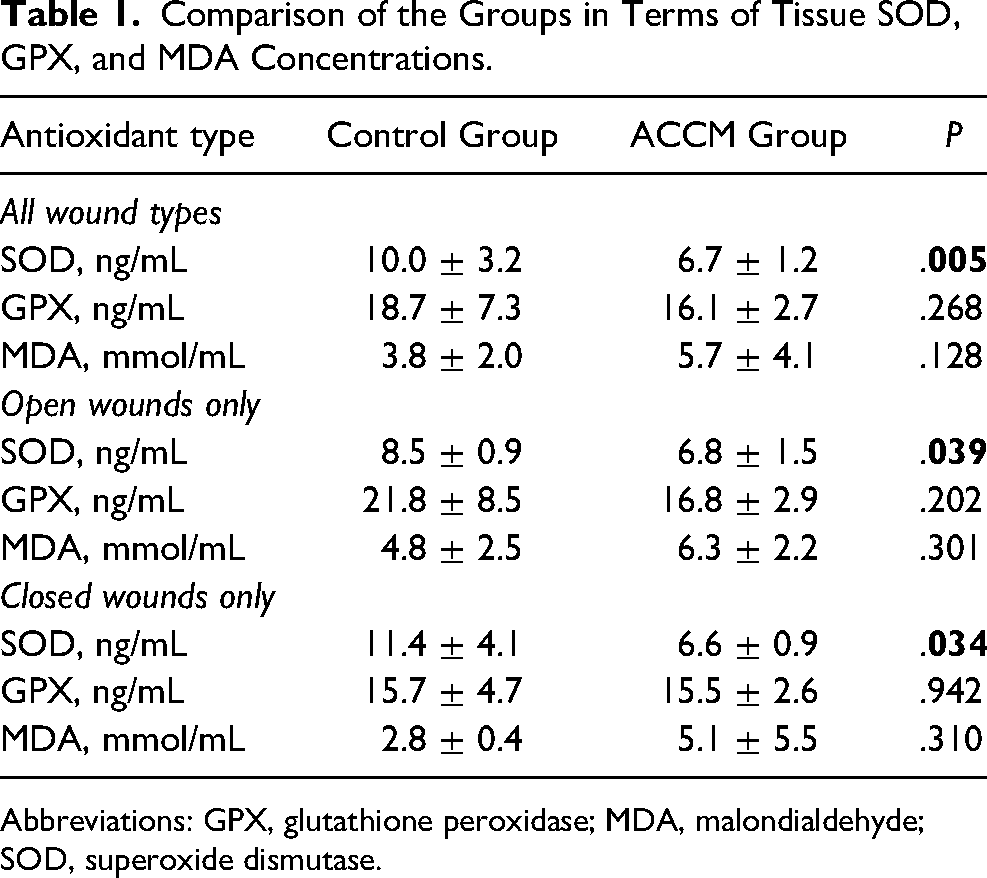
Abbreviations: GPX, glutathione peroxidase; MDA, malondialdehyde; SOD, superoxide dismutase.
Histopathological Examination
Tables 2 and 3 show the comparisons of the groups in terms of histopathological examination. For open wounds, inflammation and neovascularization were more prominent in the ACCM group at day 14. However, at day 21 neovascularization and granulation were more prominent in controls. For closed wounds, neovascularization was more prominent in controls at days 14 and 21.
Comparison of the Groups in Terms of Histopathological Examination of the Open Wounds.
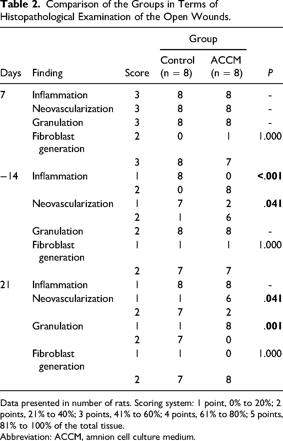
Data presented in number of rats. Scoring system: 1 point, 0% to 20%; 2 points, 21% to 40%; 3 points, 41% to 60%; 4 points, 61% to 80%; 5 points, 81% to 100% of the total tissue.
Abbreviation: ACCM, amnion cell culture medium.
Comparison of the Groups in Terms of Histopathological Examination of the Closed Wounds.
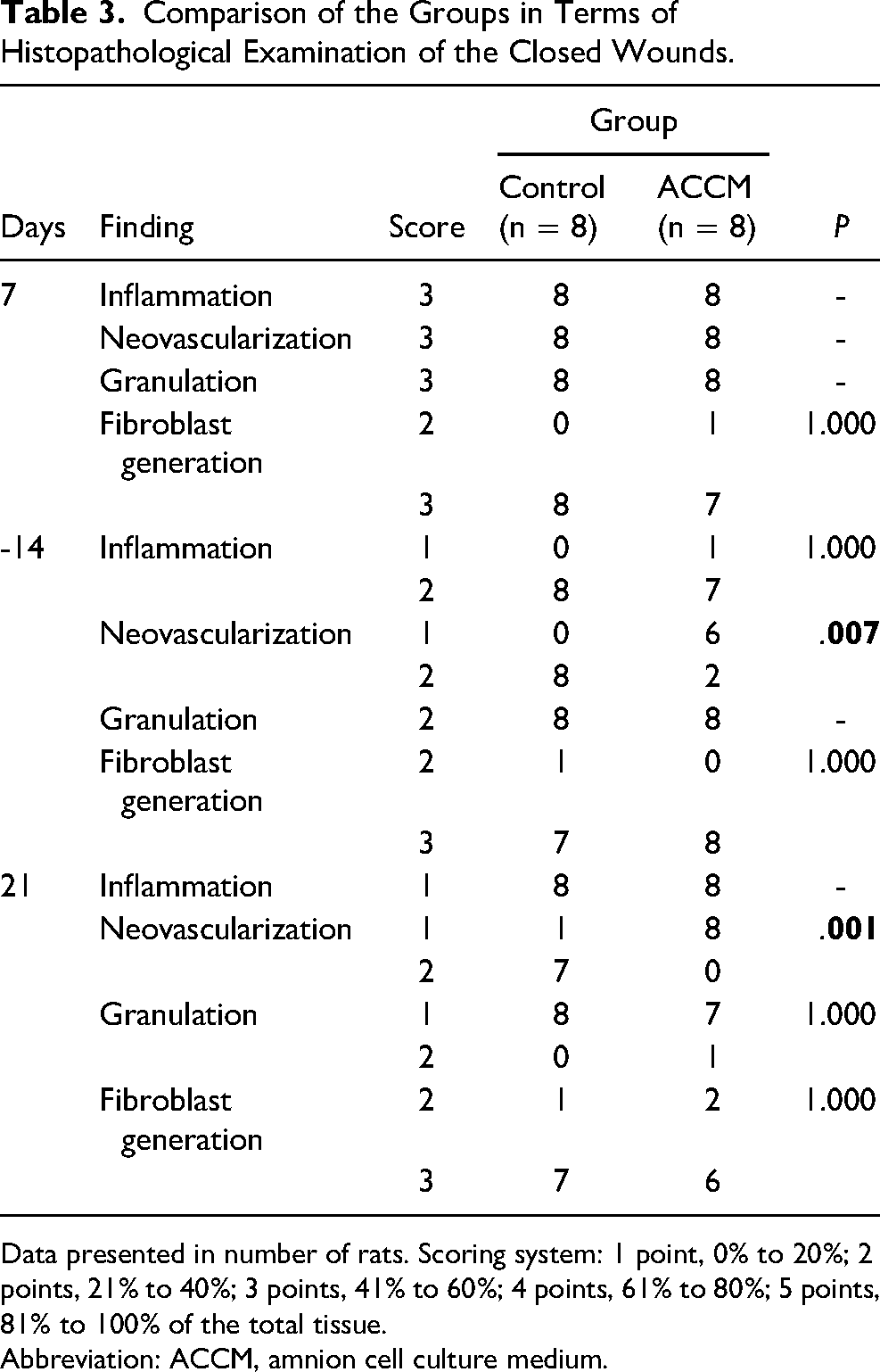
Data presented in number of rats. Scoring system: 1 point, 0% to 20%; 2 points, 21% to 40%; 3 points, 41% to 60%; 4 points, 61% to 80%; 5 points, 81% to 100% of the total tissue.
Abbreviation: ACCM, amnion cell culture medium.
Tissue Perfusion
Table 4 shows the comparison of the groups in terms of tissue perfusion at different time points. No significant difference was found between groups in all time points and for both wound types (open or closed). Similarly, Figure 1 shows the changes in tissue perfusion over time in open wounds and closed wounds, respectively. As indicated by error bars, the 2 groups did not differ in terms of tissue perfusion.
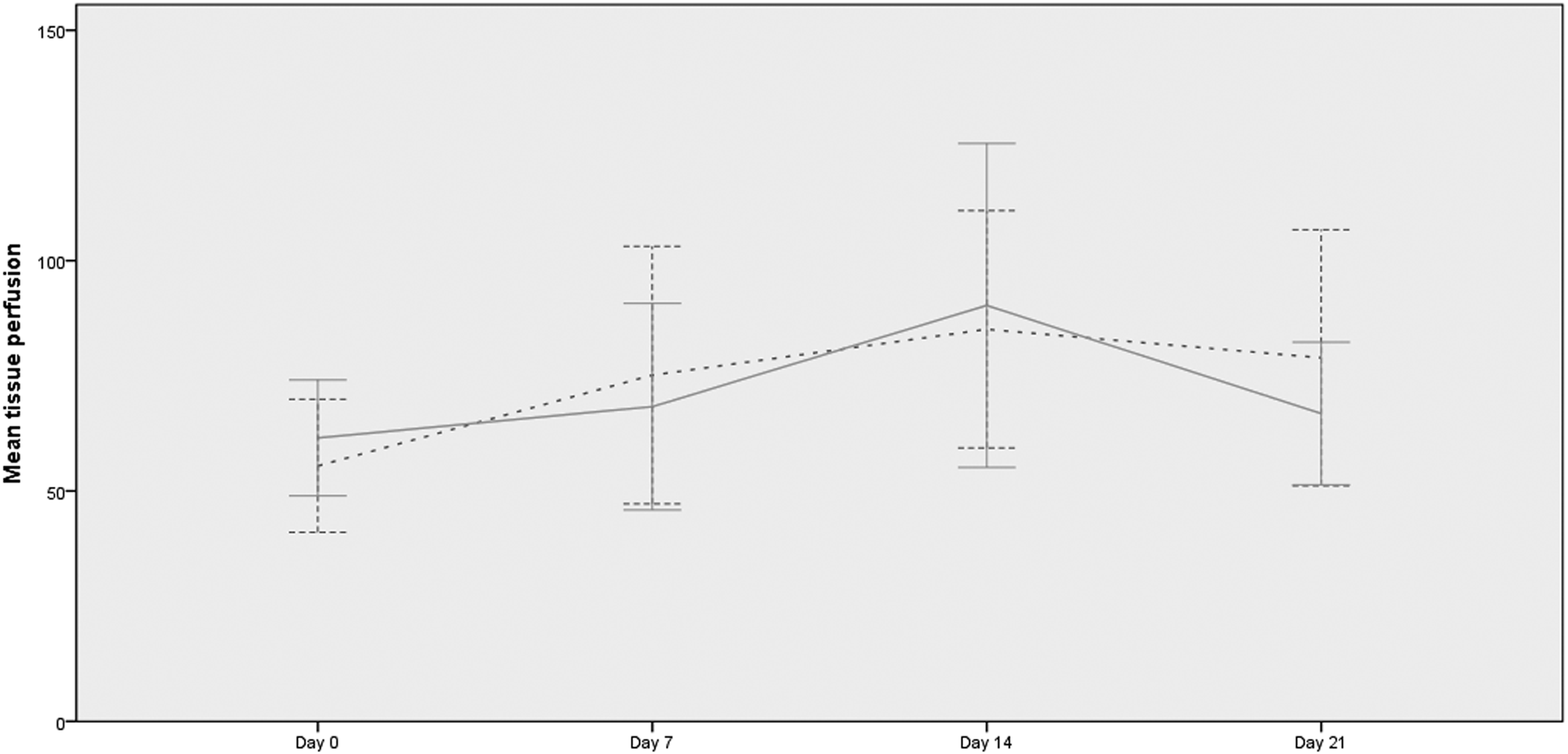
Changes in tissue perfusion of the wounds over time. (A) open wounds, (B) closed wounds. Error bars indicate 95% confidence intervals. Straight lines indicate ACCM group, dotted lines indicate controls. Abbreviation: ACCM, amnion cell culture medium.
Comparison of the Groups in Terms of Tissue Perfusion at Different Time Points.
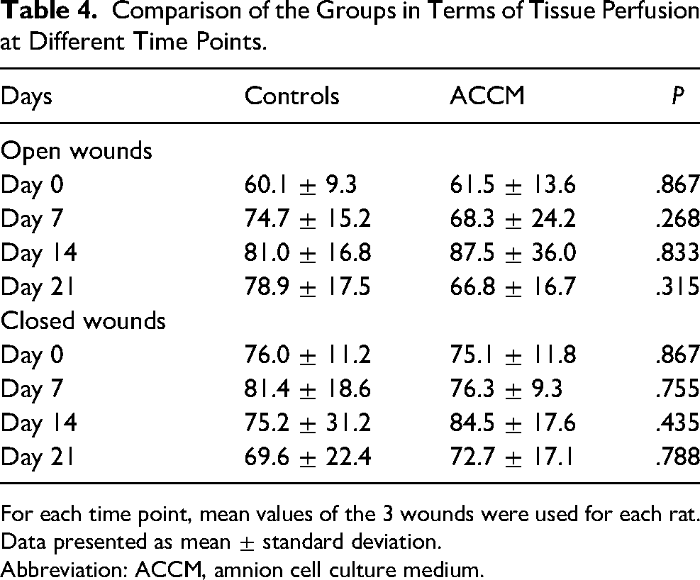
For each time point, mean values of the 3 wounds were used for each rat. Data presented as mean ± standard deviation.
Abbreviation: ACCM, amnion cell culture medium.
Discussion
This study examined any potential benefit of ACCM application on wound healing of surgically formed rat skin injury; however, did not find any benefit during 21-day period. This study is among the few studies on the effect of ACCM on wound healing, a solution used for the cultivation of amnion cells; thus, may have a composition to promote the wound healing process.
To date, several studies have examined the effect of amnion cell culture medium on wound healing. In a recent 2021 study, Mirapoglu et al 19 examined the effects of platelet rich plasma (PRP), ACCM, and controls on wound healing in an animal tracheal injury model. Morphometric, histological, and biochemical evaluation of samples showed that both ACCM and PRP has some beneficial effects on wound healing. Both ACCM and PRP had better morphometric results, higher total antioxidant levels, and lower interleukin 6 (IL-6), IL-1, and vascular endothelial growth factor levels than controls. In a 2020 study, the effect of ACCM on necrosis was examined in a McFarlane dorsal skin flap model, 18 where ACCM resulted in less skin necrosis. In addition, blood flow was higher in the ACCM group than controls at the 10th day of the application.
Potential effects of ACCM on ischemia/reperfusion injury have also been examined. In a recent 2021 study, Aydogdu et al 17 investigated the effects of ACCM on experimental rat testis ischemia/reperfusion injury model. Testicular intraparenchymal injection of ACCM 1 min after detorsion resulted in lower degree of necrosis. The authors concluded that injection of ACCM is more favorable when performed after detorsion. Ozturk et al 20 conducted an experimental study using rat ovarian torsion model to examine the effect of ACCM (applied either topical or intraparenchymal) on ischemia/reperfusion injury. Total antioxidant capacity, total oxidant capacity, oxidative stress index, IL-1B, IL-6, and tumor necrosis factor alpha levels were compared. For both ACCM application methods, oxidative stress and inflammatory marker levels were better; however, total antioxidant capacity, oxidative stress index, IL-1B, IL-6, and tumor necrosis factor alpha levels were even better for topical application. Similarly, Akyol et al 16 investigated the effect of renal ACCM injection (either subcapsular or supracapsular) using a rat kidney ischemia/reperfusion injury model. Both treatment types reduced oxidative stress and inflammation, but supracapsular therapy reduced them more than subcapsular therapy.
Abovementioned studies all found some benefit of the use of ACCM for wound healing or ischemia/reperfusion injury. However, findings of the present study are not in line with these previous studies. One possible explanation may be the use of a different brand of medium in previous studies, which may have different composition. Another explanation may be the lack of amniotic fluid components in ACCM such as amnion cells, growth factors, hyaluronic acid, and related cells and biological molecules. Prominent neovascularization and granulation in controls at the end of the study and lower SOD levels in ACCM group may even be interpreted as a potential interference or suppression by ACCM. However, it should be born in mind that several studies have found significant effects; thus, future studies with better design would shed light on the issue.
In conclusion, findings of this experimental study do not support beneficial effect of ACCM on wound healing based on perfusion, histopathology, and biochemical evaluation. Although marginal difference was found between controls and ACCM group for several parameters, they may be even interpreted as an unfavorable effect of ACCM. Larger studies are warranted.
Footnotes
Declaration of Conflicting Interests
The author(s) declared no potential conflicts of interest with respect to the research, authorship, and/or publication of this article.
Funding
The author(s) received no financial support for the research, authorship, and/or publication of this article.