
Abstract
There is accumulating evidence that magnesium, an important mineral having a pivotal role in many physiological functions, may be important in development and healing of diabetic foot ulcers (DFUs). In this non-systematic mini review, we discuss the role of magnesium in DFUs, as well as the effects of magnesium administration in DFUs. Reduced Mg levels appear to be associated with DFUs. Moreover, Mg administration may be beneficial for the outcome of DFUs. Further investigation is imperative in order to shed more light on these findings.
Diabetic foot ulcers (DFUs) are closely associated with increased risk of infection, hospitalization, amputations, and mortality.1,2 Magnesium (Mg) is the fourth most abundant mineral in the human body.3,4 It is a cofactor for more than 300 enzymatic reactions. 3 As a result, Mg intake is crucial for many biochemical pathways and for prevention of various diseases, such as chronic degenerative, cardiac, and neuromuscular disorders.3,4
It has been already demonstrated that nutritional status may have an important impact on DFUs. 5 In this context, the aim of the present mini review was to discuss the role of Mg levels and its administration in DFUs.
Search Strategy
In this non-systematic mini review, an electronic search was conducted in PubMed from July 2001 until March 2023 using combinations of the following keywords: “magnesium” “diabetic foot”, “nutrition”, “diabetic foot ulcers”. Only studies written in English were included. Additionally, references of all included articles were studied. We only included original studies evaluating the interplay between the magnesium and DFUs, as shown in Tables 1 and 2.6–13 Animal studies were excluded.
Magnesium Levels and DFUs.
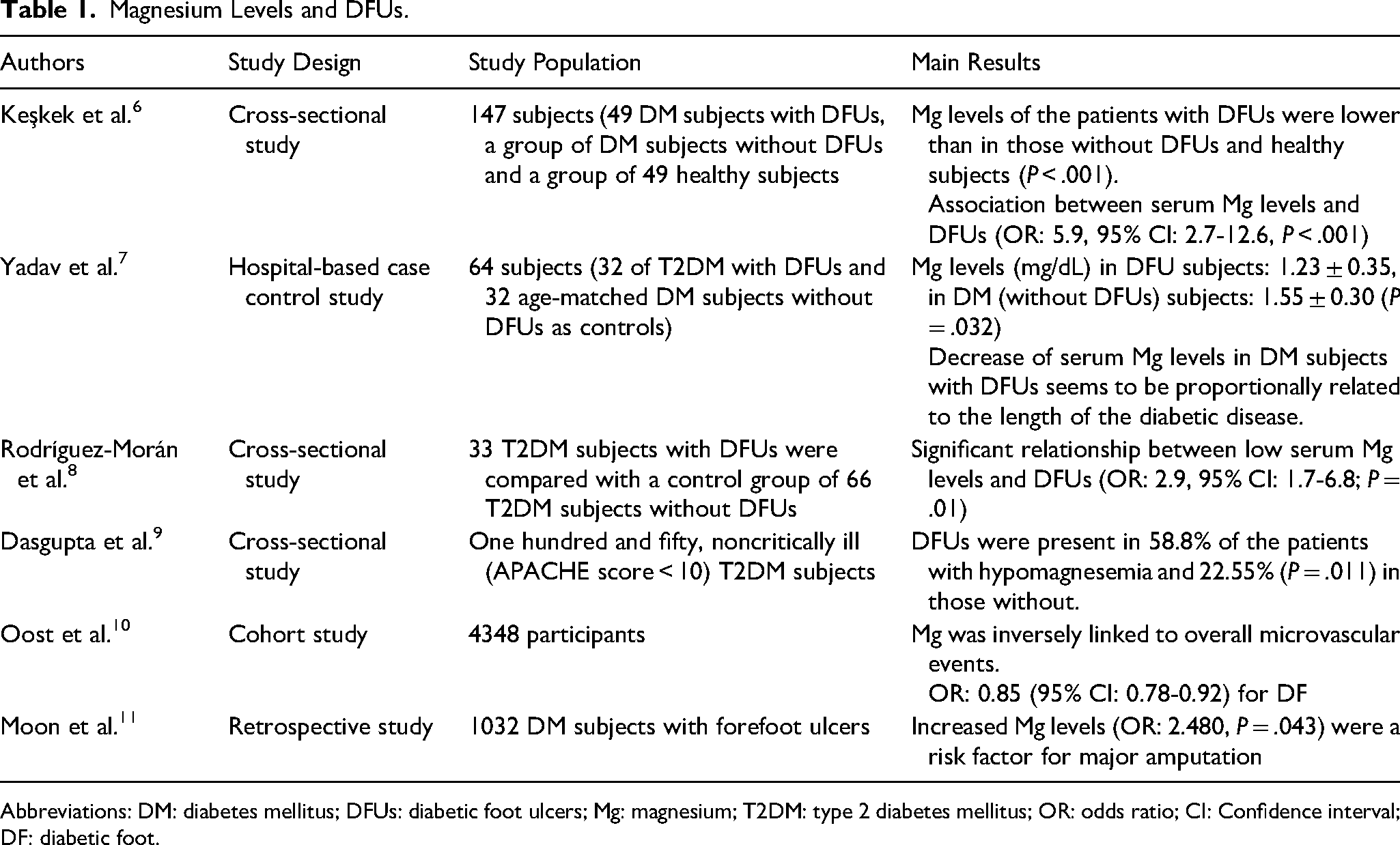
Abbreviations: DM: diabetes mellitus; DFUs: diabetic foot ulcers; Mg: magnesium; T2DM: type 2 diabetes mellitus; OR: odds ratio; CI: Confidence interval; DF: diabetic foot.
The Effect of Magnesium Administration on DFUs.
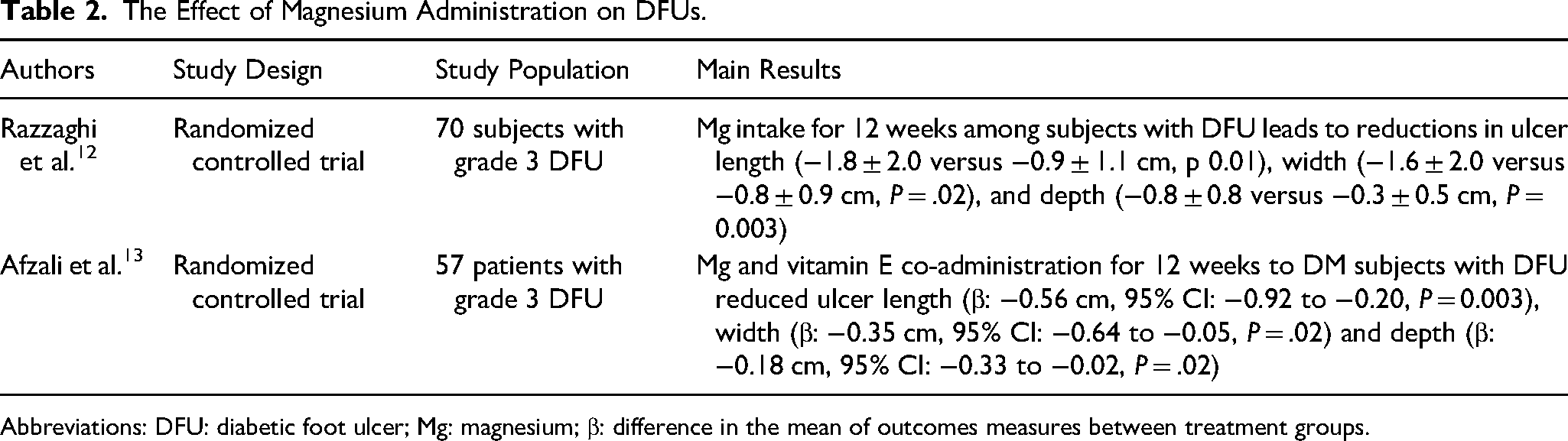
Abbreviations: DFU: diabetic foot ulcer; Mg: magnesium; β: difference in the mean of outcomes measures between treatment groups.
Magnesium: An Important Nutrient
Mg is an important and essential mineral of the human body which is crucial to maintain normal cellular and organ function. 14 The origin of the name derives from Magnesia, which is a district in Greece. 15 This nutrient is required for DNA and RNA synthesis and protein synthesis, while it is important for the regulation of blood pressure, insulin metabolism, muscular contraction, vasomotor tone, cardiac excitability, nerve transmission, and neuromuscular conduction. 3
The main source of Mg is diet, especially whole and unrefined grains, cocoa, nuts, seeds, green leafy vegetables, and almonds. 16 Reduced Mg levels are commoner than hypermagnesemia, but they only become manifest in severe deficiencies such as muscle spasms, cramps, paresthesia and arrhythmias.3,15 Critical reference values are below 0.5 mmol/L (or 1.0 mg/L) and above 2.0 mmol/L (or 4.9 mg/dL). 15 On the other hand, hypermagnesemia is a quite rare but possibly fatal electrolytic disorder. 15
Low Mg levels may be linked with numerous chronic diseases, notably Alzheimer's disease, hypertension, stroke, ischemic heart disease, myocardial infarction, type-2 diabetes mellitus (T2DM), migraine, gastrointestinal diseases, liver cirrhosis, and thyroid dysfunction.3,16
In therapy, Mg is mainly administered orally. In urgent situations, like arrhythmias or pre-eclampsia, it is administered intravenously.17,18
Levels of Magnesium and DFUs: The Potential Connection
Keşkek et al. 6 looked at the interplay between serum Mg levels and DFUs (Table 1). They included 147 subjects divided into three groups: a study group (N = 49) with DM and DFUs, a control group (N = 49) with DM without DFUs, and a control group (Ν = 49) of healthy participants. 6 These groups were similar in terms of age and sex. Mg levels were lower among DM subjects with DFUs than in the other two groups (P < .001). In addition, it seemed to be a strong relationship between the serum Mg levels and the incidence of DFUs (odds ratio [OR]: 5.9, 95% confidence interval [CI]: 2.7–12.6, P < .001). 6
Yadav et al. 7 conducted a hospital-based case control study including 64 DM participants aged 40–60 years. Among them, 32 subjects had DFUs, and 32 age-subjects had no DFUs. 11 Serum zinc, Mg, and copper levels were estimated by colorimetric methods in semi-autoanalyser. These were significantly decreased among subjects with DFUs (P < .05). 7 In addition, more severe deficiencies of these three minerals were associated with worse glycemic control. 7
Rodríguez-Morán et al. 8 investigated the interplay between low Mg levels and DFUs (<2 months duration) in T2DM. They included 33 out-patients with T2DM and DFUs (group A: 16 women and 17 men) and a control group of 66 out-patients with T2DM without DFUs (group B: 35 women and 31 men). 8 Serum Mg levels were assessed by colorimetric method and the association between serum Mg and foot ulcers was assessed by logistic regression. Low Mg levels were seen more frequently in group A (93.9% vs 73.1%, P = .02). 8 In addition, Mg levels were lower in group A than in group B 1.48 ± 0.33 versus 1.68 ± 0.32, P < .001). Lower Mg levels increased the likelihood of having DFUs (OR: 2.9, CI 95% 1.7–6.8; P = .01). 8
Dasgupta et al. 9 studied 150 non-critically ill (APACHE score < 10) T2DM subjects admitted for uncontrolled hyperglycemia and/or various diabetic complications. Hypomagnesemia (Mg < 1.6 mg/dL) was recorded in 17 (11.33%) subjects with a female: male ratio of 9:8. Mean HbA1c was lower among hypomagnesemic in comparison with normomagnesemic subjects (11.9% vs 9.8%, P = .0016). 9 Frequency of retinopathy, microalbuminuria, macroalbuminuria, foot ulceration, and neuropathy was 64%, 47%, 17.64%, 58.8%, and 82.35%, respectively, among subjects with hypomagnesemia. This compared with 45.8% (P = .118), 38.34% (P = .704), 15.03% (P = .566), 22.55% (P = .011) and 82.7% (P = .976) among subjects with normal Mg. 9
Others examined whether serum Mg was prospectively linked to macro- or microvascular complications in 4348 DM subjects. 10 Average baseline serum Mg concentration was 0.80 ± 0.08 mmol/L. Importantly, while during 5.1 years of follow-up, serum Mg was inversely linked to DFUs (OR: 0.85, 95% CI: 0.78–0.92) at 5.1 years of follow-up. 10
Another work included 1032 hospitalized DM subjects with forefoot ulcers, they tried to investigate the risk factors for major amputation. 11 The authors identified 88 risk factors of major amputations in univariate analysis. In multivariate analysis after adjustment, the OR for Mg levels was 2.480 (95% CI: 1.027-5.986, P = .043), pointing to a significant effect of increased Mg levels in amputation risk. 11
Impact of Magnesium Administration on DFUs
Razzaghi et al. 12 carried out a randomized controlled trial to examine the impact of Mg administration on wound healing and metabolic status in DM subjects with DFUs (Table 2). They included 70 subjects with grade 3 DFUs, randomly divided into two groups (35 subjects in each group): 250 mg Mg oxide supplements (group A) or placebo daily (group B) for 12 weeks. After 12 weeks, subjects in group A exhibited a significant increase in serum Mg (0.3 ± 0.3 vs −0.1 ± 0.2 mg/dL, P < .001) and significant reductions in ulcer length (−1.8 ± 2.0 versus −0.9 ± 1.1 cm, P = .01), width (−1.6 ± 2.0 versus −0.8 ± 0.9 cm, P = .02), and depth (−0.8 ± 0.8 versus −0.3 ± 0.5 cm, P = .003). Moreover, significant reductions in fasting plasma glucose (−45.4 ± 82.6 versus −10.6 ± 53.7 mg/dL, P = .04), serum insulin values (−2.4 ± 5.6 versus +1.5 ± 9.6 μIU/mL, P = .04), and HbA1c (−0.7 ± 1.5 versus −0.1 ± 0.4%, P = .03) and a significant rise in the quantitative insulin sensitivity check index (+0.01 ± 0.01 vs 0.004 ± 0.02, P = .01) were seen in group A. 12
Afzali et al. 13 examined the impact of Mg and vitamin E co-administration on wound healing and metabolic status in 57 DM subjects with grade 3 DFUs. Participants were randomized to 250 mg Mg oxide plus 400 IU vitamin E (group A, N = 29) or placebo per day (group B, N = 28) for 12 weeks. Subjects in group A exhibited reduced ulcer length (mean difference −0.56 cm, 95% CI: −0.92 to −0.20, P = .003), width (mean difference −0.35 cm, 95% CI: −0.64 to −0.05, P = .02) and depth (mean difference −0.18 cm, 95% CI, −0.33 to −0.02, P = .02). 19 They also exhibited significant reductions in fasting plasma glucose (mean difference −13.41 mg/dL, 95% CI; −20.96 to −5.86, P = .001), insulin resistance (mean difference −0.60, 95% CI: −0.99, −0.20, P = .003), and HbA1c (mean difference −0.32%; 95% CI to −0.48, −0.16, P < .003). 13
Discussion
This review has discussed the evidence on the role of Mg in DFUs. It is becoming clear that low Mg levels may be associated with DFUs. Moreover, Mg administration appears to be of some benefit in terms of DFUs healing.
However, there are important limitations. First, participant numbers were small. Moreover, most studies were single-center. Thirdly, follow-up was not long.
Thus, we need multi-center studies with more participants and longer follow-up. It would also be interesting if these results could be validated by experimental results or even in silico models. Other issues to determine are optimal Mg dosage and treatment duration, as well as the contribution of other co-administered minerals and vitamins. Answering these questions will enrich our knowledge of the conundrum of nutritional support for DFUs.19–24
In conclusion, it seems that low levels of Mg are closely linked to DFUs outcomes and can be present in DM subjects with DFUs. In addition, the administration of Mg supplements could be particularly beneficial for ulcer size and glycemic control. Nevertheless, further studies are essential in order to validate these claims.
Footnotes
Declaration of Conflicting Interests
N. Papanas has been an advisory board member of Astra-Zeneca, Boehringer Ingelheim, MSD, Novo Nordisk, Pfizer, Takeda and TrigoCare International; has participated in sponsored studies by Astra-Zeneca, Eli-Lilly, GSK, MSD, Novo Nordisk, Novartis and Sanofi-Aventis; has received honoraria as a speaker for Astra-Zeneca, Boehringer Ingelheim, Eli-Lilly, ELPEN, Galenica, MSD, Mylan, Novo Nordisk, Pfizer, Sanofi-Aventis, Takeda and Vianex; and attended conferences sponsored by TrigoCare International, Eli-Lilly, Galenica, Novo Nordisk, Pfizer and Sanofi-Aventis.
Funding
The author(s) received no financial support for the research, authorship, and/or publication of this article.