
Abstract
Treatment of chronic wounds has been shifted to traditional approaches due to surge in antibiotic resistance. Wounds that fail to heal satisfactorily may result in the amputation of the organ. In this research work, cinnamon oil (CO) and aloe vera (AV) that have been traditionally used as antibacterial agents are combined in a unique gel (COVA) and its antibacterial activity has been evaluated through in vitro and in vivo studies. Antibacterial activity was measured through disk diffusion and agar dilution method against Pseudomonas aeruginosa and Staphylococcus aureus. To check antibacterial and wound healing activity, diabetic excision wound healing rat model was used. Wound closure, wound contraction, tissue hydroxyproline content, antioxidant capacity (TAC), and malondialdehyde (MDA) level were monitored. The minimum inhibitory concentrations of CO + AV for bacterium P. aeruginosa and S. aureus were 100 and 200 µg/ml, respectively. After 14 days, the wounds covered with COVA therapy reached to nearly full wound closure (79% wound contraction) compared to control. The collagen content and level of TAC increased significantly (P < 0.05) in treated groups; therefore, 25% fast healing was observed in wounds treated with CO and AV gel combined. Reduced levels of tissue MDA were observed in all treated groups and specially wound covered with COVA (0.43 mM/mg in control vs 0.25 mM/mg in COVA). Histopathological examination also supported the outcomes. Significantly elevated increase in the level of hydroxyproline was found in rats of COVA treatment group (37.1 ± 0.44). Combination of CO and AV can be potentially used to prevent infection in wound; as these herbal agents not only inhibit the growth of pathogenic bacteria but also accelerate tissue repair.
Introduction
Many herbal plants in a form of oil and gel are used to treats many diseases in humans and animals against bacterial and fungal infections. The knowledge regarding different properties of plants came with experience and empirical lessons and then passed from generation to generation. 1 Cinnamon (Cinnamomum verum J. Presl) has been used as traditional medicine in many parts of the word.2,3 Cinnamomum verum is more popular elsewhere in the world, whereas Cinnamomum cassia native to China. In China and India, cinnamon has been used as antimicrobial 4 and analgesic 5 ; these properties improve wound healing. Due to its antibacterial activity, it has long been used as additive or flavoring agent to preserve food. Although cinnamon is used in many forms but its oil form is superior due to easy application and long shelf life. Cinnamaldehyde 55-78% is a major part in cinnamon oil (CO) which has a great role in an antibacterial activity. 6 Tropical application of COs on wound healing have revealed enhanced activities of insulin-like growth factor-1, vascular endothelial growth factor, and fibroblast growth factor (FGF). 7
The Egyptians, Romans, as well as indigenous peoples of Africa, Asia, and Americas have all used aloe vera (AV) to cure wounds and dermatological applications for over 5000 years.8–10 It is still considered a first-line treatment for burns, ulcers, and surgical wounds. AV (Aloe barbadensis Mill) belongs to the Liliaceous family which is succulent, xerophytic, shrubby, perennial plant of pea-green color.11,12 It grows mainly in dry regions of Asia, America, and Europe. AV contains almost 75 active ingredients; therefore, it has a range of applications as mentioned above. In wound healing process, active ingredients glucomannan and gibberellin are involved. Glucomannan is mannose sugar while gibberellin is a hormone. These interact with many other growth factors on fibroblast to enhance proliferation that eventually stimulate collagen synthesis. 13 Glucomannan and gibberellin not only stimulate collagen but also increase cross linking of collagen therefore strengthening the scar. 14 Higher concentration of dermatan sulfate and hyaluronic acid is found in granulation tissue after oral and topical use of AV. 13 Hyaluronic acid is a sugar present in the skin that attracts the water molecules towards collagen therefore, the skin appear hydrated, while dermatan sulfate promote the release of FGF-2 after injury. 15
The combination of active antibacterial agent with a drug of healing properties is a trending therapy for wound closure. This study suggested a strategy to replace the chemical drugs with the natural agents. The antibacterial 6 as well as greasy nature of CO when combine with AV gel having several pharmacological properties related to wound closure13,14 is likely to enhance wound healing and ease of tropical application. This research work was two pronged: first, in vitro evaluation of combined antibacterial effect of cinnamon and AV; secondly, to measure combined effect on wound healing in diabetic rat model.
Materials and Methods
Samples and Materials
Pseudomonas aeruginosa ATCC 27853 (P. aeruginosa) and Staphylococcus aureus ATCC 25923 (S. aureus) were used to evaluate antibacterial activity. Cinammon bark oil was purchased from Spectrum Chemical (CAS: 8015-91-6). Fresh mature leaves of AV were cut from plant, washed with sterile water and thick gel was collected in a sterile container after removal of outer thick layer. All other chemicals were purchased from Sigma-Aldrich.
Cinnamon oil-aloe vera gel (COVAG) was made by mixing of CO and AV gel in equal 1 : 1 ratio.
in vitro Evaluation of Antibacterial Properties
Two antibacterial assays were conducted to evaluate antibacterial properties of CO, AV gel, and its combination (COVAG). (1) The qualitative analysis was done using Kirby Bauer disc diffusion method. 16 (2) The quantitative analysis to check the minimum inhibitory concentration (MIC) was done by agar dilution method.
Disc Diffusion Method
Overnight grown bacterial culture P. aeruginosa and S. aureus were diluted to represent 1 × 10⁵ colony forming units (CFUs). Bacterial lawn was prepared on fresh Muller Hinton agar plates, and 6 mm filter paper disk soaked with 50 µl of CO, AV gel, or combination (1 : 1) was used. The petri plates were than incubated at 37 °C for 24 h. Simultaneously control plate was prepared by using 50 µl of dimethyl sulfoxide (DMSO). All steps were performed in sterilized aseptic conditions. Then zone of inhibition was measured in mm while tests were performed in triplicate.
Determination of MIC
The quantitative analysis to check the MIC was done by agar dilution method. Doses selection and methodology were based on findings of previous studies for MIC estimation of CO and its combinations.4,6,17 Brief protocol is given here. The CO, AVG, and CO-AVG were tested over a concentration range from 25 to 500 µg/ml (25, 50, 100, 200, 300, 400, 500 µg/ml). To prepare the test solutions, dilutions of the testing chemicals were made in DMSO. All tubes containing the mixtures were vortexed to ensure homogeneity; 10 µl of these tested molecules were added to microfuge tubes with molten Mueller-Hinton agar (90 µl). The microfuge tubes were vortexed and kept at 50 °C until they were dispensed into the 96-well microplate (100 µl per well). Each dilution of tested chemicals was tested in triplicate. The plate was kept at ambient temperature so medium gets solidified. The bacterial suspension of each bacterial isolate was prepared from overnight grown culture. Growth was diluted to obtain bacterial concentration of 107 CFU per ml (using 0.5 McFarland Standard) and then 2 µl was added in each well of the microtiter plate to obtain final density of approximately 104 CFU per well. After solidification of agar medium, the microplate was inoculated with 2 µl of freshly prepared inoculum. Microtiter plate was incubated at 37 °C for 16-20 h. The MIC was considered to be the minimum concentration of testing sample that showed no visible growth. The MIC was determined against the selected bacterium separately.
Animals and Experimental Groups for In vivo Study
Albino rats, weighing about 200 g-250 g were kept in animal house of institute under controlled room temperature in light and dark cycle of 12 h. Streptozotocin (50 mg/kg body weight) chemical was used to induce diabetics in all rats. 18 All experimental procedures involving animals were conducted in accordance with the international guidelines regarding the use and care of experimental animals and were approved by institutes ethical Committee. The animals were acclimatized to the standard laboratory conditions and fed with standard diet and water ad libitum during the study.
Excision Wound Model
Rats were anesthetized using intraperitoneal injection of ketamine hydrochloride (50 mg/kg). The dorsal region hairs were removed with a razor blade and antiseptic treatment (70% alcohol). An impression was made on the dorsal thoracic region 1 cm far from vertebral section and 5 cm far from ear using round seal of 1 cm breadth on the anesthetized rats. Then, the skin was excised to get the wound. Immediately after the wound infection, a suspension of P. aeruginosa in sterile phosphate-buffered saline (PBS) equivalent to 1 × 10⁵ CFUs according to McFarland standard was spread on each rat wounds.
Rats were randomly divided into groups (n = 8). After 1 h the wound was bandaged. Group O: wound of rat was bandaged with CO. Group A: wound was bandaged with AV. Group OA: wound was bandaged with the sterile gauze supplemented with both CO and AV. Group C: control wounds bandaged with the sterile gauze only.
Wound Closure
Rat's wound closure was determined by the method presented by Werner et al, in 1994. Rat's wounds were observed for 14 days. The wound area was measured on seventh and fourteenth day. Wound contraction was expressed as the percentage of area reduction from the initial wound.
Tissue Sample Collection
On seventh and fourteenth days of the experiment, four rats from each group were anesthetized. From each wound, tissue was taken. One part of tissue was used for histopathological examination and placed in 10% formalin, and the rest of the tissue was processed for the measurement of hydroxyproline assay, antioxidant capacity (TAC), and malondialdehyde (MDA). Tissue for biochemical assays was homogenized with PBS and kept at −20 °C until further use.
Hydroxyproline, Total Antioxidant Capacity (TAC), and Malondialdehyde (MDA) Measurement
Hydroxyproline is an indirect way to measure collagen level. hydroxyproline content in the tissue samples was measured and calculated through method previously described. 19 A portion of tissue was homogenized in ice-cold KCl and centrifuged at 3000×g for 10 min. From supernatants TAC evaluation was done on the basis of power reduction in ferric antioxidant assay. Lipid peroxidation levels, in turn, the MDA content of the sample cutaneous tissue was carried through thiobarbituric acid reaction. The results were presented as nmol per mg protein of the samples.
Statistical Analysis
Data analysis was performed on Graph Pad Prism 5 program. Duplicates and triplicate values were represented in mean ± SD values. One-way ANOVA followed by Tukey-Kramer Multiple Comparisons to analyze significant differences among groups for biochemical tests. Differences between groups were considered significant at P < 0.05 levels.
Results
Antibacterial Activity
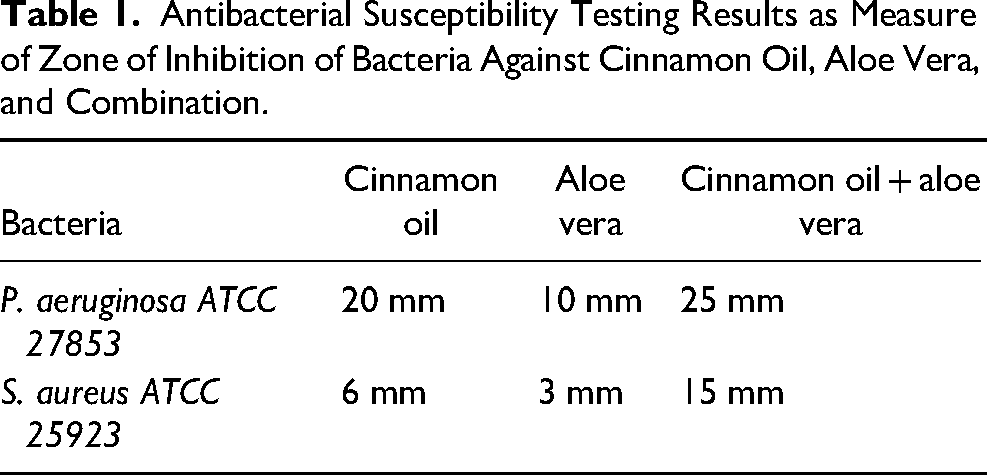
Antibacterial activity of AV gel, CO and combination (1 : 1) was evaluated on two bacterial strains. Zone diameter showed that in CO and AV when used in combination showed more activity than used alone. Inhibitory effect was more in P. aeruginosa than S. aureus (Table 1). Agar dilution method antibacterial activity for MIC determination was also observed in dose-dependent manner. The MIC of CO + AV for bacterium P. aeruginosa and S. aureus were 100 and 200 µg/ml, respectively.
Antibacterial Susceptibility Testing Results as Measure of Zone of Inhibition of Bacteria Against Cinnamon Oil, Aloe Vera, and Combination.
Wound Area Measurement
The wound area was measured on seventh and fourteenth day in wounds infected with P. aeruginosa and S. aureus and then given different treatments. In all cases, the wound area in CO group was decreased as compared to AV group while it was decreased significantly in treatment group CO + AV (Table 2).
Comparison of Wound Area In Different Groups. Values are Given as Mean ± SD.

Wound Contraction
There was a significant increase in contraction percentage in all treated groups as compared to untreated control (Figure 1). However, the most significance rise was observed in combinational therapy on fourteenth day.
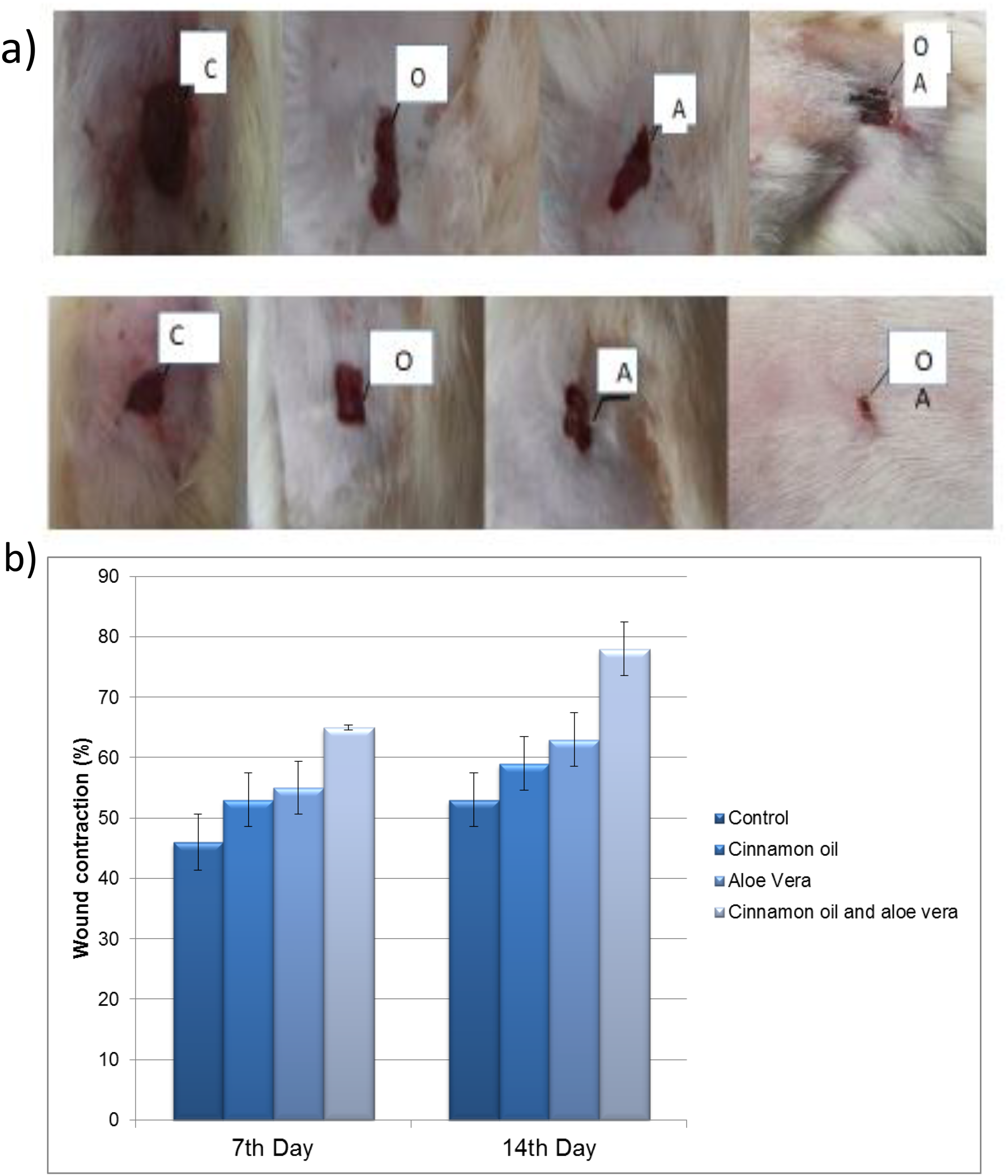
Wound contraction (a) wound area at seventh day (upper panel) and fourteenth day (lower panel); group C (control), O (cinnamon oil), A (aloe vera), and OA (cinnamon oil and aloe vera). (b) Bar graph representing mean ± SD of wound area in rat at day 7 and day 14 in different treated groups.
Histopathological Study
Histopathological examination of tissue from wound side after fourteen days was done and features of different groups have been shown in Figure 2. C (Diabetic control) group: Epidermis damage, hypodermis is not formed, hair follicles are not developed (in stress condition), no collagen present. A (AV) group: Epidermis damage start forming, hair follicles not developed but are still in stress condition), collagen start forming, hypodermis formed, dermis formed. O (CO) group: Epidermis damage start forming, proliferation of numerous hairs shafts, hypodermis formed, dermis formed. OA (CO and AV) group: Epidermis formed, prominent hair shafts, hypodermis formed, dermis formed, collagen present, sweat glands present.
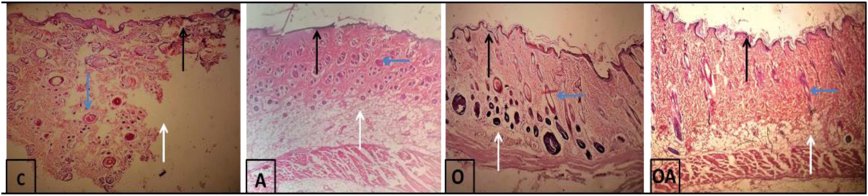
Histology analysis of skin tissue at 40×; C (diabetic injured control), A (aloe vera), O (cinnamon oil), OA (cinnamon oil and aloe vera).
Antioxidant Assessment
As compared to the untreated group, TAC levels in wound tissue were significantly (P < 0.05) increased in all treated groups after 14 days of wound. Drastic effect was observed in AV and CO combined treatment tissue (Figure 3). Same pattern was observed in MDA assay; level was significantly low in all treated rats, in comparison with control (P < 0.05). The lipid peroxidation level was effectively suppressed and the antioxidant enzymes level was improved in the experimental animal.
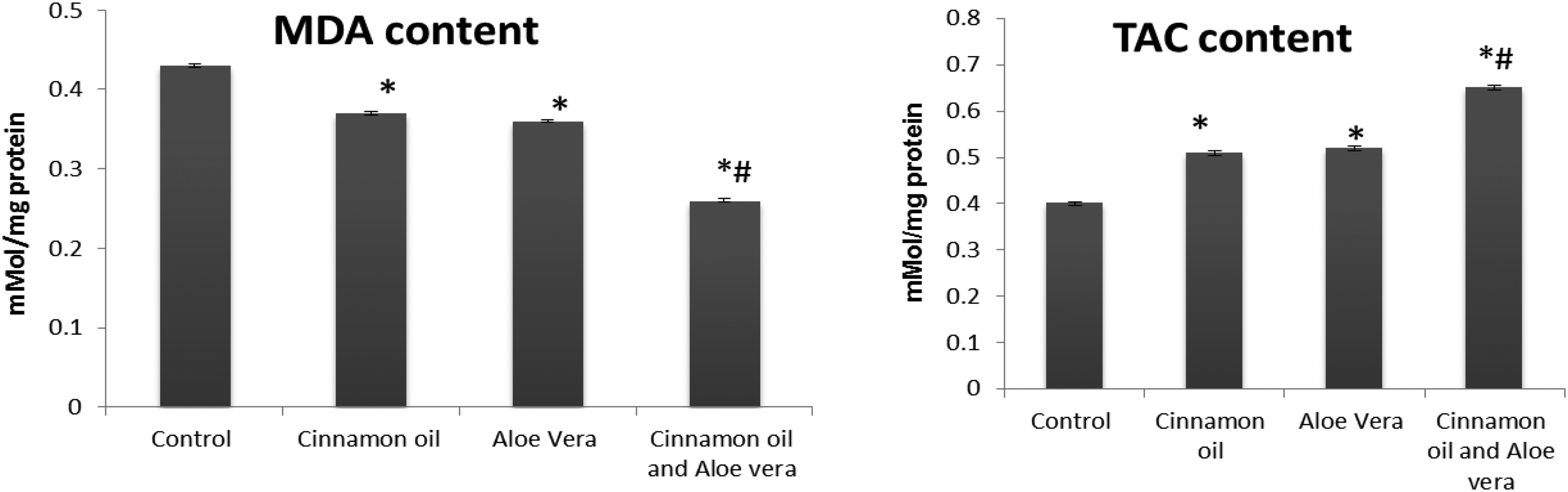
Graph representing MDA and TCA levels in tissue of different rat groups. The values are presented as mean ± SD of replicates. The significance level was calculated by one-way ANOVA subsequently Newman Keuls test. *P < .05 (cinnamon oil/aloe vera gel/combine cinnamon and aloe vera vs control). #P < .05 (cinnamon oil/aloe vera gel vs combine cinnamon and aloe vera). MDA, malondialdehyde.
Hydroxyproline Assay
Collagen turnover is considered to be an important phenomenon in the tissue repair mechanism. Hydroxyproline assay determines the levels of hydroxylproline that can be used as an indicator of collagen content. Hydroxyproline was estimated from the tissue in excision wound (Figure 4). It was observed that there was an increase in hydroxyproline content in CO group (34.2 ± 0.7) and AV group (32.6 ± 0.23) as compared to non-treated control rats (29.5 ± 0.81). But significantly elevated rise was found in rats of CO + AV treatment group (37.1 ± 0.44).
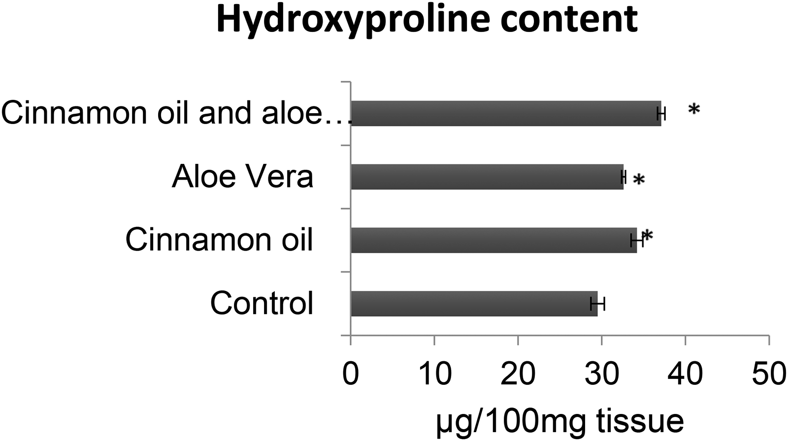
Bar graph representing tissue hydroxyproline content in control, aloe vera, cinnamon oil, and combination of cinnamon oil and aloe vera. Significant difference as compared to non-treated control group ∗P < 0.05.
Discussion
A wound can be defined to be an interruption in the normal anatomical shape and undisturbed functions of the skin. The procedure of wound healing comprised of four stages namely hemostasis, inflammation, proliferation, and tissue remodeling. These stages bring about the reconstruction and revascularization of skin as these steps allow the formation of active dermis and epidermis. The main objectives in treating the wounds are quick wound closure and formation of an active and satisfactory scar. To achieve this purpose, covering the wound is important in wound management. 20 Treatment of wound with infection almost always requires some antimicrobial therapy. If left untreated, infected wound become chronic or non-healing. Thus, it is strongly advised to use antibacterial agent. 21 There are two major routes through which treatment can be given are as follows. (1) Systemically: antimicrobial delivery to the whole body via the oral or parenteral or intravenous or intramuscular, (2) topically ie, through application creams, gels, and ointments. Use of tropical medicine for wound healing is still a debatable issue. For treatment of most superficial infections, topical antimicrobial therapy alone is useful. Controversies occur while considering deep wounds infections that weather topical antimicrobial agents can be beneficial or not. There are different types of antibacterial agents that can be used chemical antibiotic, metal particles, cationic organic agents, and natural extracts.22,23 Antibiotics are becoming useless against many commonly isolated pathogens. So, there is need of alternative treatments for infections. In spite of the fact that many alternatives as of now exist in nature but the challenge is to execute them for clinical purpose. One such alternative is the use of plant extract as an antibiotic agent. Historically plants and their extracts have been using for treating infectious diseases in many nations. 1 Due to overdose of antibiotics the resistance is increasing day by day against bacteria. CO finds its application in a wide range of clinical settings due to its availability, cost-efficiency, and easy application.
It was observed in control groups that skin is disturbed by injury while treatment after seventh and fourteenth day improved the skin, CO and AV and combination of CO and AV treatment, indicating significant improvement in skin. The mechanism action of essential oils involves the breakdown the bacterial cell wall by attacking on the phospholipids and lipids molecules which eventually disrupt the major cellular functions like ATP production, transcription, and translation. 24
The combination of CO and AV can be affected in the recovery of wound. This combination grafting is less expensive. This combination is better than use of excess amount of drugs with potential side effects. For future, it is suggested that more experiments should be conducted for use in conjugation with other antibiotics for healing wound.
Conclusion
In conclusion, CO and AV combination was found to be more effective than separate CO and AV grafts. The combine use resulted in not only faster wound closure but provided protection from bacterial infection as well. Further pharmacological studies could be done to find optimum and stable formulation.
Footnotes
Author Contributions
Sana Khurshid: conceptualization; formal analysis, supervision, and writing—review & editing. Ume-Farwa: conceptualization, investigation, methodology, and writing—original draft. Zaryab Mazhar: methodology and formal analysis. Yasmeen Ishaq: investigation, visualization, and writing—review & editing. Hamna Naeem: validation and formal analysis.
Declaration of Conflicting Interests
The authors declared no potential conflicts of interest with respect to the research, authorship, and/or publication of this article.
Ethics Approval
All experimental procedures involving animals were conducted in accordance with the international guidelines regarding use and care of experimental animals and were approved by the ethical Committee of IMBB (reference IMBB/UOL/20/137).
Funding
The authors received no financial support for the research, authorship, and/or publication of this article.