
Abstract
The wound healing process is really interesting, dynamic, and complex, captivating researchers for a long time. With the growing worldwide concern regarding the prevalence of wounds and the associated healthcare challenges, efforts to expedite this natural process have intensified. Fortunately, with a particular focus on improving wound dressings, significant advancements have been made in wound care management including using of nanoparticle-based delivery systems. These nanoparticles, similar to molecular messengers, purchase vast promise for revolutionizing wound treatment. Among them, chitosan nanoparticles stand out as remarkable candidates. Their safety profile, biocompatibility, and bioactivity make them particularly appealing for wound care. In this article, we will delve into the intricacies of wound healing and then discuss the wound-healing properties of chitosan nanoparticles, supported by comprehensive study results. Current evidence highlights the wound-healing effects of chitosan nanoparticles, which can be considered independent agents for wound management. In conclusion, the utilization of chitosan nanoparticles for wound healing presents significant opportunities and potential.
Graphical abstract
Introduction
Wounds can arise from pathological processes inside or outside the body. They may result from accidental events, intentional actions, or underlying medical conditions and diseases. 1 The global prevalence of wounds is highly concerning and has consequently imposed a significant financial burden on healthcare systems worldwide.2,3 Despite the routine implementation of optimal wound care practices in clinical settings today, challenges persist due to factors such as resource limitations and rising costs, particularly in developing countries. 4
In conclusion, there is a significant demand for wound care products and new treatment options that can both reduce treatment costs and recovery time while accelerating wound healing. To achieve this goal, numerous studies have examined various pharmaceutical formulations. 5 Creating effective pharmaceutical treatments is a complex process but, to achieve these pharmaceutical formulations, researchers have utilized advanced technologies and biomaterials with special properties to create sophisticated wound dressings. One of the emerging fields in wound healing is using nanotechnology, which has made incredible advancements in wound improvement and is being used more and more in both the medical and pharmaceutical fields. 6
Nanoparticles, a defining feature of this innovative technology, possess unique properties due to their high surface-to-volume ratio.
7
Consequently, they find applications across diverse fields. In the realm of wound healing, two main types of nanoparticles are utilized:
Intrinsic Healing Nanoparticles: have intrinsic wound healing properties and contribute to wound closure. Drug Carrier Nanoparticles: These nanoparticles serve as carriers for therapeutic agents.
8
The objective of this study is to explore the effects of using of chitosan nanoparticles (CNPs) for wound healing by investigating their intrinsic properties and potential mechanisms. Widely reported results indicate that these nanoparticles accelerate wound improvement. CNPs are employed either independently or in combination with other compounds, including antibiotics, natural substances, and peptides. The formulation, based on the minute size of the chitosan nanoparticles, enhances their penetration into the wound environment.
Wound Healing
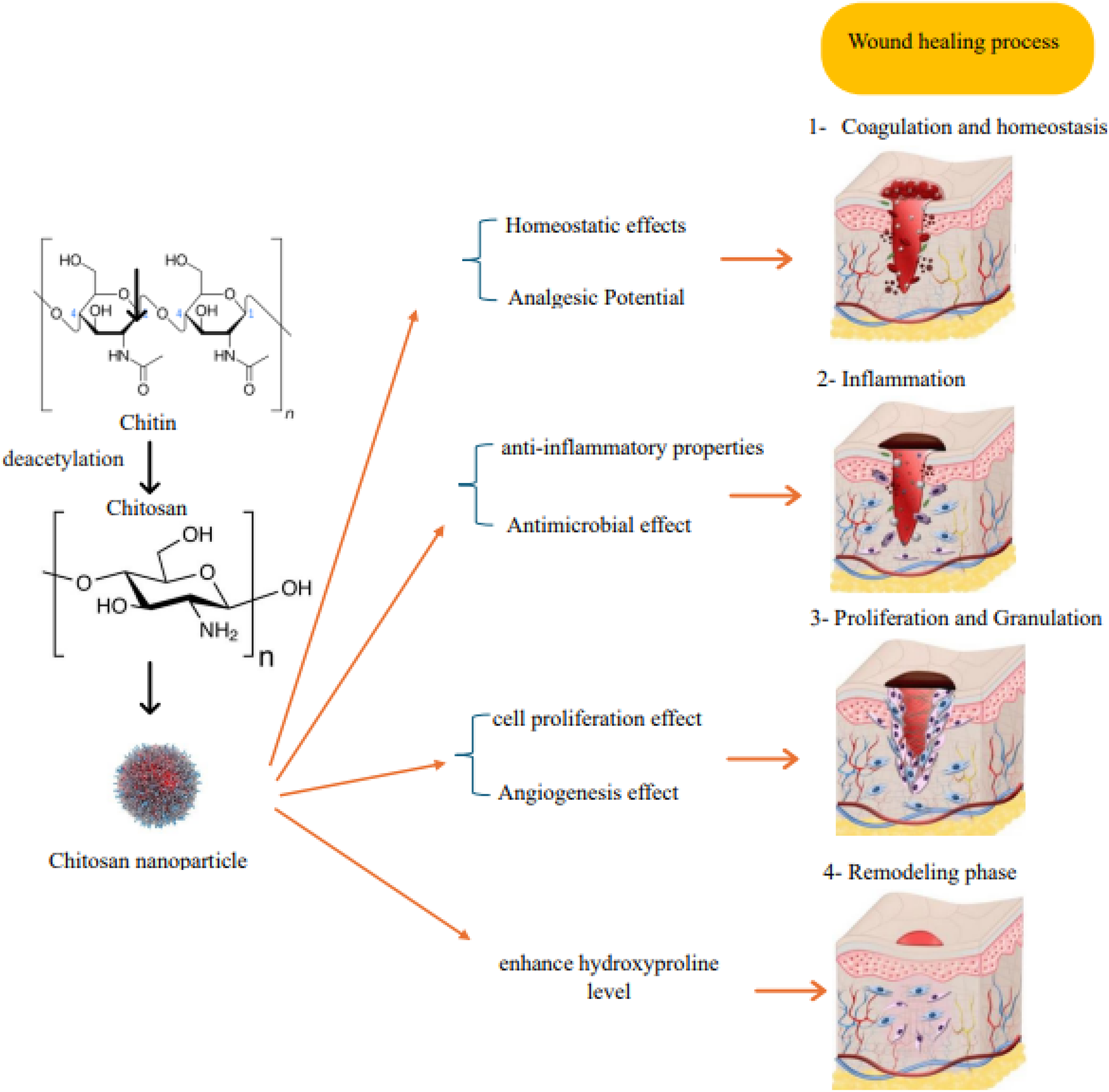
The skin, as a vast and the largest organ of the body covering, is a resilient shield against external threats. If this vital barrier is compromised, it can lead to a myriad of health complications. Fortunately, in moments of injury and turmoil, when wounds disrupt its surface, the skin reveals its remarkable capacity to regenerate and restore itself with Conspicuous efficacy. 9 Wound healing is a complex and dynamic process that can be divided into overlapping stages that are intricately linked to each other. 10 Let's explore these stages: Now we explore these stages, Hemostasis: the initial phase where immediate actions are taken to prevent excessive blood loss. Adjacent blood vessels constrict and platelets aggregate to form a platelet plug. Then, thrombosis occurs as part of the coagulation cascade. During the inflammatory phase, immune cells infiltrate the wound site, with neutrophils being the first responders, followed by macrophages. Their presence helps clear debris and prevents infection.11–13
In the phase of granulation tissue formation, the wound undergoes a remarkable transformation, generation of new granulation tissue, and initiates re-epithelialization. Simultaneously, the intricate process of angiogenesis begins the intricate process of new blood vessel formation. It is within this delicate process that fibroblasts emerge as key players, meticulously fabricating essential constituents of the extracellular matrix (ECM) such as collagen III, fibronectin, and hyaluronic acid. In addition to this vital role, myofibroblasts aid in wound contraction with their deft action. 14
Advancement into maturation and remodeling phase: during the progression of wound healing, collagen type III is gradually replaced by collagen type I. Meanwhile, matrix metalloproteinases (MMPs) degrading any unnecessary ECM components that may impede progress toward complete restoration. In this stage, newly formed leaky blood vessels, are replaced by more mature, perfused vessels. 15
Complete recovery becomes evident after wound closure, as observed by the physical appearance on the external surface. This is followed by the reconstruction phase begins, which may continue for several months or even years after complete wound closure. This phase involves the maturation of subdermal tissue and the emergence of scar tissue. 16
Chitosan
Chitin or “the last biomass”, after cellulose, is the most abundant polysaccharide that exists in the exoskeleton of crustaceans and insects.17,18 Several billion tons of chitin are manufactured annually by marine copepods alone. 19 The structural composition of chitin is characterized by its lengthy, unbranched nature, akin to cellulose derivatives. 20 Notably, it exhibits a resemblance to glycosaminoglycans in terms of functional characteristics, attributed to its inherent structural properties. 21
Chitosan (CS), or (1,4)-2-amino-2-deoxy-beta-D-glucan, is an unrivaled compound obtained through the deacetylation of chitin. 22 The emergence and discovery of Chi originate from 1859 when Roget first reported its formation during the boiling of chitin in a concentrated potassium hydroxide solution. 23 Fundamental research on CS began in earnest about a century later. 24 In 1934, Rigby patented two inventions, one for the production of CS from chitin and the other for the production of films and fibers from CS. 25 The mechanism of CS production from chitin is illustrated in Figure 1.
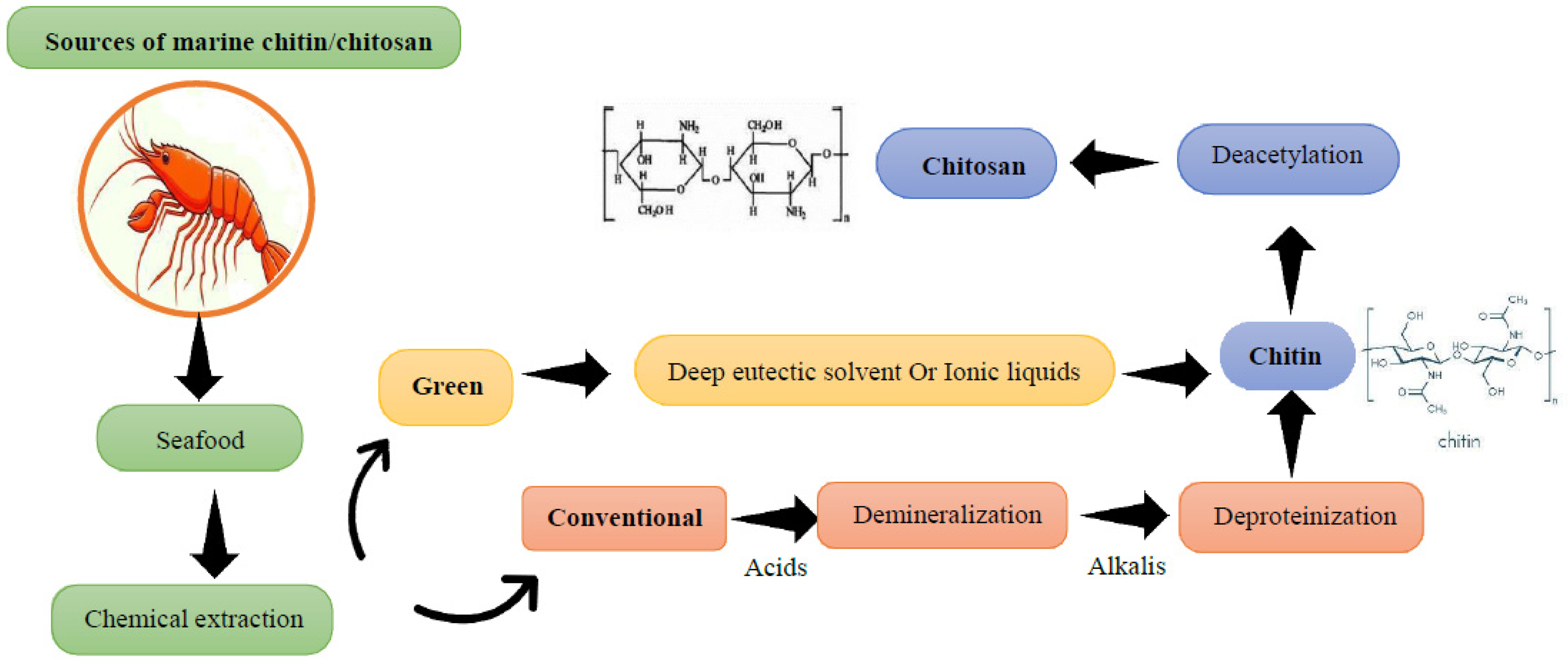
Chitosan Synthesis Pathways.
Unlike chitin, CS is easily soluble in aqueous solutions, many mineral and organic acids. 26 CS has more chemical and biochemical reactions than chitin due to the presence of free primary amino groups that are regularly distributed in its molecular chain. 27 CS has no allergic or stimulant effects and is biocompatible with human skin. 28
CS with immune-boosting, blood-thinning, antibacterial, and antifungal qualities, CS has found widespread use as a catalyst for the healing of surgical wounds. 29
This miracle substance, derived from nature as a plentiful polysaccharide, boasts attributes of being non-toxic and biodegradable.13,30 Unlike oil and coal, CS is a natural renewable source (eg crab and shrimp shells) that can be increased by artificial cultivation. 21 Chitin and CS have been reported to be present in the cell wall of fungi. 31
Chitosan Nanoparticles
CS is the most widely studied polymer for the preparation of nanoparticles due to its special properties mentioned above. 32 Its cheapness, hydrophilic nature, and solubility make nanoparticles highly capable of controlling and releasing drugs. 33
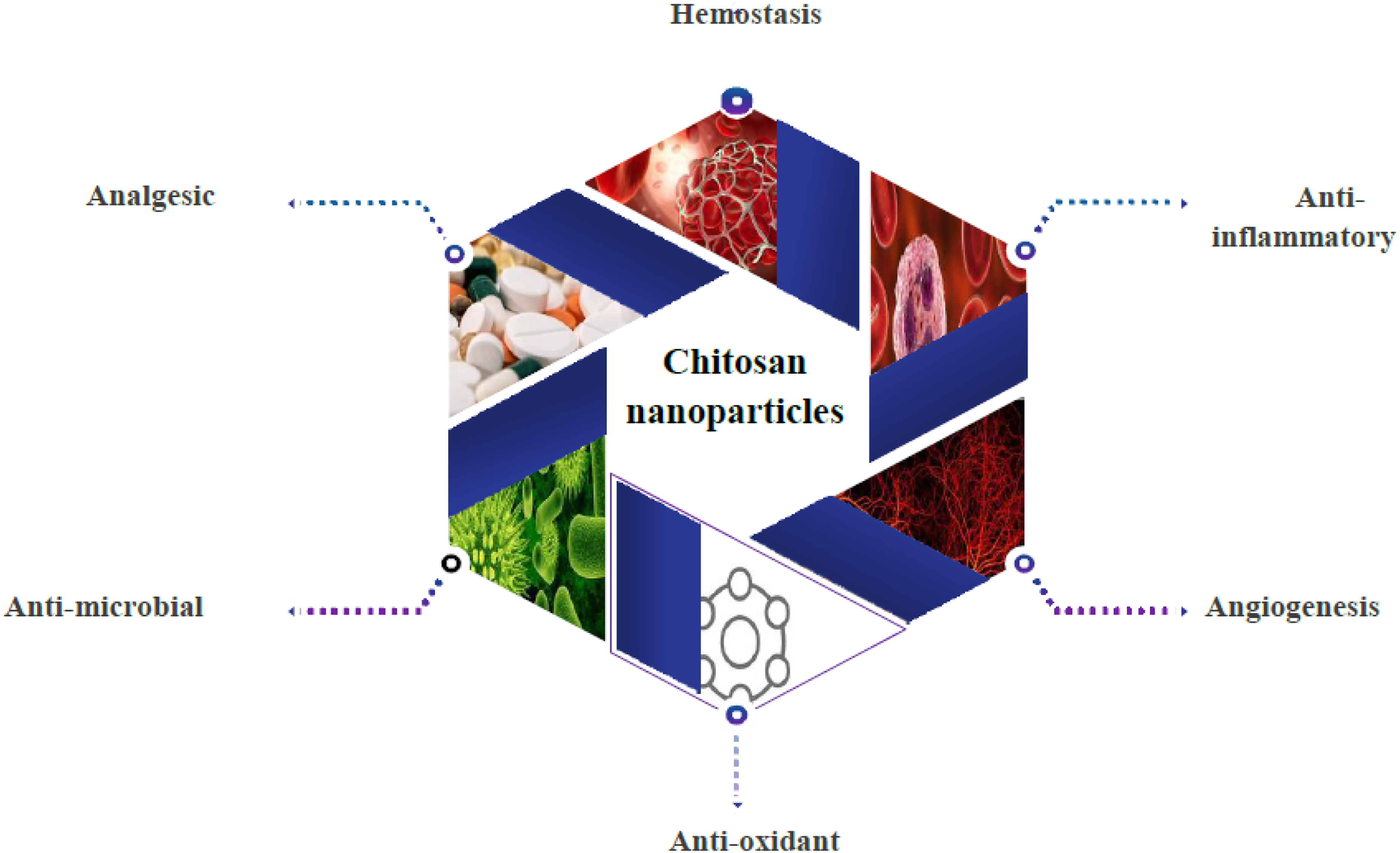
Among the new drug delivery systems, chitosan micro / nanoparticles have shown great promise in oral, injectable, topical, and inhalation applications. 34 In the polymer field, the size and surface properties of CNPs play an important role in their transport through cell membranes. 35 These particles can be used for controlled drug delivery. 36 Due to the mucosal nature of CS, these nanoparticles have the ability to improve drug uptake and bioavailability by prolonged drug contact with the mucosal layer and high surface-to-volume ratio and may enhance this effect. 37 The development of CNPs has maximized the benefits of CS as a polymeric drug carrier by increasing water solubility, systemic adsorption, bioavailability and drug stability. 38 CNPs with minimal toxicity can be a desirable non-viral vector for gene therapy. 39 The use of chitosan-based dressings has also been investigated for wound healing applications. 40 Some applications and properties of CNPs are presented in Figure 2.
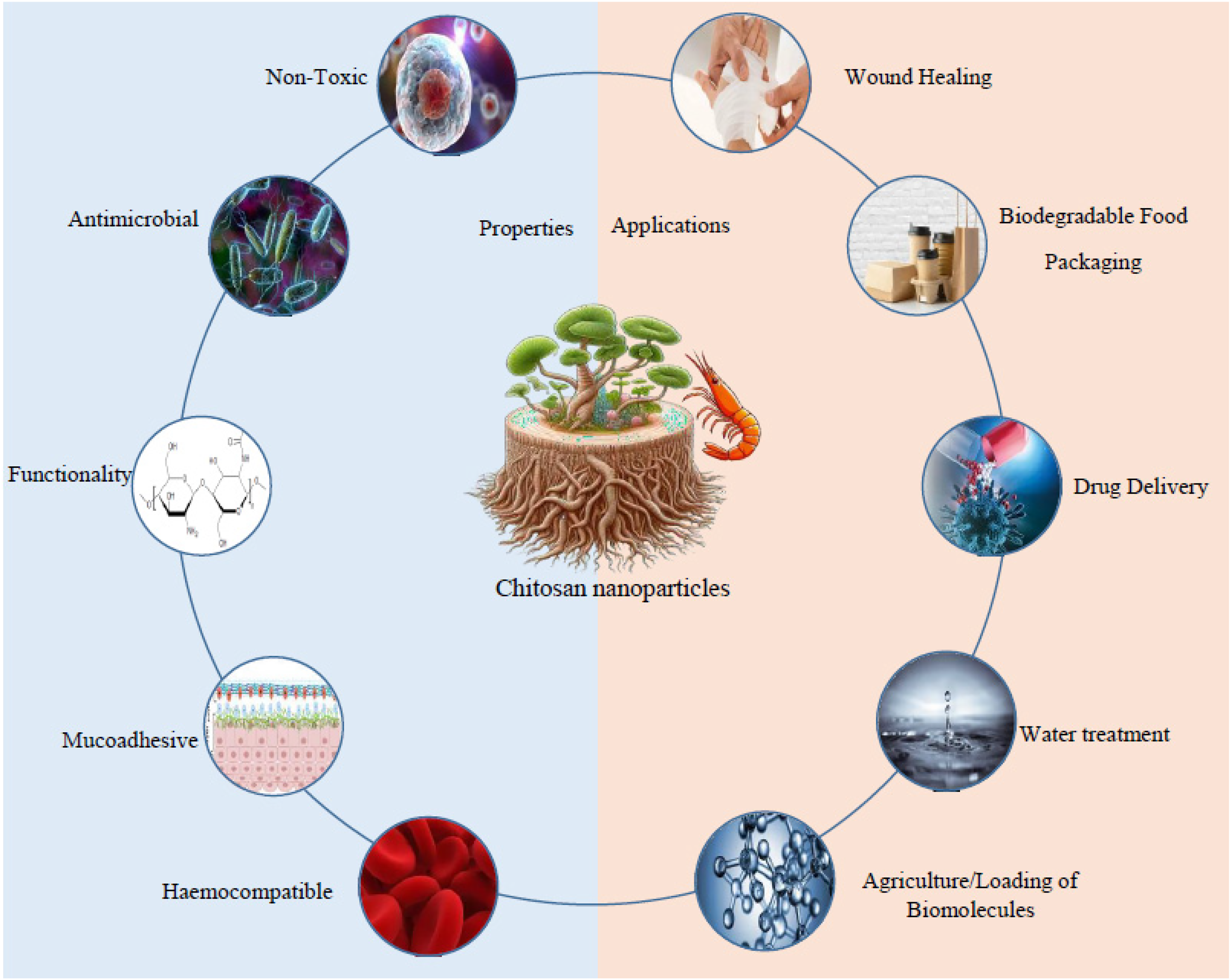
Properties and Applications of Chitosan nanoparticles.
Mechanism of Action of CNPs
Coagulation and Homeostasis
Homeostasis
Hemostatic agents are important in pharmaceutical fields because homeostasis is the first phase in wound healing. 41 Due to their high levels of porosity and surface-to-volume ratio, nanoparticles have attracted broad attention in homeostatic applications. 42 In this regard, CNPs has been developed as a highly biocompatible textile with antimicrobial and homeostatic properties. 43 The positive charge of CS is due to D–glucosamine residues in amino groups, which characterize the mechanism promoting hemostasis. 44
The intrinsic cationic characteristics of CNPs enable them to be involved in the creation of polyelectrolyte complexes with biomolecules possessing a negative charge, thereby facilitating interactions with cell membranes to induce platelet aggregation and promote hemostasis. 45 Research has presented evidence indicating that these CNPs prompt the agglutination of red blood cells in rabbits. This is due to the interaction between the CNPs and the erythrocyte membrane. 46
The study by Santosh S. Biranje et al showed that the use of CNPs in wound dressing accelerates hemostasis activity for efficient and rapid wound healing. 47 In a study that examined the antimicrobial and hematological effects of chitosan-copper oxide nanocomposites and CNPs, it was determined that the CS-CuO-NPnanocomposite demonstrated high sensitivity to all tested microorganisms. On the other hand, CNPs acted as a coagulating agent for both healthy and diabetic blood samples. 48
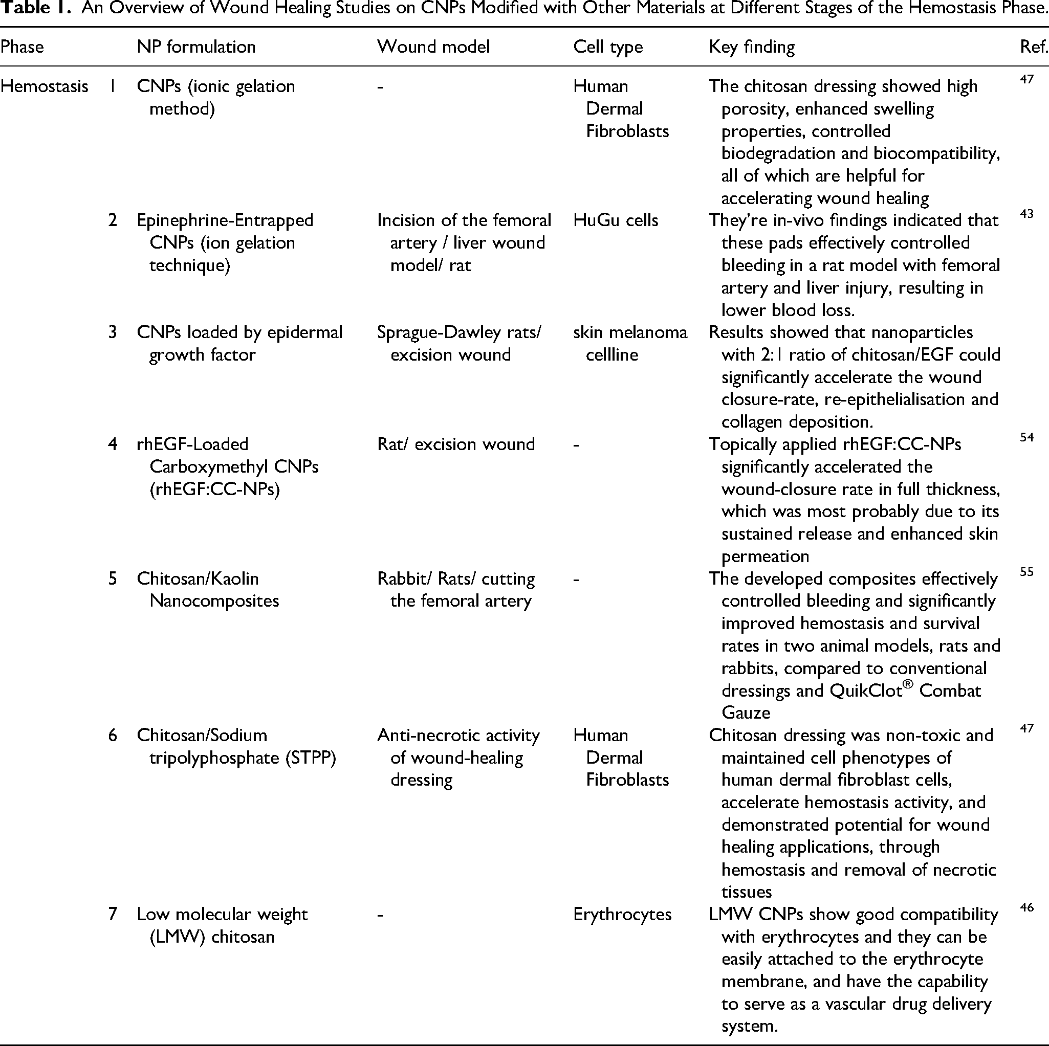
Based on the mentioned content, it appears that CNPs have an advantage over CS polymers in enhancing hemostasis. This is because the smaller sizes of CNPs provide them with a greater surface-to-volume ratio, which enables better agglutination, platelet aggregation, and plasma protein absorption. Additionally, this particle size and consequently higher surface charges, leading to a greater total surface-to-volume ratio and absorption of negatively charged red blood cells, improving hemostatic effects. Several studies that have confirmed the hemostatic effects of CNPs are summarized in Table 1.
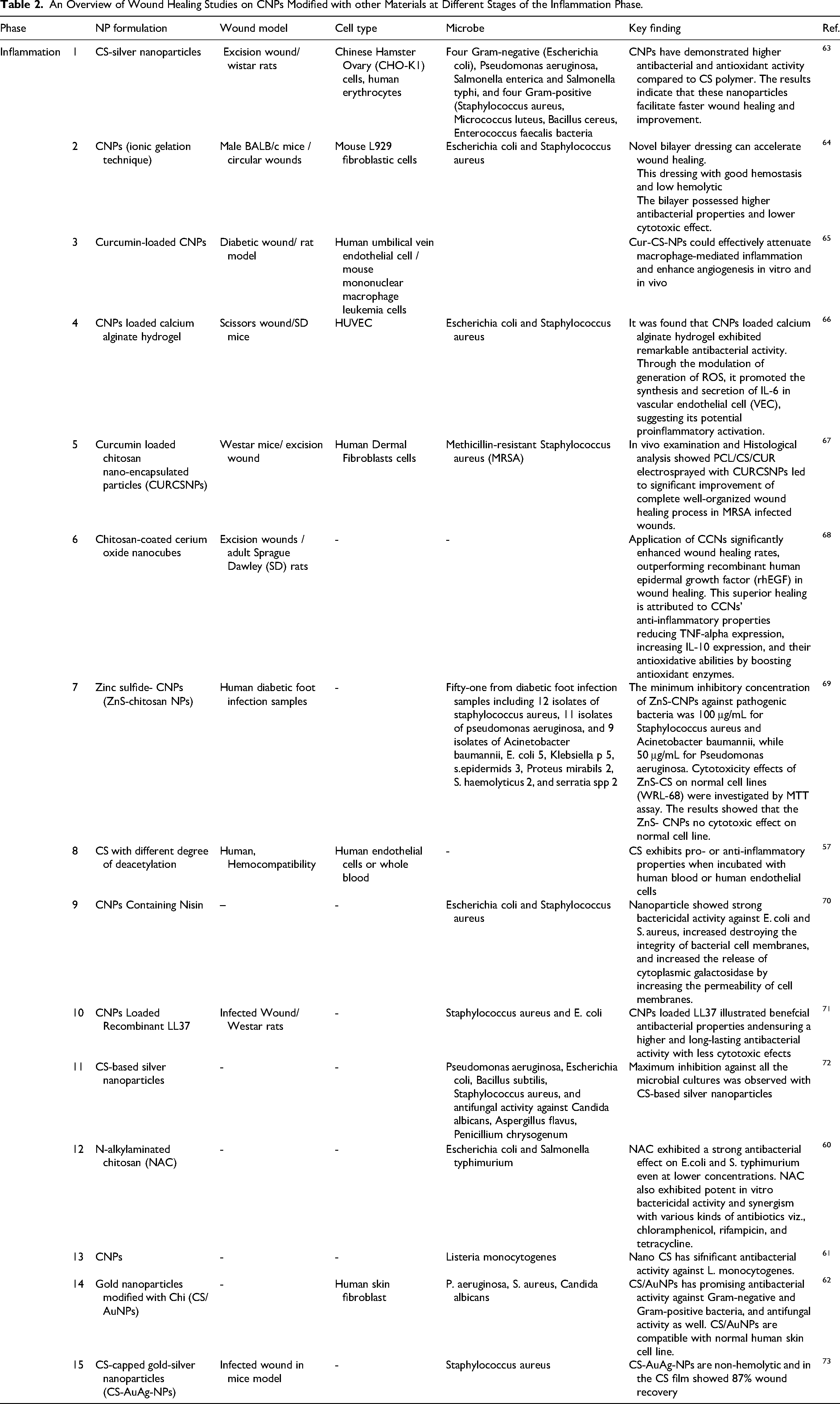
An Overview of Wound Healing Studies on CNPs Modified with Other Materials at Different Stages of the Hemostasis Phase.
Analgesic Potential
CNPs exhibit analgesic effects through their modulation of inflammatory responses, immunomodulatory properties, and antibacterial effects. These nanoparticles hold promise for various applications in pain management, including drug delivery, wound dressings, and hydrogels.
CNPs with immunomodulatory effects can contribute to reducing inflammation and pain. 49 The immunological effects of CNPs add another dimension to their potential for pain relief. 50
CNPs can also exert their analgesic effects through modulation of inflammatory responses. CNPs can inhibit the release of pro-inflammatory cytokines and chemokines, reducing inflammation and subsequent pain. 51 Additionally, CNPs can interact with cell membranes, triggering cellular responses that modulate pain perception. 52
CNPs possess inherent antibacterial properties, which can be beneficial in pain management. Bacterial infections can cause pain. CNPs can inhibit bacterial growth and biofilm formation. 53 As a result, they can alleviate pain associated with bacterial infections by preventing or treating them. Further research is needed to explore the mechanisms underlying the analgesic effects of CNPs and optimize their application in specific pain-related conditions.
Inflammation
CNPs have been extensively investigated for their anti-inflammatory properties and have shown promise in a range of applications, including targeted therapy for cutaneous pathogens. 56 A study by Nolte et al has shown that CNPs stimulate white blood cells to release inflammatory cytokines such as IL-18, IL-1β, and granulocyte colony-stimulating factor. 57 A study found that when a combination of bone marrow-derived mast cells (BMMCs) and chitosan biofilm was introduced to wound sites, it accelerated the infiltration process and granulation tissue formation by reducing inflammation response and promoting angiogenesis. This suggests that BMMCs act as growth factor carriers that only degranulate in situ without inducing inflammatory response. 58 Additionally, CNPs can regulate immune cell function, promoting an anti-inflammatory environment. These effects contribute to the suppression of chronic inflammation and the modulation of immune responses within the tissue environment.50,59
Antimicrobial Effect
N-alkylaminated chitosan (NAC) is a derivative of CS that can be effectively used as a cationic antimicrobial agent either on its own or in conjunction with antibiotics, particularly against Escherichia coli and Salmonella typhimurium. 60
A study was conducted to evaluate the antimicrobial effect of CNPs on Listeria monocytogenes bacteria isolated from pregnant women. The results revealed that CNPs exhibit significant antibacterial activity against L. monocytogenes. 61
Chitosan-based gold nanoparticles (CS/AuNPs) have demonstrated significant antimicrobial properties against both bacteria and fungi. The most pronounced inhibitory effect was observed against Pseudomonas aeruginosa, showcasing their potential in wound healing applications. The combined effects of gold nanoparticles (AuNPs) and CS work together to disrupt bacterial membranes, leading to their antimicrobial activity. This dual-action approach has proven effective against a wide range of bacteria, including both Gram-negative and Gram-positive strains, with Pseudomonas aeruginosa being particularly susceptible. Additionally, these nanoparticles have shown antifungal activity against Candida albicans. 62
The bimetallic CS-capped gold-silver nanoparticles (CS-AuAg-NPs) demonstrated a minimal bactericidal concentration and proved to be more effective against multidrug-resistant Acinetobacter baumannii (MDR AB) and multidrug-resistant Citrobacter freundii (MDR CF) when compared to the standard antibiotic clindamycin. In a mouse model, these nanoparticles exhibited an impressive 87% wound recovery after just 7 days. Furthermore, the nanoparticles are considered to be safer due to their non-hemolytic properties and have shown increased efficacy against MDR bacteria, while also promoting skin regeneration in infected wounds. 73
Utilizing biocompatible molecules in conjunction with nanoscience and natural polymers is crucial in combating bacterial infections and antibiotic resistance. CS-based silver nanoparticles have exhibited antimicrobial properties and are non-toxic to human cells, making them well-suited for wound healing purposes. These nanoparticles can effectively manage topical wound infections by impeding the growth of harmful pathogens such as S. aureus and E. coli, while also stimulating cell growth in the affected area. Furthermore, studies have confirmed the non-toxicity of these nanoparticles to mammalian cells and their effectiveness in enhancing the wound healing process. 74
A study was conducted on CNPs loaded with recombinant LL37 antimicrobial peptide. The study showed that encapsulating LL37 in CSLL37NPs had a more efficient effect on wound healing progress and the elimination of MRSA infection compared to free LL37. 71 CS-based silver nanoparticles showed effective antibacterial and antifungal activity. 72
The antibacterial efficiency of ChNP depends on the molecular weight of CS and the pH of the medium at 5.0. Numerous studies have reported the antimicrobial activities of ChNP against bacteria, fungi, yeasts, and algae, both in vivo and in vitro settings. In pharmaceutical applications, ChNP has been utilized as an antimicrobial coating to enhance wound healing, prevent infections, and address the growing threat of infectious diseases. 75
A study was conducted to assess the antibacterial effect of CNPs loaded with Nisin against Escherichia coli and Staphylococcus. The study demonstrated that the integrity of the cell membranes of the bacteria was compromised when treated with the nanoparticles. 70 The study examined the use of dressing coated with silver sulfadiazine (SSD) loaded CNPs (CSNPs) for the controlled release of SSD into burn wounds to manage bacterial growth. CNPs serve as drug carrier systems and an effective fabric coating material, making this novel antimicrobial silver sulfadiazine dressing a promising solution for wound care. 45 A summary of various studies that have demonstreated the anti-inflammatory properties of CNPs can be found in Table 2.
An Overview of Wound Healing Studies on CNPs Modified with other Materials at Different Stages of the Inflammation Phase.
Proliferation and Granulation
CNPs also play a role in the granulation phase of wound healing. Granulation is the process of forming new blood vessels and connective tissue in the wound bed. CNPs have been shown to promote angiogenesis (formation of new blood vessels) and stimulate the production of collagen, a key component of the extracellular matrix.
Angiogenesis Effect
In an experimental study by diabetic mice by Masayuki Ishihara et al, FGF-2-containing CS hydrogels induced angiogenesis and lateral circulation in diabetic rats with healing disorders and in ischemic organs. 76
A 2009 study by Jessica Guzman-morales et al found that adding CS to the collagen matrix improved the properties of the collagen matrix and its ability to support endothelial cells and angiogenesis for use in cardiac tissue engineering applications.
It is suggested that CNPs have the potential to enhance and improve wound healing. This is achieved by promoting the proliferation of fibroblast cells and angiogenesis, while also mitigating inflammation. 77
A study has demonstrated that the combination of CNPs and photothermal nanoparticles can accelerate wound healing and angiogenesis. Additionally, it has been found to possess antimicrobial effects in the treatment of infected diabetic wounds. 78
Research has demonstrated the positive impact of CNPs on both angiogenesis and wound healing. Histological analysis has consistently revealed enhanced epithelialization, dermal differentiation, collagen deposition, and angiogenesis as a result of CNPs treatment. 79
CNPs possess the potential to deliver angiogenic growth factors to target cells, thereby enhancing angiogenesis in tissue engineering applications. A hydrogel composed of Gelatin methacryloyl (GelMA) and CNPs has proven to be highly effective in delivering angiogenic growth factors. This innovative combination not only stimulates cell proliferation but also ensures a sustained release of bFGF (basic fibroblast growth factor). 80
CNPs and Their Effect of Cell Proliferation
CNPs can stimulate the migration and proliferation of fibroblasts, keratinocytes, and endothelial cells, as well as the synthesis and deposition of collagen, elastin, and glycosaminoglycans during the proliferation phase. 81 CNPs have been selected as an ideal choice for drug delivery due to their ability to accelerate the skin remodeling process without any toxic effects. A study has shown that CS-embedded recombinant human epidermal growth factor (rh-EGF) nanoparticles exhibit superior results in terms of cellular proliferation and migration when compared to free rh-EGF. Additionally, these nanoparticles have been found to upregulate the expression of key genes such as TGF-β, VEGF, and PDGF, which play a crucial role in wound healing and skin remodeling. 82 CS-coupled silver nanoparticles (Ch-AgNPs) have demonstrated efficacy in wound healing assays by promoting cell proliferation in the wound area. 74 Research has shown that N-acetyl glucosamine, a monomer unit of CS, promotes hemostasis, enhances cell proliferation, and speeds up the process of wound healing. 67
CS-stabilized selenium nanoparticles, known as Ch-SeNPs, have demonstrated a remarkable ability to target mitosis by partially inhibiting the expression of cyclin-dependent kinase 1 (Cdk1). 83 N-succinyl CS and N-succinyl CNPs film have been shown to accelerate wound healing by promoting remarkable cell proliferation in human dermal fibroblast cells. 84
The use of curcumin-loaded CNPs (CS-NPs) has been shown to accelerate the wound healing process by transitioning from the inflammation phase to the proliferation and remodeling phases. This is achieved through the expression of proteins that are associated with cell proliferation and migration in HG-HUVECs. Additionally, these nanoparticles promote the proliferation and migration of endothelial cells both in vivo and in vitro. 65
CS is commonly used as a wound dressing material because of its anti-inflammatory properties, biocompatibility, ability to retain fibroblast growth factors, and stimulation of human skin fibroblast activities. CS-based gold nanoparticles have shown promise in promoting the proliferative phase of the wound-healing process. 62
CS is known to activate macrophages, enhancing their ability to combat tumors. Additionally, it stimulates cell proliferation and tissue organization. As it gradually breaks down into N-acetyl-β-D-glucosamine, CS plays a crucial role in initiating fibroblast proliferation, promoting the orderly deposition of collagen, and stimulating the natural synthesis of hyaluronic acid at the wound site. 85
In vivo studies conducted on skin wounds in mice demonstrated that CNPs loaded with calcium alginate hydrogel enhanced the proliferation and migration of vascular endothelial cells (VECs) by inducing the production of reactive oxygen species (ROS). This resulted in accelerated skin wound healing and re-epithelialization. 66
CNPs have the ability to enhance fibroblasts by promoting adhesion, proliferation, and secretion of proteins that are crucial for tissue repair mechanisms and progressive remodeling. 86 A new layer of skin, known as the epidermal layer, starts to develop on the surface of a wound as keratinocytes multiply and move during the re-epithelialization process. CS plays a crucial role in promoting the growth of granulation tissue by stimulating the proliferation of dermal fibroblasts. 87 Table 3 provides an overview of different studies that have shown the proliferative effect of CNPs.
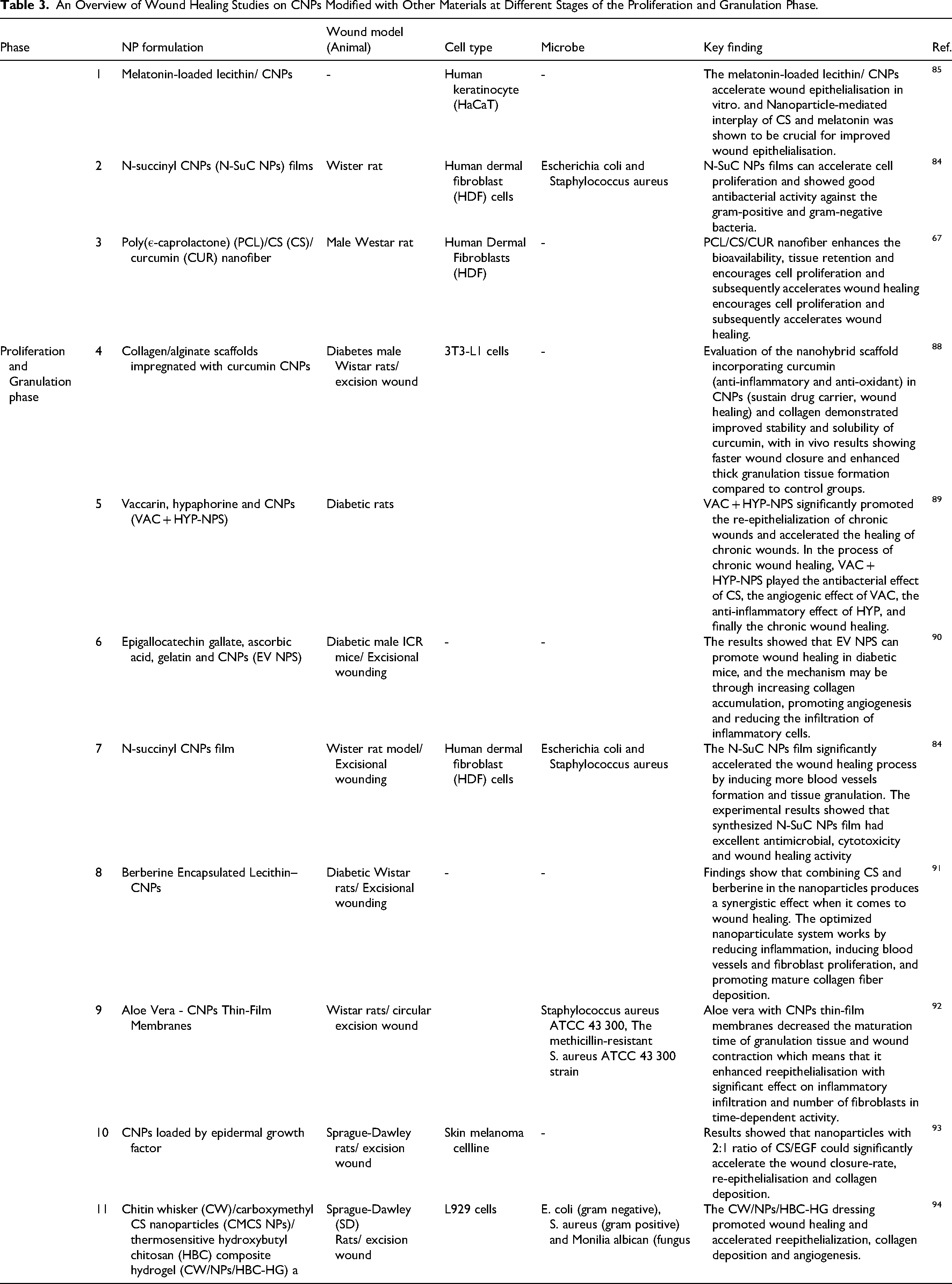
An Overview of Wound Healing Studies on CNPs Modified with Other Materials at Different Stages of the Proliferation and Granulation Phase.
Remodeling Phase
CNPs exhibit immunomodulatory effects essential for wound healing. 95 They can regulate the immune response, promoting the migration of immune cells to the wound site and stimulating the stimulating of growth factors and cytokines crucial for tissue regeneration. 96
CSNP can also regulate the balance between matrix metalloproteinases and tissue inhibitors of metalloproteinases, as well as influence the alignment and cross-linking of collagen fibers during the tissue remodeling process. 97
The reconstruction and reorganization of ECM are two separate but intertwined processes that are essential for facilitating the successful regeneration and healing of wounds. Collagen plays a significant role in maintaining the structural and dynamic integrity of the ECM, serving as a scaffold for various biological processes. Throughout the proliferation phase of wound healing, type III collagen is more abundant, while during the remodeling phase, scar tissue is predominantly composed of Type I collagen. Hydroxyproline, an amino acid, acts as an indicator for estimating collagen levels within tissues. Studies have noted elevated levels of hydroxyproline in wounds treated with CNPs.
In a study, Amit Kumar and colleagues found that hydroxyproline levels were higher in the CS-treated group compared to the control group. This finding suggested a beneficial impact on the healing of skin wounds. Furthermore, the study revealed that that higher hydroxyproline levels enhanced maturity and collagen production during the healing process. 98
Additionally, Seyed Mansour Alamshah and colleagues explored the potential of hydroxyproline as a marker for improving wound healing in diabetic tissue. They investigated hydroxyproline levels in diabetic foot ulcers throughout the healing phase. The study revealed that lower hydroxyproline levels in diabetic patients with foot ulcers may delay the healing process. 99
In a recent study conducted by Ranjbar and colleagues, the effects of combining Artemisia dracunculus with CNPs biofilm were explored. The study focused on excisional wounds infected with MRSA (methicillin-resistant Staphylococcus aureus). The findings revealed significant differences between the treatment group using the specified biofilm and the control groups. Notably, the study observed microbiological transformations, wound surface reduction, and alterations in hydroxyproline level in the treatment group compared to the control group. 100 Another study demonstrated that Chitosan/Nanoselenium biofilm can significantly enhance the activity of fibroblasts, promote collagen formation, and accelerate epithelialization. 101
In another recent investigation, researchers explored the effects of a biofilm containing CNPs and Ferula assa-foetida on MRSA-infected wounds in diabetic patients. The findings revealed that this biofilm could be beneficial due to its ability to enhance hydroxyproline level and inhibit the proliferation of bacteria. 102 Table 4 summarizes various studies that have demonstrated the remodeling effects of CNPs.
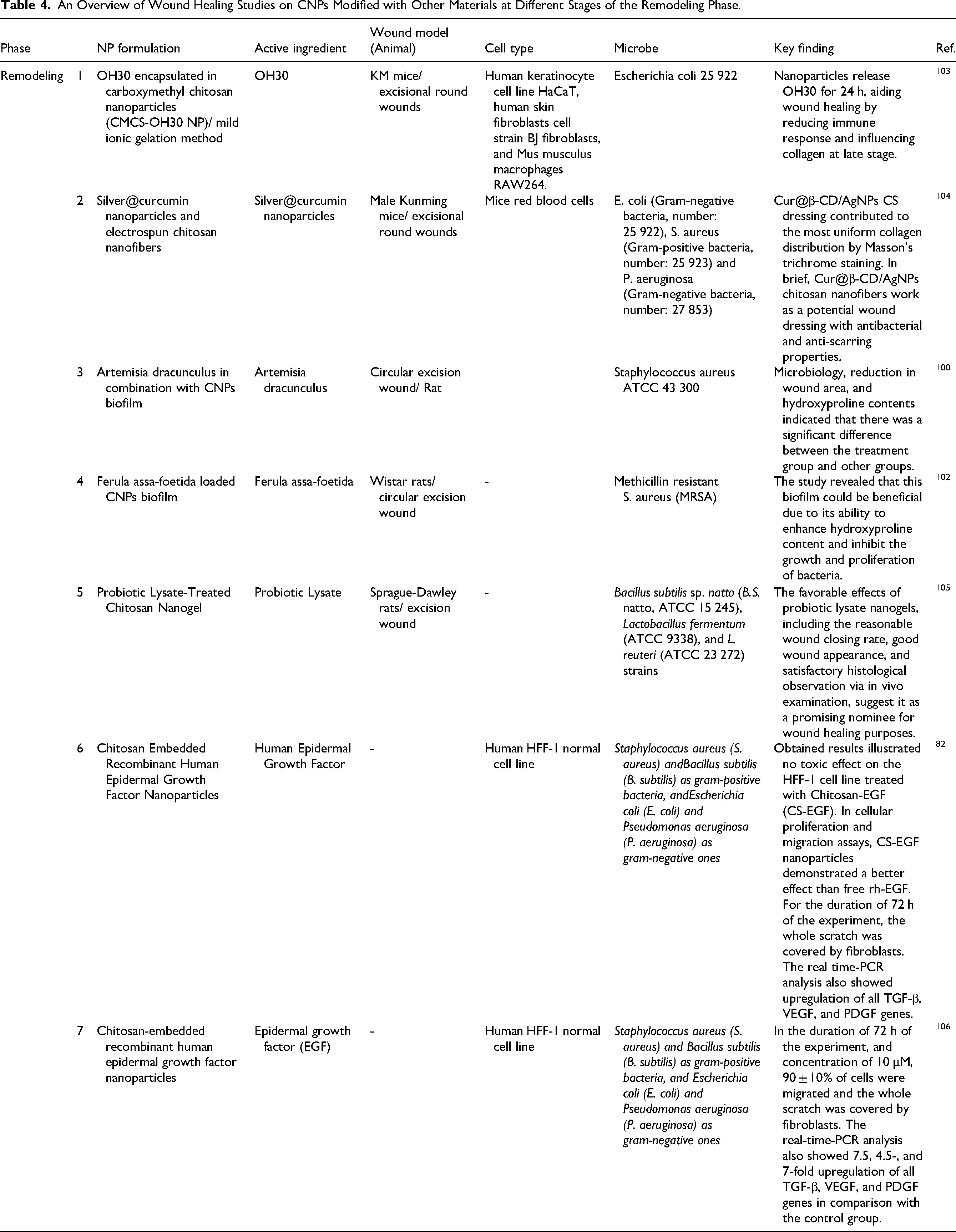
An Overview of Wound Healing Studies on CNPs Modified with Other Materials at Different Stages of the Remodeling Phase.
Wound Healing Properties
Utilizing various forms of CS as a wound dressing on open wounds of animals does not leave scars due to its biocompatibility, biodegradability and, accelerates wound healing process. 107
CS monomers aid in the regular deposition of collagen and increase the amount of natural hyaluronic acid synthesis at the wound site. 108 Additionally, CS provides a cellulose matrix to facilitate skin tissue regeneration and macrophage activation to inhibit abnormal growth activity, which helps faster healing of full-thickness wounds and prevent scarring.79,109
CNPs combined with Epidermal Growth Factor (EGF) have demonstrated the capability to accelerate wound healing process by promoting faster wound closure, aiding re-epithelialization, and enhancing collagen deposition. 93 A study has revealed that the combination of silver nanoparticles and CS has a synergistic effect, which accelerates wound healing and improving wound healing capacity. 74 Chitosan nanosheets (CNSs) have shown exceptional wound healing capabilities, such as reduced inflammatory cell infiltration, increased fibroblast activity, enhanced epithelium thickness, enlarged granulation area, and higher density of collagen fibers. 110 Chitosan nanocomposites exhibit no cytotoxicity and have been shown to enhance tissue regeneration. Furthermore, they can reduce inflammation and possess antibacterial properties. 77 The mechanical characteristics of chitosan-based nanoparticles can be adjusted to broaden their potential applications in tissue engineering when combined with fillers like hydroxyapatite. CNPs possess multitude of advantageous characteristics, including antibacterial, antioxidant, and hemostatic properties, making them suitable for diverse procedures such as wound treatment and the development of novel drug delivery systems. 111 The wound healing properties of CNPs are shown in Figure 3.
Future Perspective
The potential applications of CNPs for addressing inflammation have been widely studied, in disorders such as arthritis, dermatitis, and digestive tract. These nanoparticles exhibit promising potential for various pain management applications. They have the ability to deliver analgesics to specific pain sites and provide stable drug release, and providing prolonged pain relief. Additionally, these nanoparticles can also be incorporated into hydrogels to create localized analgesic effects. Consequently, it can be concluded that CNPs have significant potential in pain management.
Furthermore, CNPs have potential applications in tissue engineering for the preparation of scaffolds to stimulate angiogenesis and promote wound healing process, highlighting the potential of these nanoparticles in medical advancements. One of the limitations in the field of using CNPs is that most of the studies has conducted on the effects of CNPs alone or conducted with metals on normal and infectious wounds, so expanding these treatments to more complex wounds unveil a new perspective in this field.
Lack of clinical trials on CNPs is an additional issue. Currently, numerous preclinical animal studies have been conducted on diverse wound types, which can provide sufficient knowledge for investment in clinical models in a suitable environment. In addition, the use of novel technologies and techniques such as nanotechnology can facilitate the development of new dressings applications.
Poor solubility in neutral and alkaline solutions is another limitation of these nanoparticles, which impeding their efficacy. Moreover, many studies have examined the toxicity and biocompatibility of these nanoparticles, but more investigation is needed for efficient use of them. Finally, there is a need for a strong evidence-based database for the development of dressings that improve wound healing results with less cost and time. Based on this database, necessary actions, including financial resources and investments, are taken with more precision and lead to an improvement in in this research area.
Conclusion
Based on the research analyzed in this article, it can be concluded that chitosan nanoparticles, owing to their properties that enhance hemostasis, exhibit antimicrobial and anti-inflammatory effects, provide analgesic potential, promote granulation tissue formation, stimulate angiogenesis, and accelerate cellular proliferation, can be utilized as a standalone medication for wound healing. Some of these properties have not been clearly demonstrated, but it remains a very promising medication. These nanoparticles can be employed in various forms, including drug delivery systems, wound dressings, or hydrogels. Furthermore, the design of these dressings, utilizing chitosan nanoparticles, offers superior advantages in penetration and functionality due to their smaller size and higher surface-to-volume ratio compared to chitosan polymer. This underscores the inherent capabilities of chitosan nanoparticles as a therapeutic agent in wound repair.
Summary of the wound healing properties of chitosan nanoparticles.
Footnotes
Declaration of Conflicting Interests
The authors declared no potential conflicts of interest with respect to the research, authorship, and/or publication of this article.
Funding
The authors received no financial support for the research, authorship, and/or publication of this article.