
Abstract
This research examined the in vitro antibacterial, potential antioxidant, and antidiabetic activities of Matricaria chamomilla extract (MCE), Anastatica hierochuntica extract (AHE), and their combination against selected foodborne pathogens. Agar well diffusion, minimum inhibitory concentration (MIC), and minimum bactericidal/fungicidal concentration (MBC/MFC), as well as time-kill effect experiments, were applied to evaluate the antimicrobial characteristics. The combined extract showed better antibacterial qualities than the individual extracts. Results revealed that the tested microbes, including Pseudomonas aeruginosa, Klebsiella pneumoniae, Bacillus cereus, Staphylococcus aureus, and Candida albicans, were among the studied pathogens, with inhibition zones ranging from 24 to 31 mm. By contrast, the inhibition zones of MCE and AHE were 20–26 mm and 19–23 mm, respectively. Furthermore, compared with MCE (150–275 µg/mL; 175–300 µg/mL) and AHE (200–350 µg/mL; 225–375 µg/mL), the combined extract showed reduced MIC (100–200 µg/mL) and MBC/MFC (125–225 µg/mL), indicating even greater antimicrobial efficacy. With an IC50 of 24.44 µg/mL, the combined extract exhibited the greatest radical-scavenging activity in the DPPH experiment, whereas the corresponding values of IC50 for MCE and AHE were 38.19 and 54.49 µg/mL, respectively. The combined extract showed the greatest inhibitory activity against both α-glucosidase and α-amylase. The combined extract showed the strongest and fastest bactericidal action in time-kill tests. Overall, the combined extract demonstrated enhanced, reliable, and repeatable in vitro antibacterial activity, as well as potential antioxidant and antidiabetic properties.
Keywords
Introduction
The global prevalence of metabolic disorders, oxidative stress-mediated diseases, and microbial infections represents a major health care challenge in the 21st century (Thomaz et al., 2024). A metabolic disorder known as diabetes mellitus (DM) develops gradually due to insufficient insulin synthesis or defective insulin action; its hallmark is hyperglycemia, or consistently elevated blood sugar levels (Lancet, 2023). This disease is a huge global health problem since it affects people of all ages, genders, and locations, and it is acknowledged as one of the leading causes of sickness and mortality worldwide (Młynarska et al., 2025). Type 2 DM (T2DM), microbial resistance, and chronic inflammatory disorders are some of the most serious health problems that affect people all over the globe (Parker et al., 2024). Likewise, T2DM, which accounts for roughly 90% of diabetes cases, may be caused by environmental factors such as being overweight, not eating well, and not getting enough exercise. Genetics also plays a role (Mizukami and Kudoh, 2022). In type 1 DM, on the other hand, the main cause of absolute insulin insufficiency is the autoimmune destruction of pancreatic β-cells (Morran et al., 2015). Another kind, known as gestational DM (GDM), develops during pregnancy and increases the risk of problems during delivery for the mother and the unborn child (Alam et al., 2021). Although established diagnostic criteria exist for the two types of diabetes, the worldwide impact of the disease continues to grow (Sun et al., 2022). Remarkably, postprandial hyperglycemia is an important treatment goal for people with T2DM, and blocking carbohydrate-hydrolyzing enzymes such as α-glucosidase and α-amylase is a proven way to reduce glucose absorption (Panunzi et al., 2016).
An additional significant issue in health care is the fast spread of bacteria and viruses that are resistant to several drugs (Wagenlehner and Dittmar, 2022). This is happening at the same time as metabolic illnesses like diabetes are on the increase across the world (Friedman et al., 2016). The development of antimicrobial resistance (AMR) is accelerated by the overuse and abuse of antibiotics in both the medical and agricultural sectors, making many conventional medicines less effective (Hailu et al., 2021). Infections caused by multidrug-resistant (MDR) pathogens, including methicillin-resistant Staphylococcus aureus, carbapenem-resistant Enterobacteriaceae, and Pseudomonas aeruginosa, are associated with prolonged hospitalization, increased health care costs, and elevated mortality rates (Antochevis et al., 2025). Furthermore, the management of infections in immunocompromised individuals and patients with diabetes is particularly challenging, as fungal pathogens such as Candida albicans are increasingly exhibiting reduced susceptibility to conventional antifungal therapies (Kolbe-Busch et al., 2025; Loh and Lam, 2023).
Consequently, the advancement of plant-based treatments that integrate effectiveness with enhanced safety profiles has attracted significant interest in recent years (Jabeen et al., 2024; Siddiqui et al., 2024). Furthermore, adding potential antioxidants and antimicrobials to these kinds of formulations also makes them more useful as medicines, as oxidative stress and infections are commonly involved in the development and consequences of diabetes (Salehi and Rashidinejad, 2025). Traditionally, medicinal plants have been a significant source of bioactive chemicals, providing treatment options for several ailments (Chaachouay and Zidane, 2024; Rasheed et al., 2025). Their different phytochemical composition, which contains phenolic acids, flavonoids, terpenoids, alkaloids, and coumarins, helps with a number of biological functions, such as fighting bacteria, diabetes, and inflammation (Agidew, 2022).
Chamomile (Matricaria chamomilla L.), a member of the Asteraceae family, is among the most widely served medicinal herbs (Dai et al., 2022). Likewise, chamomile has a significant position in both traditional and contemporary herbal therapy, celebrated for its many pharmacological uses (Mao et al., 2016). Generally, it has been used to address many health issues, including metabolic illnesses such as diabetes and chronic inflammatory diseases (El Joumaa and Borjac, 2022; Aksoy and Sözbilir, 2012).
Anastatica hierochuntica, often referred to as kaff-e-Maryam, is an herb of medicinal value that inhabits arid environments and is classified in the Brassicaceae family. While it proliferates globally, the preponderance of its habitats is situated in Arab nations (Oman, Saudi Arabia, Iraq, and Kuwait) (Yusof et al., 2016). It is also cultivated successfully in numerous regions in Asia, Europe, and Africa. Further, A. hierochuntica has great therapeutic potential and should be utilized to manage a wide range of health issues, such as DM, and to help with birthing (Zin et al., 2017). Further, depression, bronchial asthma, mouth ulcers, arthritis, epilepsy, gastrointestinal problems, and malaria are among the many illnesses and aches that it is used to treat (Kim Sooi and Lean Keng, 2013). According to recent research, A. hierochuntica has a number of bioactive substances that support its pharmacological actions, including flavonoids (naringin and kaempferol), phenolic acids (caffeic acid and gallic acid), and sesquiterpene lactones (AlGamdi et al., 2011). Notably, its role in modulating glucose metabolism and inhibiting carbohydrate-digesting enzymes has been reported, though studies are limited and not fully conclusive (Hassan and Tariq I, 2020).
In this context, medicinal plants have attracted considerable attention as alternative sources of bioactive compounds with multifunctional biological activities, including antimicrobial, potential antioxidant, and enzyme-inhibitory effects. However, most existing studies have primarily focused on individual plant extracts, with limited investigation into the potential benefits of combining different plant-derived bioactive compounds. Importantly, the combining plant extracts can lead to enhanced or additive biological activities, potentially offering more potent therapeutic effects than using individual extracts (Álvarez-Martínez et al., 2021). While both chamomile and A. hierochuntica have demonstrated bioactivity individually, the scientific literature lacks comprehensive studies investigating their combined effects, particularly regarding enhanced action against key metabolic and microbial targets. Combining plant extracts with complementary phytochemical profiles may enhance therapeutic efficacy through enhanced or additive interactions, in which compounds act on multiple pathways simultaneously (Thotathil et al., 2023).
Notably, combined plant-extract formulations may offer enhanced or complementary biological effects by interacting among diverse phytochemicals that simultaneously target multiple pathways. Despite this potential, there remains a significant gap in the literature regarding the systematic evaluation of combined plant extracts, particularly against clinically and food-relevant pathogenic microorganisms.
Therefore, the present study aims to investigate the in vitro antimicrobial, potential antioxidant, and antidiabetic activities of M. chamomilla L. and A. hierochuntica L., both individually and in combination, with a specific focus on their effectiveness against foodborne pathogens. This work seeks to provide a comprehensive evaluation of their potential as natural agents for food safety applications and for the management of metabolic disorders.
Materials and Methods
Selection of plant materials
Fresh aerial parts of chamomile (M. chamomilla) and A. hierochuntica were obtained from a local herbal shop in Iraq. The plant materials were meticulously cleaned with distilled water to remove contaminants and then shade-dried at ambient temperature (25 ± 2°C) for 2 weeks to maintain their phytochemical integrity. After they had completely dried, an electric grinder crushed the materials into a uniform powder. The powdered botanical components were then preserved in sealed containers at 4°C until future extraction processes.
Preparation of plant extracts
Each plant powder of M. chamomilla (MC) and A. hierochuntica (AH), weighing 50 g, was separately extracted by maceration in 500 mL of ethanolic water (70%) at ambient temperature with agitation (120 rpm) for 72 h. The extracts of MCE and AHE were filtered through Whatman No. 1 filter paper, and the solvents were evaporated under reduced pressure using a rotary evaporator at 40°C. The combined extract (MCE + AHE) was created by blending the desiccated extracts of MCE and AHE in a 1:1 (w/w) ratio. All extracts were preserved at −20°C for subsequent analysis. Prior to biological evaluation, the dried extracts were reconstituted in dimethyl sulfoxide (DMSO) to prepare stock solutions. The final concentration of DMSO in all assays did not exceed 1% (v/v), and appropriate solvent controls were included to ensure that DMSO had no effect on microbial growth.
Determination of total phenolic content
The total phenolic content (TPC) of MCE and AHE extracts was quantified using a modified Folin–Ciocalteu assay. Briefly, 200 µL of each extract (1 mg/mL) was mixed with 1 mL of 10% Folin–Ciocalteu reagent and incubated for 5 min. Then, 800 µL of 7.5% sodium carbonate solution was added, and the mixture was left to react at room temperature for 30 min in the dark. The absorbance was recorded at 765 nm using a UV–Vis spectrophotometer. Gallic acid was applied as the standard, and TPC was expressed as mg gallic acid equivalent (GAE)/g extract (Siddiqui et al., 2017).
Determination of total flavonoid content
The total flavonoid content (TFC) of MCE and AHE was determined by an aluminum chloride-based colorimetric assay. For each sample, 500 µL of extract (1 mg/mL) was mixed with 500 µL of 2% aluminum chloride solution and 3 mL of methanol. The mixture was left to stand at room temperature for 30 min. The absorbance was then measured at 415 nm. Quercetin was used to generate the standard curve, and results were reported as milligrams of quercetin equivalents (mg QE) per gram of extract (Shraim et al., 2021).
Chromatographic analysis of plant extracts
Gas chromatography–mass spectrometry analysis of extracts
The gas chromatography–mass spectrometry (GC-MS) using a Perkin-Elmer Clarus 680 system that had an electron ionization source and an Elite-5MS capillary column (30 m × 0.25 mm, 0.25 µm film thickness) was applied to identify the phytochemical components of MCE and AHE. The carrier gas was helium, which flowed at a steady rate of 1 mL/min. Before being injected, samples were diluted and filtered. 1 µL was injected in split mode. The injector and detector temperatures were both set to 280°C. The oven was set to start at 50°C and stay there for 2 min. The temperature went up by 5°C every 1 min until it reached 300°C, and it stayed there for 10 min (Dąbrowski, 2020).
High-performance liquid chromatography analysis
Through the application of a Vanquish™ Core HPLC system (Thermo Fisher Scientific, USA) fitted with a C18 reversed-phase column (125 × 4.60 mm, 5 µm), the phenolic content of MCE and AHE was investigated. Methanol (solvent A) and acetic acid in water (1:25 v/v) (solvent B) were used in a gradient elution. After 3 min of 100% solvent B, the program switched to 50% solvent A for 5 min, increased to 80% solvent A for 2 min, and then went back to 50% solvent A for 5 min. The column temperature was maintained at 25°C, the flow rate was 1.0 mL/min, and detection took place at 250 nm. Both extracts were injected at a volume of 25 µL, facilitating the identification and quantification of the key phenolic components (Kuntić et al., 2007).
Antimicrobial properties of plant extracts
Determination of zone of inhibition
The microbial strains used in this study included Pseudomona aeruginosa (ATCC 27853), Klebsiella pneumoniae (ATCC 13883), Bacillus cereus (ATCC 10876), Staphylococcus aureus (ATCC 25923), and Candida albicans (ATCC 10231). All strains were obtained from a recognized microbial culture collection and maintained under standard laboratory conditions. Bacterial strains were cultured on Mueller–Hinton agar, while C. albicans was cultured on Sabouraud dextrose agar (SDA). Prior to each experiment, microbial suspensions were adjusted to a 0.5 McFarland standard (1.5 × 10⁸ colony-forming unit [CFU]/mL) to ensure consistency.
The agar well diffusion technique was used to evaluate the initial antibacterial efficacy. Microbial suspensions were calibrated to 0.5 McFarland and inoculated onto Mueller–Hinton agar (MHA) for bacterial cultures and SDA for fungal cultures. Wells with a diameter of 6 mm were filled with 100 µL of each extract at a concentration of 200 mg/mL. Plates were incubated at 37°C for 24 h for bacterial cultures and at 30°C for 48 h for fungal cultures. The diameter of the inhibition zones (IZ) in mm was measured (Mahdi et al., 2024). Standard antibiotics, gentamicin (10 µg/disc) for bacterial strains and nystatin (100 U/disc) for fungal strains, were used as positive controls (Klimko et al., 2020).
Determination of MIC and MBC
The broth microdilution technique was performed in 96-well microplates to determine the minimum inhibitory concentration (MIC) and the minimum bactericidal/fungicidal concentration (MBC/MFC). Serial dilutions of the extracts, ranging from 25 to 500 µg/mL, were prepared in Mueller–Hinton broth for bacterial cultures and in Sabouraud dextrose broth for fungal cultures. Following inoculation and incubation, the MIC was determined as the lowest concentration that completely suppressed observable microbial growth. To find the MBC or MFC, which is the lowest concentration that inhibits colony formation, wells exhibiting no discernible microbial growth were subcultured onto agar plates (Rodríguez-Melcón et al., 2022).
Time-kill kinetics assay
The time-kill assay was performed to evaluate the bactericidal and fungicidal activities of MCE, AHE, and the combined extract against selected pathogenic microorganisms. Overnight cultures of each tested microbe were grown in appropriate growth media and adjusted to approximately 1.5 × 10⁶ CFU/mL. Aliquots of the standardized microbial suspensions were exposed to MCE, AHE, and the combined extract at concentrations equivalent to their respective MICs. Untreated microbial suspensions served as the control. At predetermined time intervals (0, 10, 25, 50, 75, 100, 125, and 150 min), samples were withdrawn, serially diluted in sterile phosphate-buffered saline, and plated on agar media suitable for each microorganism. Plates were incubated under optimal growth conditions, and viable colonies were counted to determine CFU/mL. All experiments were performed in triplicate (n = 3), and results were expressed as mean ± standard deviation (SD). Time-kill curves were constructed by plotting log CFU/mL against time.
Potential antioxidant activities of the tested plant extracts
The DPPH radical-scavenging assay of MCE, AHE, and the combined extract was applied with a few adjustments. In brief, a 0.1 mM DPPH solution was prepared in methanol and kept in the dark to prevent decomposition. The extracts were evaluated at concentrations ranging from 15.6 to 1000 µg/mL. After that, 100 µL of extract was mixed with 100 µL of DPPH solution in each well of a 96-well plate, and the mixture was allowed to stand in the dark at room temperature for 30 min. A microplate reader measured the absorbance at 517 nm (Lorenz et al., 2017). The positive control was

In vitro antidiabetic activity
Determination of α-glucosidase inhibition
The α-glucosidase inhibitory activity of MCE, AHE, and their combined extract was assessed using the p-nitrophenyl-α-

Determination of α-amylase inhibition
The α-amylase inhibitory activity of MCE, AHE, and their combined extract was evaluated using the starch-iodine technique. Plant extracts (15.6–1000 µg/mL) were combined with α-amylase (1 U/mL) and preincubated at 37°C for 10 min. Following the addition of 1% soluble starch, the reaction persisted for 15 min and was then halted by the introduction of 1 M hydrochloric acid and iodine reagent (5 mM I2/5 mM KI). Absorbance was recorded at 620 nm, and the percentage inhibition and IC 50 values were determined in a manner similar to the α-glucosidase experiment (Jini et al., 2022).
In silico molecular docking of bioactive compounds
In silico molecular docking was performed as an exploratory approach to predict potential interactions between selected bioactive compounds identified by GC-MS analysis and relevant biological targets associated with antimicrobial, antioxidant, and antidiabetic activities. The docking analysis was intended to provide theoretical support for the experimentally observed in vitro results, rather than to serve as definitive evidence of biological activity. The ligand structures were energy-minimized using the MMFF94 force field in Avogadro 1.2.0 (Hanwell et al., 2012) after being imported from the PubChem database in SDF format. This guarantees optimum configurations. Swiss Target Prediction (Gfeller et al., 2014) was used to predict antidiabetic and potential antioxidant targets, while PharmMapper (Liu et al., 2022) was utilized to find prospective biological targets by recognizing pharmacophore-based targets relevant to bacterial species. The protein structures Alox5 (P12527), Gcgr (P30082), Hmgcr (P51639), Hmox1 (P06762), Escherichia coli allC (P77425), and Bacillus spp. licT (P39805) were sourced from the UniProt database. Binding sites were identified from the literature and then validated using CB-DOCK2 (Liu et al., 2022). AutoDock Tools 1.5.7 was used to prepare the protein by assigning Gasteiger charges, including polar hydrogens, and eliminating water molecules. AutoDock Vina was used to conduct molecular docking simulations (Morris et al., 2009). The exhaustiveness value was established at 8, and grid boxes were centered on the projected active spots. The standard scoring system was used to evaluate the binding conformations and affinities. Finally, the docked complexes were visualized and evaluated using BIOVIA Discovery Studio Visualiser 2020 (Al-Nema et al., 2020). This facilitated the analysis and documentation of significant chemical interactions, including hydrogen bonding and hydrophobic contacts, as well as binding affinities (ΔG values).
Statistical analysis
All experiments were performed in triplicate (n = 3), and results are expressed as mean ± SD. Data were statistically analyzed using two-way analysis of variance, followed by Tukey’s post hoc test to determine significant differences between groups (p < 0.05) using GraphPad Prism (version 8.0.1).
Results and Discussion
Phytochemical profiling of extracts
To assess the phytochemical composition of the tested extracts (MCE and AHE), a comprehensive phytochemical analysis was performed. The amounts of the TPC and total TFC were quantitatively measured and tabulated in Supplementary Table S1. The results revealed that the TPC of MCE was 101 ± 2.5 mg GAE/g extract, while AHE exhibited a significantly higher TPC of 188.6 ± 4.1 mg GAE/g extract. The combined extract (MCE + AHE, 1:1 w/w) showed a TPC of 153.7 ± 2.8 mg GAE/g extract, suggesting a potential additive effect.
Similarly, the TFC of MCE was 76.8 ± 2.2 mg QE/g extract, AHE showed 92.5 ± 1.77 mg QE/g extract, and the combined extract exhibited 85.1 ± 2.4 mg QE/g extract. Previously, Haghi et al. (2014) stated that the 70% aqueous ethanol extract of chamomile exhibited a TPC of 49.5 mg GAE/g extract and a TFC of 24.3 mg QE/g extract. In a previous study, AHE was reported to contain 67.49 mg GAE/g of TPC and 49.78 mg QE/g of TFC (Almundarij et al., 2021).
Identification of phytochemical compounds using GC-MS analysis
The GC-MS analysis was performed to identify the phytochemical components of MCE and AHE. The GC-MS analysis of MCE identified 15 phytochemicals, all of which contribute to the extract’s diverse medicinal uses. The findings in Table 1 show that MCE has a varied chemical composition, with sesquiterpenes, fatty acids, esters, alkaloids, and coumarin derivatives accounting for the majority. These components all contribute to the traditional and current therapeutic and biomedical applications of chamomile. Prominently, the MCE included several compounds such as (Z)-2-(hexa-2,4-diyn-1-ylidene)-1,6-dioxaspiro[4.4]non-3-ene (9.53%), 9-octadecenoic acid (Z)—(8.63%), and 9,12-octadecadienoic acid (Z,Z)—(7.01%). The potential antioxidant and antidiabetic properties of these fatty acids, including their ability to prevent lipid peroxidation and improve insulin sensitivity, have been extensively studied.
Phytochemical Constituents of MCE Identified by GC-MS
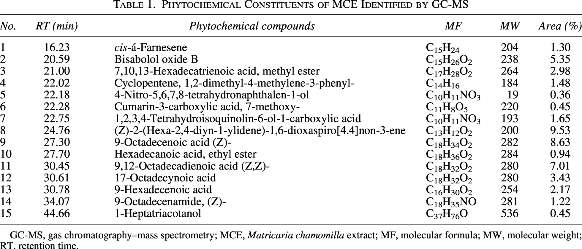
GC-MS, gas chromatography–mass spectrometry; MCE, Matricaria chamomilla extract; MF, molecular formula; MW, molecular weight; RT, retention time.
The presence of 5.35% bisabolol oxide B, a proven antibacterial and anti-inflammatory agent, is consistent with chamomile’s noted benefits for wound healing. Other minor terpenes, phenolic compounds, and cis-α-Farnesene (1.30%) all contribute to the extract’s broad-spectrum antibacterial effectiveness. Because of compounds such as 17-octadecynoic acid (3.43%) and 9-octadecenamide (Z-) (1.22%), which have been linked to hypoglycemic and metabolic regulatory effects, the extract may have antidiabetic applications. Furthermore, trace amounts of isoquinoline alkaloids and coumarin derivatives enhance the extract’s potential for antioxidant and antibacterial synergism.
The GC-MS analysis of the ethanolic extract from chamomile (M. chamomilla) has revealed a complex mixture of bioactive compounds, each contributing to the plant’s well-documented medicinal properties. The results consistently identify sesquiterpenes such as α-bisabolol and chamazulene, which are renowned for their anti-inflammatory and calming effects (Lu et al., 2024). Flavonoids like apigenin and quercetin, as well as coumarins and phenolic acids, were also prominent, supporting the extract’s potential antioxidant and antimicrobial activities (Al-Shuhaib and Al-Shuhaib, 2025). The detection of volatile oils, such as camphor and 1,8-cineole, further underscores the extract’s aromatic and antimicrobial potential (El Mihyaoui et al., 2022).
These findings align with previous research, which demonstrates that ethanol is an effective solvent for extracting both polar and nonpolar phytochemicals, thereby offering a comprehensive chemical profile of chamomile (Putra et al., 2025). The broad spectrum and significant number of these compounds support the traditional use of chamomile in herbal remedies, cosmetics, and dietary supplements. They also show how important extraction methods are for figuring out the phytochemical yield and health benefits of plant-based products (Dai et al., 2022).
On the other hand, GC-MS analysis was used to identify phytochemical compounds in AHE. Results presented in Table 2 indicate that the AHE contains phytoconstituents belonging to various classes of bioactive compounds, including terpenoids, fatty acids, esters, alcohols, and oxygenated hydrocarbons. These components are widely recognized for their diverse biological properties, particularly in antimicrobial, potential antioxidant, and antidiabetic applications. The major compound identified was 2H-pyran-3-ol, tetrahydro-2,2,6-trimethyl-6-(4-methyl-3-cyclohexen-1-yl)- at 23.14%, a monoterpene derivative known for its potential antioxidant and free radical-scavenging properties, as well as possible anti-inflammatory effects.
Phytochemical Constituents of AHE Identified by GC-MS
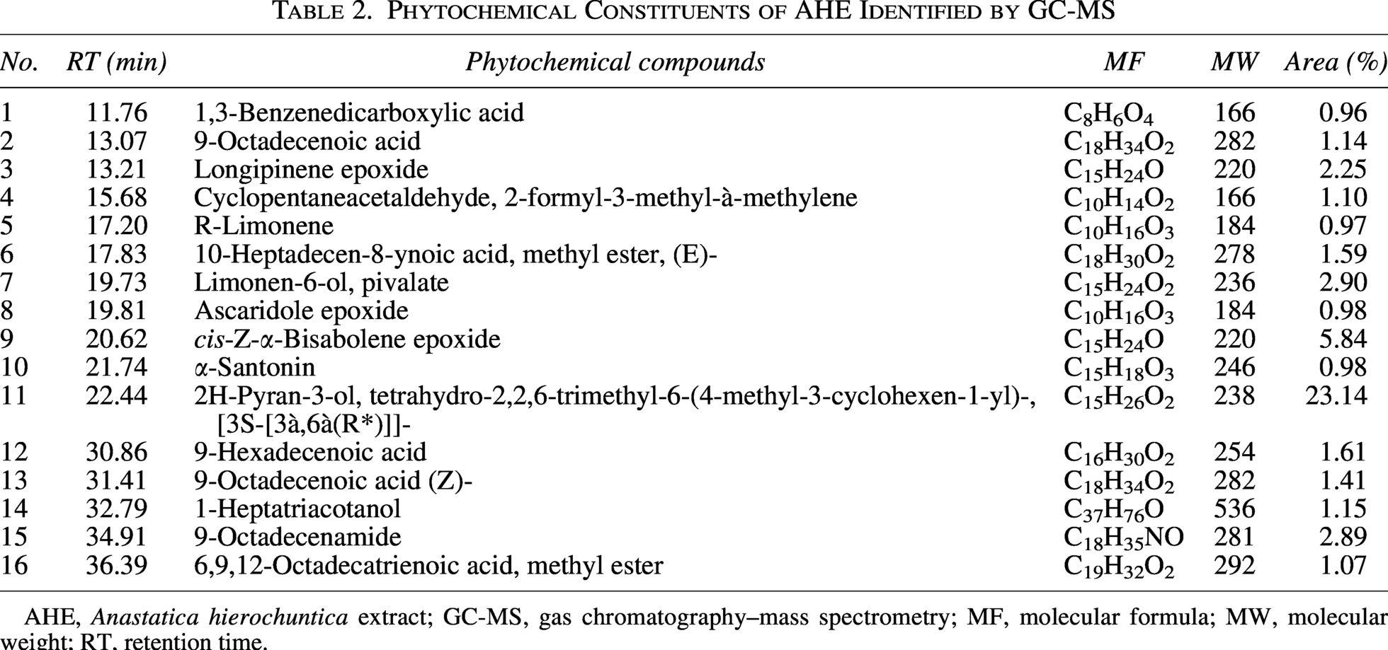
AHE, Anastatica hierochuntica extract; GC-MS, gas chromatography–mass spectrometry; MF, molecular formula; MW, molecular weight; RT, retention time.
Another prevalent molecule was cis-Z-α-bisabolene epoxide (5.84%), a sesquiterpene oxide known for its antibacterial, antifungal, and anti-inflammatory properties, which support the plant’s historical use in treating infections. Similarly, longipinene epoxide (2.25%) and limonene-6-ol, pivalate (2.90%) are oxygenated terpenes with reported potential antioxidant and antimicrobial activities. The extract also contained several unsaturated fatty acids and their derivatives, such as 9-octadecenoic acid (Z)- (1.41%), 9-hexadecenoic acid (1.61%), and 6,9,12-octadecatrienoic acid methyl ester (1.07%), all of which have demonstrated potential antioxidant, antidiabetic, and lipid-modulating effects. These compounds are known to improve insulin sensitivity and protect against oxidative stress, thereby supporting the extract’s potential for managing metabolic disorders.
The identification of ascaridole epoxide (0.98%) and α-santonin (0.98%) adds further pharmacological relevance due to their antimicrobial and antiparasitic properties, while R-limonene (0.97%) is a well-documented monoterpene with potential antioxidant, antimicrobial, and antidiabetic activities.
The diverse phytochemicals identified in A. hierochuntica, including a rich array of flavonoids, phenols, tannins, and terpenoids, provide a strong scientific basis for its traditional therapeutic applications (Thotathil et al., 2023). Similarly, large concentrations of flavonoids like quercetin and kaempferol as well as phenolic components like sinapic and syringic acids contribute to its robust potential antioxidant qualities. Together, these substances reduce oxidative stress in cells and eliminate free radicals. It has been used for a long time to avoid organ damage, mostly because of its protective action. The plant’s broad-spectrum antibacterial properties, which function by disrupting the integrity of microbial cells and preventing vital enzymes from functioning, are also derived from these same bioactive components (Rameshbabu et al., 2024).
Quantification of phenolic compounds in extracts using HPLC analysis
HPLC-DAD was used to quantify phenolic and flavonoid components in MCE and AHE. Table 3 and Figures 1 and 2 display the findings. In the case of MCE, seven phenolic acids and seven flavonoids were detected and quantified at levels that could be perceived (Table 3 and Fig. 1a,b). For AHE, seven phenolic acids and five flavonoids were identified. The phenolic profile of AHE included caffeic acid, gallic acid, cinnamic acid, syringic acid, ferulic acid, catechol, and benzoic acid, whereas the flavonoids comprised naringin, rutin, myricetin, quercetin, and kaempferol. These findings highlight the rich and varied phytochemical composition of both extracts, supporting their potential antioxidant, antimicrobial, and antidiabetic activities (Table 3 and Fig. 2a,b). The complementary profiles of MCE and AHE suggest a possible enhanced therapeutic effect when combined.
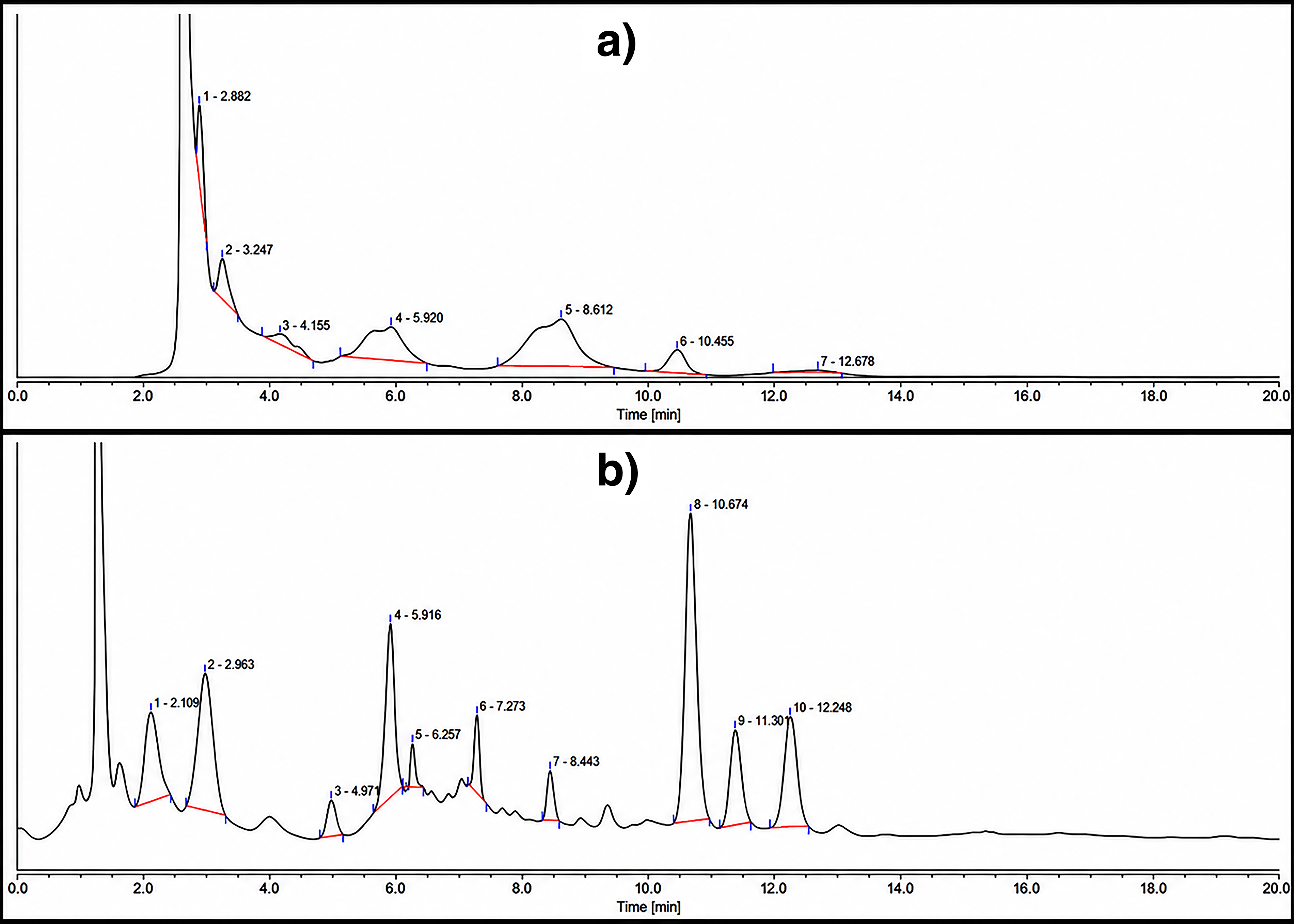
HPLC-DAD chromatogram and major peaks of identified phenolic compounds:
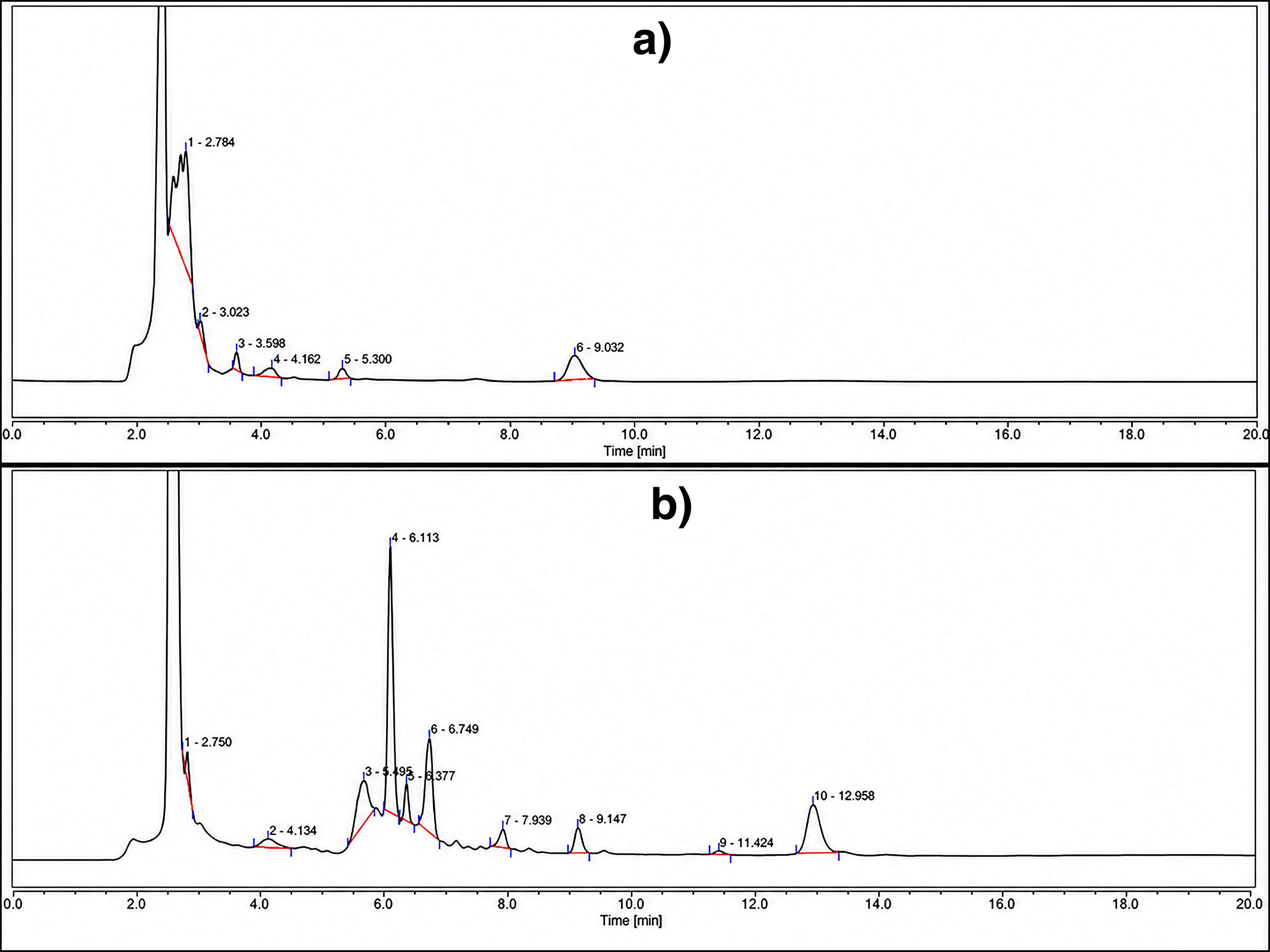
HPLC-DAD chromatogram and major peaks of identified phenolic compounds:
Quantitative Analysis of Phenolic and Flavonoid Compounds Identified in the Tested Plant Extracts (MCE and AHE) Using HPLC-DAD
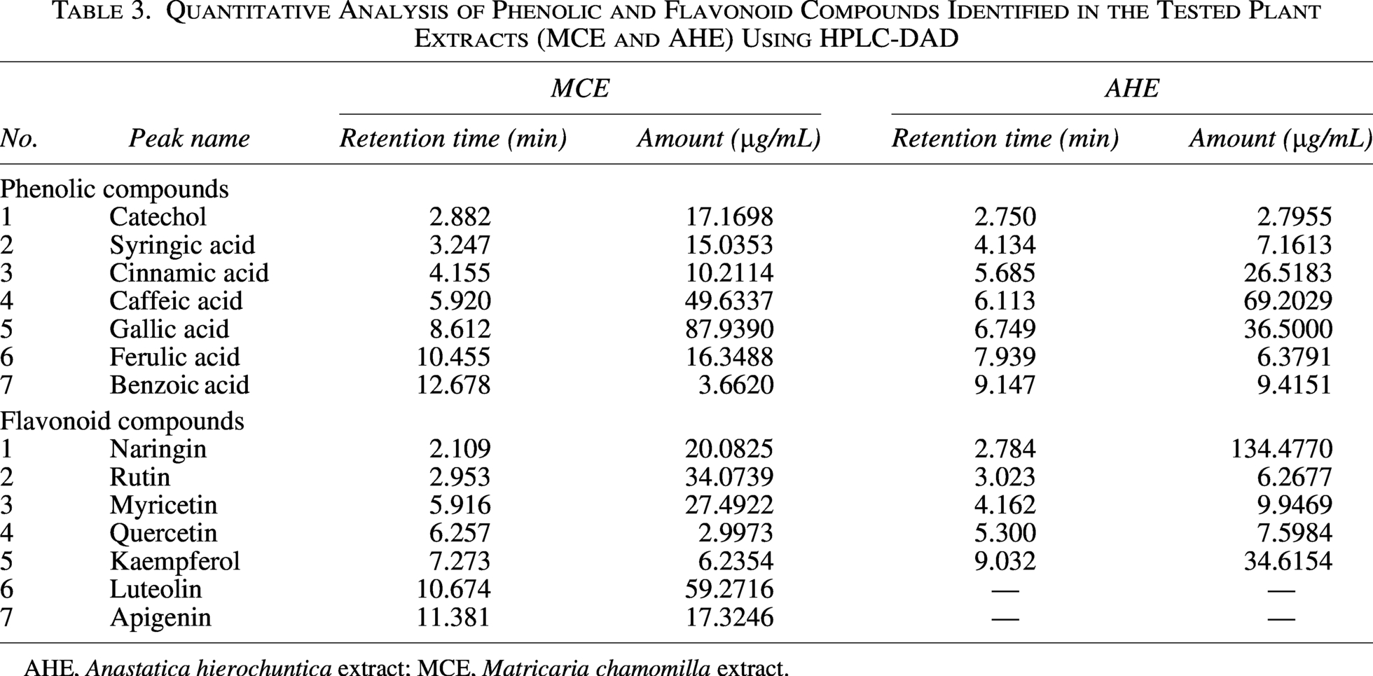
AHE, Anastatica hierochuntica extract; MCE, Matricaria chamomilla extract.
Results indicate that gallic acid was the major phenolic component in MCE (87.93 µg/mL), significantly enhancing its antioxidant potential, while AHE had a lower, yet notable concentration (36.50 µg/mL). Caffeic acid, a recognized potential antioxidant and antibacterial compound, was identified in both extracts, with a greater quantity in AHE (69.20 µg/mL) than in MCE (49.63 µg/mL). Cinnamic acid was present in greater quantities in AHE (26.51 µg/mL) compared with MCE (10.21 µg/mL), indicating a possible contribution to the antibacterial and metabolic-regulating properties of AHE.
Further, syringic acid, ferulic acid, and catechol were identified in both extracts, with MCE typically exhibiting higher levels, especially of catechol (17.17 µg/mL), known for its free radical-scavenging capabilities. Benzoic acid was detected in both extracts, with AHE exhibiting a higher concentration (9.41 µg/mL), suggesting a potential contribution to antibacterial effectiveness. Naringin was highly prevalent in AHE (134.47 µg/mL), indicating a substantial contribution to its antidiabetic and potential antioxidant properties, whereas MCE exhibited lower concentrations (20.08 µg/mL). In contrast, rutin, myricetin, and quercetin were present in greater quantities in MCE, corroborating its established anti-inflammatory and anticancer characteristics. Furthermore, the kaempferol was predominantly present in AHE (34.61 µg/mL), whereas luteolin and apigenin were detected only in MCE at 59.27 and 17.32 µg/mL, respectively, underscoring the unique anticancer and potential antioxidant potential of MCE. The findings indicate that the combination of MCE and AHE has complementary bioactive properties, with MCE serving as a substantial source of potential antioxidant and anticancer agents, while AHE provides notable antidiabetic and antibacterial flavonoids.
Evaluation of antimicrobial properties
Measurements of zones of inhibition
The findings of the current research display that the therapeutic and biological potential of the extracts is directly related to their rich and diverse phytochemical makeup, which includes phenolic compounds, flavonoids, tannins, and other bioactive metabolites. The antimicrobial properties of MCE, AHE, and the combined extract (MCE + AHE) were assessed against a number of harmful pathogens, such as P. aeruginosa, K. pneumoniae, B. cereus, S. aureus, and C. albicans, as well as both Gram-positive and Gram-negative bacteria. The antimicrobial performance was evaluated by measuring the zones of inhibition (ZOI) in millimeter. The findings were then compared with those of traditional antibiotics, such as gentamicin for bacteria and nystatin for fungi.
Results illustrated in Table 4 and Figure 3 show that the combined extract produced the largest inhibition zone of 31 ± 3.26 mm against P. aeruginosa, whereas MCE and AHE produced zones of 26 ± 1.97 mm and 23 ± 3.61 mm, respectively. A significantly smaller zone of 18 ± 1.43 mm was observed with the common antibiotic gentamicin; however, nystatin had no impact on these bacteria. Regarding the effect of these tested extracts on K. pneumoniae, results showed a similar trend, with the combined extract achieving a ZOI of 28 ± 0.58 mm, surpassing the individual activities of gentamicin (19 ± 2.62 mm), AHE (22 ± 1.56 mm), and MCE (25 ± 3.14 mm). For B. cereus, the combined extract resulted in a ZOI of 25 ± 1.07 mm, while MCE and AHE produced inhibition zones of 23 ± 0.86 mm and 20 ± 0.74 mm, respectively. It is important to note that the combined extract outperformed gentamicin, which showed a ZOI of 14 ± 2.16 mm. In the case of S. aureus, the combination extract recorded a ZOI of 26 ± 2.69 mm, compared with 24 ± 2.93 mm for MCE, 21 ± 2.43 mm for AHE, and 17 ± 0.95 mm for gentamicin. Regarding fungal inhibition, C. albicans was highly sensitive to the combined extract, which achieved a ZOI of 24 ± 2.15 mm, outperforming both MCE (22 ± 3.41 mm) and AHE (19 ± 1.09 mm). Notably, the combined extract also surpassed the standard antifungal nystatin, which produced a significantly smaller ZOI of 14 ± 1.62 mm.
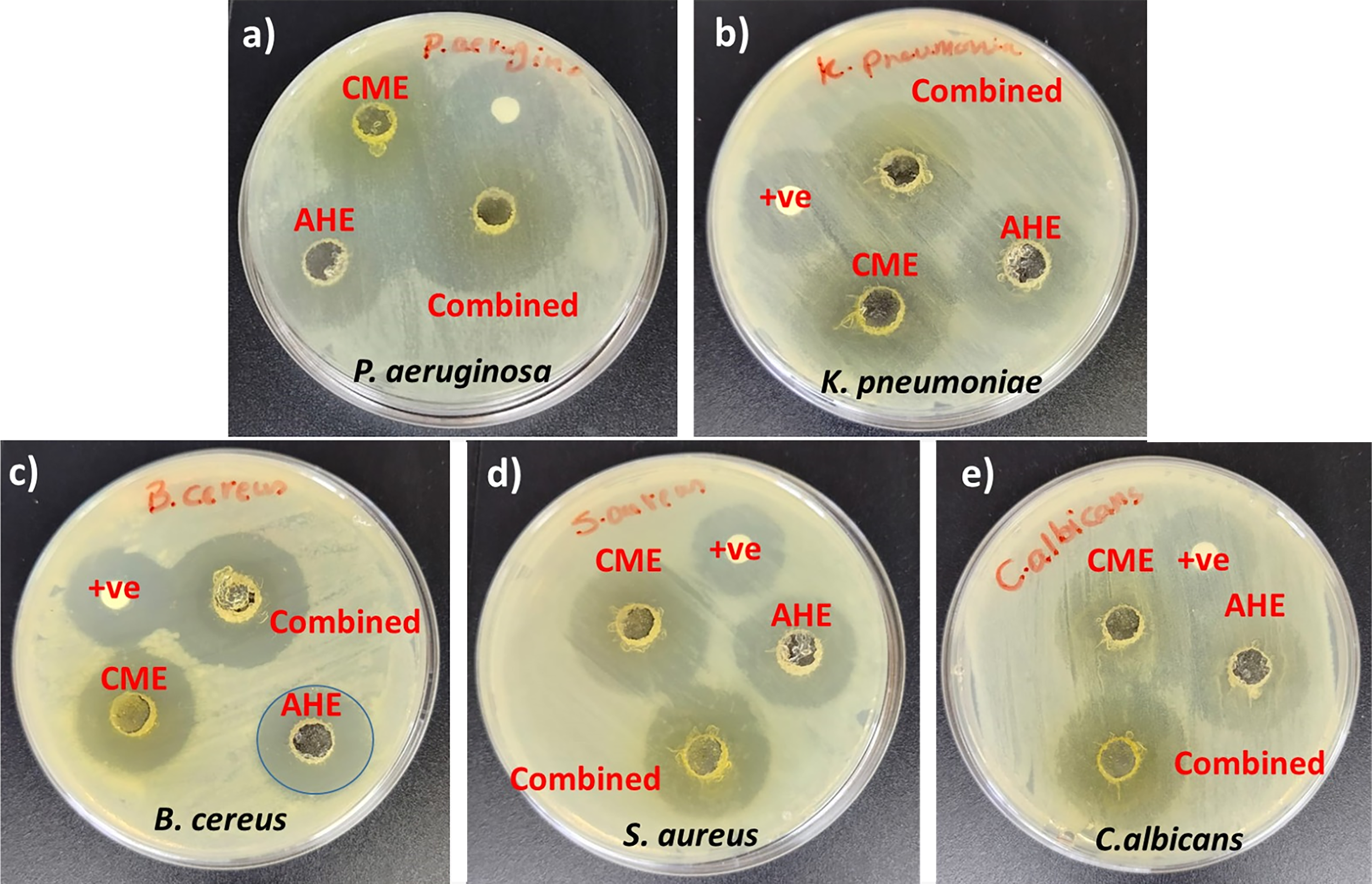
Digital images of Petri dishes of the clear zone of three different extracts, including MCE, AHE, and combined extract against five pathogenic microbes, including
Antimicrobial Activity of MCE, AHE, and Combined Extract Against Targeted Pathogenic Microorganisms Using the Zone of Inhibition Method
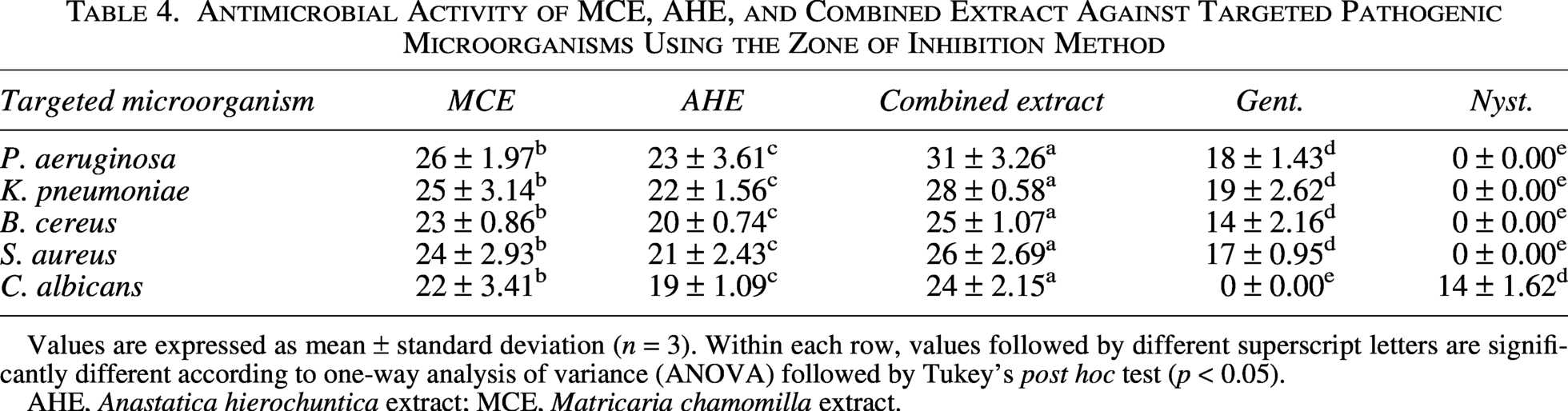
Values are expressed as mean ± standard deviation (n = 3). Within each row, values followed by different superscript letters are significantly different according to one-way analysis of variance (ANOVA) followed by Tukey’s post hoc test (p < 0.05).
AHE, Anastatica hierochuntica extract; MCE, Matricaria chamomilla extract.
The extraction solvent significantly influences the antibacterial activity of each extract by determining the efficiency and selectivity of phytochemical separation. Polar solvents, such as methanol and aqueous ethanol, are known to extract higher concentrations of phenolic and flavonoid compounds, which are primarily responsible for antibacterial efficacy (Gil-Martín et al., 2022). Consequently, maximizing the biological activity of plant-derived formulations depends on optimizing the extraction process, which also directly influences the formulations’ potential for use in pharmacological and therapeutic applications (Lee et al., 2024). These findings highlight a clear enhanced effect when MCE and AHE are combined, likely due to the complementary antimicrobial mechanisms of their bioactive constituents. The presence of bisabolol oxide B, cis-α-farnesene, and unsaturated fatty acids in chamomile, along with cis-α-bisabolene epoxide, longipinene epoxide, limonene derivatives, and ascaridole epoxide in AHE, may collectively target various microbial pathways, thereby enhancing inhibition of microbial proliferation (Park et al., 2022).
Determination of MIC and MBC/MFC values
The MIC and MBC/MFC values of MCE, AHE, and combined extract (MCE + AHE) were determined against five pathogenic microorganisms and tabulated in Table 5. The results were compared with standard antimicrobials: gentamicin for bacteria and nystatin for fungi. The results demonstrated that the combined extract enhanced antibacterial efficacy, as evidenced by consistently lower MIC and MBC/MFC values across all tested microbial strains. In comparison with MCE (MIC: 150 ± 8.25 µg/mL; MBC: 175 ± 6.02 µg/mL) and AHE (MIC: 200 ± 5.85 µg/mL; MBC: 225 ± 2.41 µg/mL), the combined extract demonstrated a MIC of 100 ± 7.02 µg/mL and an MBC of 125 ± 2.63 µg/mL against P. aeruginosa. Furthermore, the results disclose that K. pneumoniae showed a similar trend, with the combined extract surpassing MCE (MIC: 175 ± 1.93 µg/mL; MBC: 200 ± 7.32 µg/mL) and AHE (MIC: 225 ± 0.75 µg/mL; MBC: 250 ± 1.25 µg/mL) with a MIC of 100 ± 1.85 µg/mL and an MBC of 150 ± 2.14 µg/mL.
MIC and MBC Values of Three Different Extracts, Including MCE, AHE, and Combined Extract, Against Some Pathogenic Microbes, Including P. aeruginosa, K. pneumoniae, B. cereus, S. aureus, and C. albicans
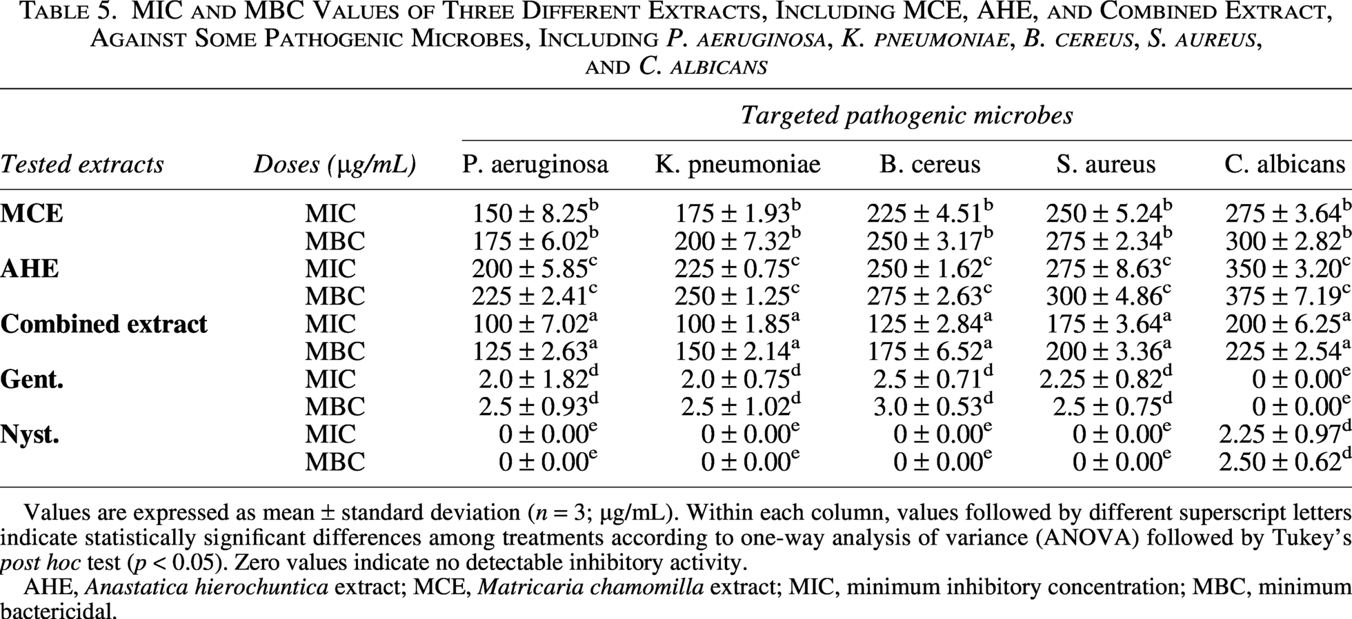
Values are expressed as mean ± standard deviation (n = 3; µg/mL). Within each column, values followed by different superscript letters indicate statistically significant differences among treatments according to one-way analysis of variance (ANOVA) followed by Tukey’s post hoc test (p < 0.05). Zero values indicate no detectable inhibitory activity.
AHE, Anastatica hierochuntica extract; MCE, Matricaria chamomilla extract; MIC, minimum inhibitory concentration; MBC, minimum bactericidal.
On the other hand, the combined extract demonstrated better effectiveness against Gram-positive bacteria. Both MCE (MIC: 225 ± 4.51 µg/mL; MBC: 250 ± 3.17 µg/mL) and AHE (MIC: 250 ± 1.62 µg/mL; MBC: 275 ± 2.63 µg/mL) were much less effective than B. cereus, which had MIC and MBC of 125 ± 2.84 µg/mL and 175 ± 6.52 µg/mL, respectively. In the same way, the combined extract had a MIC of 175 ± 3.64 µg/mL and an MBC of 200 ± 3.36 µg/mL for S. aureus, whereas MCE and AHE had MICs of 250 ± 5.24 µg/mL, 275 ± 2.34 µg/mL, and 300 ± 4.86 µg/mL, respectively.
The combined extract exhibited remarkable antifungal efficacy against C. albicans, with MIC of 200 ± 6.25 µg/mL and MFC of 225 ± 2.54 µg/mL. The MIC and MFC for MCE are 275 ± 3.64 µg/mL and 300 ± 2.82 µg/mL, respectively, whereas for AHE, they are 350 ± 3.20 µg/mL and 375 ± 7.19 µg/mL, respectively, indicating significantly worse performance than for MCE. The standard antifungal drug nystatin had an MFC of 2.50 ± 0.62 µg/mL and a MIC of 2.25 ± 0.97 µg/mL, indicating extremely powerful action as anticipated. Compared with the standard antibiotic gentamicin, which exhibited MIC values of 2.0–2.5 µg/mL and MBC values of 2.5–3.0 µg/mL for the bacterial strains, the plant extracts required higher concentrations, as expected for crude natural products.
Time-kill kinetics of plant extracts against pathogenic microorganisms
Figure 4a–e demonstrates that all tested plant extracts (MCE, AHE, and the combined extract) exerted a time-dependent antimicrobial effect against the examined pathogenic microorganisms. In contrast, the control groups maintained relatively stable CFU counts throughout the experiment, confirming that the observed reductions in microbial viability were attributable to the treatments rather than natural cell death.
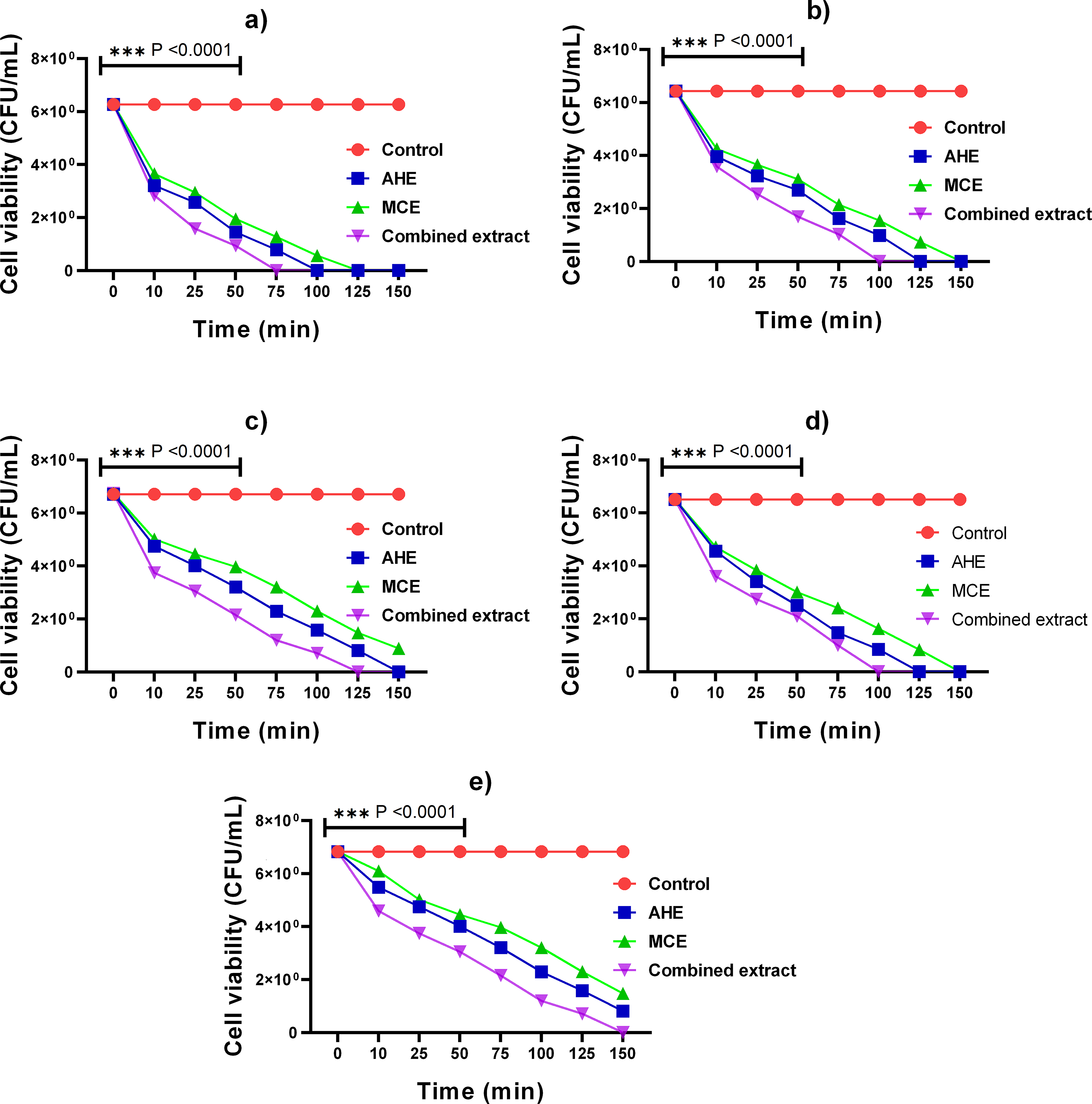
Time-killing effect of MCE, AHE, and combined extract against five pathogenic microbes, including
Among all treatments, the combined extract consistently exhibited the strongest killing kinetics, resulting in a more rapid and pronounced reduction in viable cell counts compared with the individual extracts. This enhanced activity suggests an additive or synergistic antimicrobial effect resulting from the combination of bioactive compounds in both plant extracts. At later time points (125–150 min), complete microbial inhibition was observed for most tested strains, indicating that prolonged exposure to the extracts, particularly the combined formulation, leads to effective microbial eradication. Overall, these findings demonstrate that the antimicrobial efficacy of the extracts is both time-dependent and treatment-dependent, supporting their potential application as natural antimicrobial agents against pathogenic microorganisms.
Potential antioxidant activity assessment
As depicted in Figure 5a–e, all tested samples exhibited concentration-dependent DPPH radical-scavenging activity, with increased DPPH inhibition observed at higher concentrations. The results in Figure 5a show that the combined extract demonstrated much greater antioxidant activity than the separate plant extracts (MCE and AHE) at lower doses (15.6–62.6 µg/mL). In particular, the combined extract exhibited a DPPH radical-scavenging activity of 66.20% at 62.6 µg/mL, well above the inhibition observed with MCE (43.17%) and AHE (32.70%). The combined extract maintained a strong scavenging capacity at 125 µg/mL, reaching 73.70%, which was very close to that of
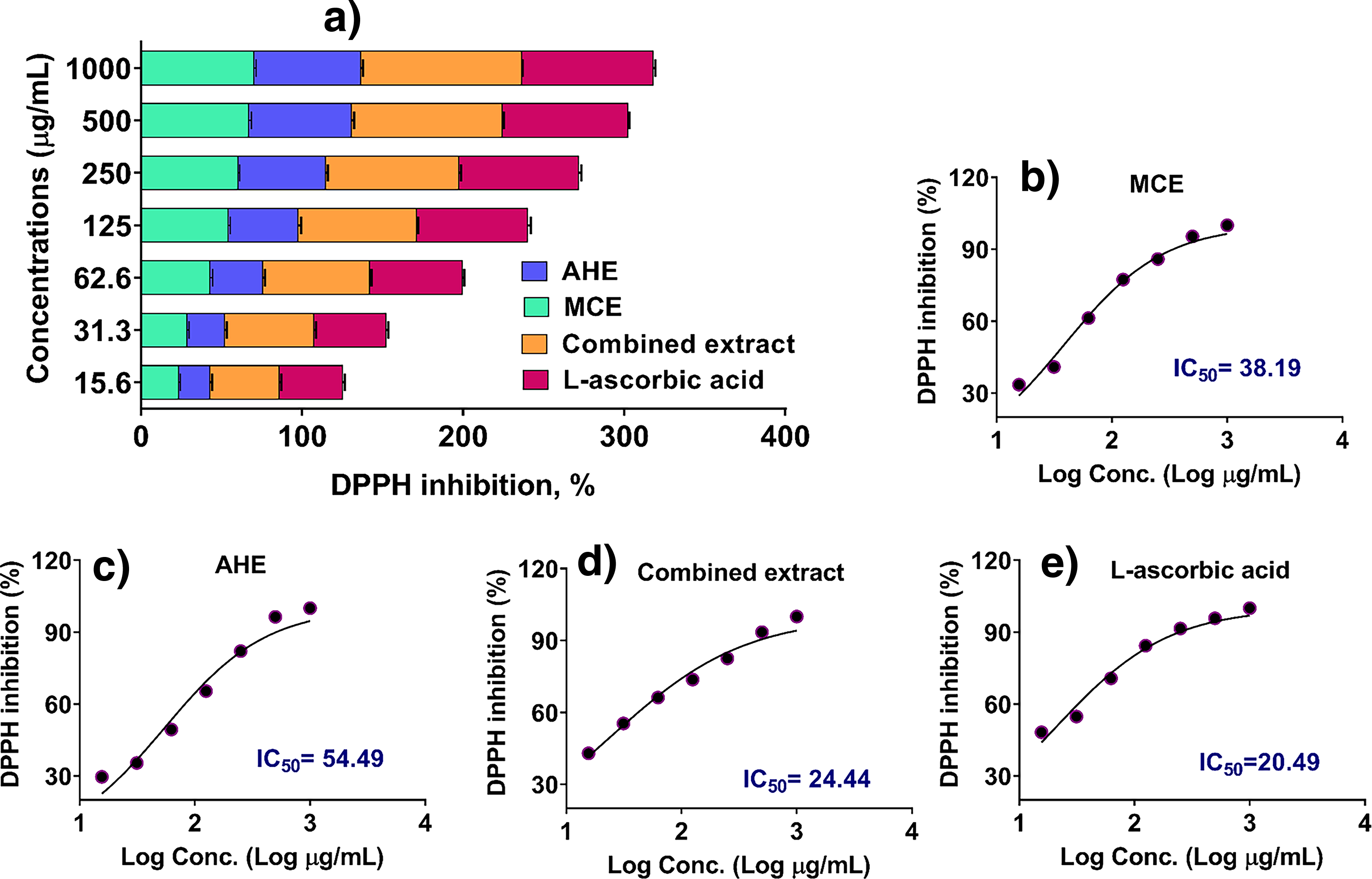
Moreover, the combined extract maintained enhanced DPPH radical-scavenging activity at elevated concentrations (250–1000 µg/mL). The results indicate that the inhibition percentage (93.50%) at 500 µg/mL was much higher than that of MCE (67.20%) and AHE (63.80%), and it greatly surpassed that of
Moreover, the IC50 values further confirm the potent DPPH radical-scavenging activity of the combined extract (Fig. 5b–e). The combined extract exhibited the lowest IC50 value of 24.44 µg/mL, indicating the highest radical-scavenging efficiency. In contrast, the IC50 values for MCE and AHE were 38.19 and 54.49 µg/mL, respectively. The reference compound,
In certain chamomile extracts, phenolic acids and flavonoids are the principal potential antioxidants, as they neutralize free radicals and reduce oxidative stress (Sotiropoulou et al., 2020). Caffeic acid, gallic acid, quercetin, and apigenin neutralize ROS and reduce oxidative damage in living beings. The ethyl acetate (EtOAc) and butanol (BuOH) components of chamomile effectively eliminated DPPH radicals, with IC50 values of 12.7 ± 3.8 and 13.8 ± 0.4 µg/mL, respectively, in an earlier investigation (Bouyahya et al., 2024). In this prior research, the EtOAc and BuOH fractions of chamomile exhibited strong DPPH radical-scavenging activity, with IC50 values of 12.7 ± 3.8 and 13.8 ± 0.4 µg/mL, respectively. In contrast, the dichloromethane (DCM) extracts displayed weaker activity (IC50: 279–290 µg/mL), which is consistent with the lower phenolic content typically found in nonpolar extracts (Mailänder et al., 2022). The significant antioxidant potential of chamomile roots is largely attributed to their content of caffeoylquinic acids and coumarins, particularly 4,5-dicaffeoylquinic acid, which has been identified as a potent radical scavenger (Mailänder et al., 2022). In comparison, the current study demonstrated a potential antioxidant effect when chamomile was combined with A. hierochuntica, resulting in a DPPH IC50 of 24.44 µg/mL for the combination, compared with higher IC50 values for the individual extracts. The IC50 value was 26.7 µg/mL for A. hierochuntica extract of chamomile flowers, underscoring the substantial impact of solvent selection on the potential antioxidant efficacy of chamomile extracts (Al-Dabbagh et al., 2019).
Evaluation of antidiabetic properties
To further evaluate the potential applicability of these extracts in the management of metabolic disorders, an in vitro antidiabetic study was conducted. The study assessed the ability of each extract to inhibit key carbohydrate-hydrolyzing enzymes through α-glucosidase and α-amylase inhibition assays, which are relevant targets for controlling postprandial hyperglycemia.
Supplementary Figure S1a shows the concentration-dependent evaluation of the α-glucosidase inhibitory activity of the conventional medication acarbose, and the tested extracts, including MCE, AHE, and the combined extract. Results disclose that the combined extract produced an inhibition of 37.6% at the lowest tested concentration (16.5 µg/mL), which was significantly greater than that of MCE (28.7%) and AHE (23.8%) and nearly equal to that of acarbose (31.9%). The combined extract continued to function better as the concentration rose to 62.2 µg/mL, achieving 54.1% inhibition, while MCE and AHE showed 48.9% and 38.0% inhibition, respectively. Similarly, the combined extract outperformed MCE (55.9%) and AHE (43.5%) at 125 µg/mL, achieving 68.2% inhibition.
Likewise, the combined extract’s strong α-glucosidase inhibitory activity persisted at higher concentrations (250–1000 µg/mL), further demonstrating its increased bioactivity. The combination achieved 76.2% inhibition at 250 µg/mL, while MCE and AHE achieved 61.5% and 53.4%, respectively. Interestingly, the combined extract showed 81.5% inhibition at 500 µg/mL, which was similar to that of acarbose (81.5%), while MCE and AHE achieved 69.5% and 59.4% inhibition, respectively. The combined extract showed an inhibition rate of 89.6% at the highest tested concentration (1000 µg/mL), which was close to that of acarbose (81.9%) and higher than the individual extracts (74.6% for MCE and 67.3% for AHE).
Moreover, among the plant-based therapies, the results in Supplementary Figure S2 show that the combined extract had the lowest IC50 value (108.2 µg/mL), indicating greater inhibitory efficacy than MCE (114.9 µg/mL) and AHE (139.4 µg/mL). This implies an enhanced or additive effect when the two extracts are combined, most likely due to the complementary effects of their bioactive components.
Supplementary Figure S1b presents the α-amylase inhibitory activities of MCE, AHE, combined extract, and acarbose, evaluated across a concentration range of 16.5–1000 µg/mL. The results demonstrate a clear dose-dependent inhibition pattern for all tested samples. Results indicate that the combined extract at the lowest concentration (16.5 µg/mL) exhibited an inhibition of 26.7%, outperforming both MCE (18.6%) and AHE (13.9%) and comparable with acarbose (22.2%). As the concentration increased to 62.2 µg/mL, the combined extract maintained higher inhibitory efficacy (46.2%) compared with MCE (38.1%) and AHE (31.5%), while acarbose showed 43.8% inhibition at this concentration. At 125 µg/mL, the combined extract reached 54.5% inhibition, whereas MCE and AHE reached 46.2% and 36.4%, respectively. Notably, the combined extract closely approached acarbose (54.6%), highlighting its competitive potency at this mid-range dose.
Conversely, the combined extract’s enhanced inhibitory action persisted at higher doses (250–1000 µg/mL). The combined extract demonstrated 75.9% inhibition at 500 µg/mL, outperforming both MCE (69.4%) and AHE (48.6%) and almost equal to acarbose (69.4%). Additionally, MCE and AHE demonstrated 65.2% and 57.5% inhibition, respectively, while the combined extract (1000 µg/mL) achieved 82.2% inhibition. In the same circumstances, acarbose demonstrated 73.8% inhibition.
Moreover, the dose–response curves presented in Supplementary Figure S3 demonstrate the inhibitory effects of MCE, AHE, combined extract, and acarbose on α-amylase activity. The calculated IC50 values were 142.2 µg/mL for MCE, 160.3 µg/mL for AHE, and 131.1 µg/mL for the combined extract, compared with 87.2 µg/mL for acarbose, the standard reference. The complementary profiles of flavonoids and phenolic acids, such as caffeic acid, gallic acid, quercetin, and naringin, which are known to modify enzymes involved in the metabolism of carbohydrates, may be the reason for the enhanced or additive interaction between the bioactive compounds of MCE and AHE (Mahdi et al., 2024). The results substantiate the effectiveness of integrating AHE extracts with MCE as a plausible natural antidiabetic therapy.
In silico multitarget analysis
The growing prevalence of diabetes and oxidative stress-related disorders has accelerated the search for novel therapeutics from natural sources. Traditional molecular docking studies often focus on single-target interactions, neglecting the complex multitarget potential of bioactive compounds. Furthermore, bacterial resistance mechanisms and their potential overlap with metabolic disease pathways remain underexplored in computational drug discovery. This study addresses this gap by evaluating the multitarget binding capabilities of natural compounds against both pathogenic bacterial proteins and key metabolic enzymes involved in diabetes and oxidative stress regulation.
Docking simulations revealed that potential antioxidant targets, particularly Hmox1 (P06762) and Alox5 (P12527), consistently showed higher binding affinities across all tested ligands than antidiabetic targets (Supplementary Table S2). Bisabolol oxide B showed the strongest interactions with Hmox1 (−8.0 kcal/mol) and Alox5 (−7.9 kcal/mol), indicating a preferential fit to potential antioxidant enzymes, likely due to its oxygen-rich sesquiterpene structure that promotes hydrogen bonding. In contrast, the antidiabetic targets Gcgr (P30082) and Hmgcr (P51639) showed moderate binding, with α-Bisabolone oxide A displaying the best affinity (−7.8 kcal/mol) for Gcgr. This suggests that while these natural compounds have broad-spectrum bioactivity, their primary therapeutic application may be in potential antioxidant defense, indirectly benefiting diabetic patients by reducing oxidative stress.
Detailed molecular interaction analysis revealed specific hydrogen bonds and hydrophobic contacts that stabilize the binding of the compounds to target proteins (Supplementary Table S3). Multiple hydrogen bonds were established between bisabolol oxide B and ARG136 in Hmox1, indicating possible regulation of heme oxygenase-1, an essential enzyme in cellular potential antioxidant pathways. In Alox5, the chemical formed persistent hydrogen bonds with ARG370 and hydrophobic interactions with LEU448 and ALA453, indicating its potential for anti-inflammatory and antioxidant treatment. Bisabolol oxide B had the greatest affinity for E. coli allC (P77425, −6.2 kcal/mol) among antimicrobial targets, mostly via hydrophobic interactions, indicating potential disruption of bacterial metabolic activities. To visualize and validate the binding modes of the key bioactive compounds, both 2D and 3D interaction analyses were performed (Fig. 6). The results demonstrate that bisabolol oxide B and α-Bisabolone oxide A formed stable complexes with multiple target proteins, including potential antioxidant, antidiabetic, and bacterial enzymes.
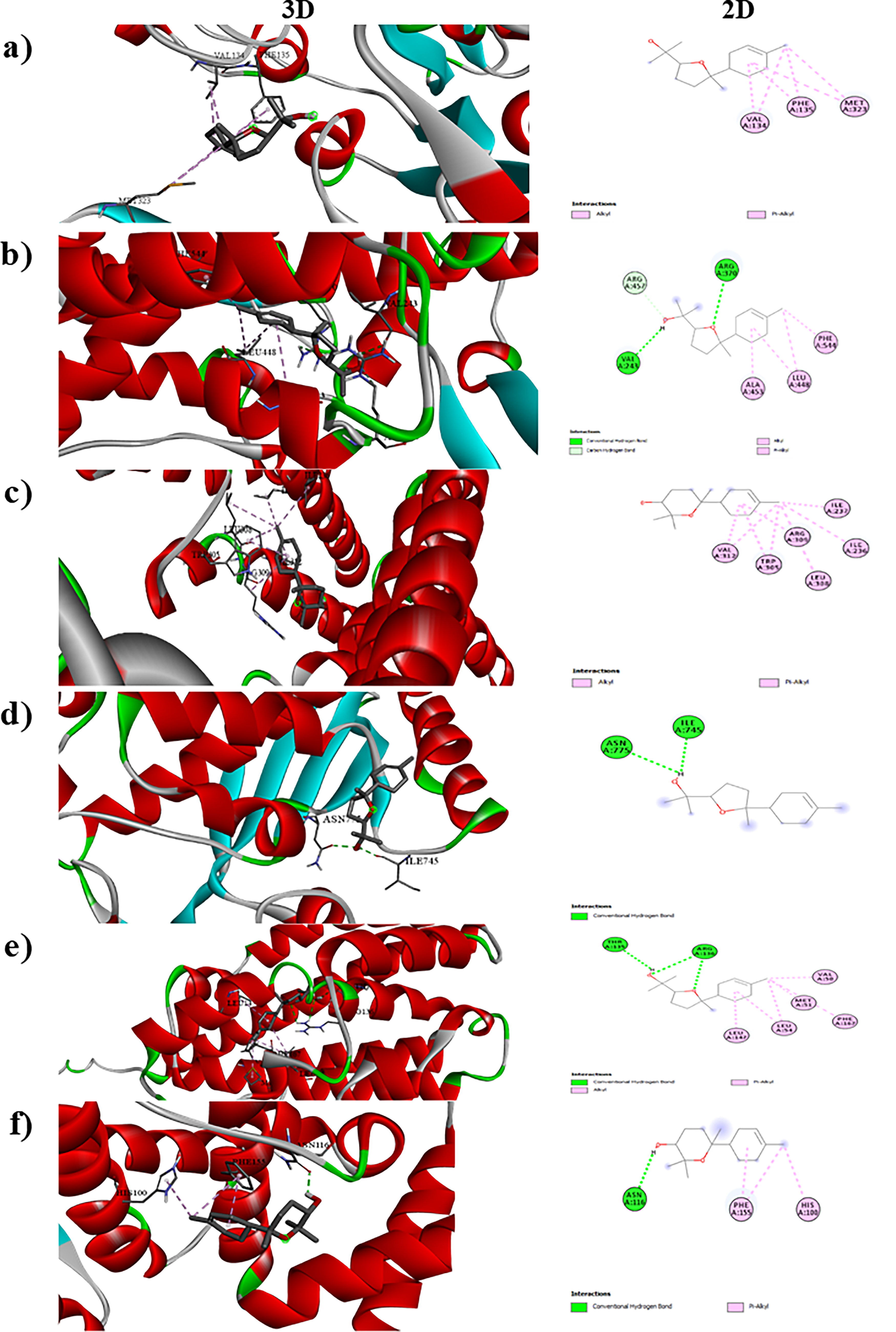
Molecular docking analysis and 3D and 2D interaction profiles of major bioactive compounds, including
The results obtained emphasize a distinct structure–activity link. Sesquiterpene oxides, especially those containing epoxide and hydroxyl functional groups, have consistently shown enhanced binding to prospective antioxidant and microbial targets owing to their ability to engage in hydrogen bonding and hydrophobic interactions. Fatty acid derivatives showed moderate affinity, with a preference for potential antioxidant targets, suggesting their role in membrane-related potential antioxidant mechanisms rather than direct enzymatic inhibition. Additionally, the spirocyclic compound (Z)-2-(hexa-2,4-diyn-1-ylidene)-1,6-dioxaspiro[4.4]non-3-ene displayed versatile binding across multiple targets, highlighting the potential of rigid, conjugated cyclic frameworks as lead structures for multifunctional drug development. This study demonstrates that natural compounds, particularly sesquiterpene oxides, possess promising multitarget bioactivities spanning potential antioxidant, antidiabetic, and antimicrobial domains (Al-Shuhaib and Al-Shuhaib, 2024). These findings provide mechanistic insights into their structure–activity relationships and establish a foundation for further in vivo and clinical investigations into their therapeutic applications.
Conclusion
The present study demonstrates that the combined ethanolic extracts of A. hierochuntica and M. chamomilla have a multifunctional bioactivity that is greater than the sum of the effects of the individual extracts. The synergistic effects of the various phytochemicals in the extracts account for the increased efficacy. The phytochemical composition is notable, especially those containing flavonoids and phenolic acids. Compared with individual extracts, the combined extract showed consistent, broad antimicrobial activity against microorganisms associated with clinical and foodborne pathogens. It also showed significant antimicrobial inhibition zones and lower MIC/MBC values. Time-kill kinetics confirmed the results, demonstrating increased bactericidal and fungicidal activity with a rapid, long-lasting antimicrobial effect. Results revealed that the combined extract demonstrated potential antioxidant activity and the ability to reduce oxidative stress. The antidiabetic evaluation revealed that the combined extract effectively inhibited both α-glucosidase and α-amylase enzymes, closely aligning with the pharmaceutical reference drug acarbose. These results show that MCE and AHE together may be used as a natural, multifunctional approach to address oxidative stress, microbial infections, and post-meal hyperglycemia. The findings support the extract’s potential to manage microbial infections and oxidative stress, and to enhance antidiabetic properties.
Ethical Approval
The current research does not involve any investigations with human or experimental animals.
Authors’ Contributions
I.S.G.: Conceptualization, data curation, formal analysis, methodology, investigation, writing—review and editing, and writing—original draft. S.A. and A.M.A.Z.: Methodology, investigation, data curation, writing—review and editing, and writing—original draft. A.A.A.: Investigation, methodology, writing—original draft, and writing—review and editing. D.J.J.: Conceptualization, formal analysis, writing—review and editing, and writing—original draft. E.M.H. and N.M.O.: Visualization, methodology, investigation, writing—review and editing, and writing—original draft. M.H.M.O.: Visualization, data analysis, writing—review and editing, and writing—original draft. A.A.Z. and H.A.A.: Supervision, formal analysis, writing—review and editing, and writing—original draft.
Footnotes
Acknowledgments
The authors are grateful to King Saud University, Riyadh, Saudi Arabia, for funding this work through the Ongoing Research Funding program—Research Chairs (
Disclosure Statement:
The authors declare no conflict of interest.
Funding Information
This research was funded by King Saud University, Riyadh, Saudi Arabia, through the Ongoing Research Funding program—Research Chairs (
Supplemental Material
References
Supplementary Material
Please find the following supplemental material available below.
For Open Access articles published under a Creative Commons License, all supplemental material carries the same license as the article it is associated with.
For non-Open Access articles published, all supplemental material carries a non-exclusive license, and permission requests for re-use of supplemental material or any part of supplemental material shall be sent directly to the copyright owner as specified in the copyright notice associated with the article.