
Abstract
The WW domain-containing oxidoreductase (WWOX) gene, encodes a tumor suppressor located on 16q23.1, spanning FRA16D, one of the most active common fragile sites in the human genome, that is altered in numerous types of cancer. WWOX’s alteration in these myriad cancers is due to disparate mechanisms including loss of heterozygosity, homozygous deletion and epigenetic changes. In vitro, WWOX has been found to be reduced or absent in numerous cancer cell lines and WWOX restoration has been found to inhibit tumor cell growth and invasion. Wwox knockout mice developed femoral focal lesions resembling osteosarcomas within one month of their life and aging Wwox heterozygous mice have an increased incidence of spontaneous lung and mammary tumors as well as B-cell lymphomas. We herein review WWOX’s role that has been unearthed thus far in different types of malignancies, its clinical significance and future implications.
Keywords
Introduction
The WW domain-containing oxidoreductase (WWOX) gene encodes a 46 kDa tumor suppressor that is altered in myriad types of cancer. WWOX is located on 16q23.1, spanning FRA16D, one of the most active common fragile sites in the human genome. The protein which it encodes is comprised of 2 WW domains and a short chain dehydrogenase reductase domain. 1 WWOX was initially discovered as researchers were interested in mapping the 16q region, an area commonly affected by allelic loss in breast cancer. Upon mapping this genomic region, WWOX (also known as FOR) was identified and cloned.2,3 Chang and colleagues also isolated and characterized WWOX (referred as WOX1) as an effector of hyaluronidase that increases cancer cell sensitivity to tumor necrosis factor (TNF) cytotoxicity. 4 WWOX was recently identified as one of the most significant somatic copy number alterations (SCNA) undergoing deletions in Beroukhim et al.’s high resolution analysis of a plethora of cancer specimens. 5 Some reports, however, documented increased levels of WWOX in breast, gastric and prostate carcinomas suggesting a complex regulation of WWOX in cancer.6,7 Nevertheless, the vast majority of observations support loss of WWOX expression in cancer and indicate a tumor suppressive role of its protein product as shall be discussed here.
WWOX’s alteration in these myriad cancers is due to disparate mechanisms including loss of heterozygosity (LOH), homozygous deletion, hypermethylation of the regulatory region and, in one study due to ubiquitination. These WWOX alterations have been associated with poorer prognoses in different types of malignancies (below). These facts have exciting therapeutic implications.
In vitro, WWOX has been found to be reduced or absent in numerous cancer cell lines and WWOX restoration has been found to inhibit invasion and to suppress tumorigenicity in xenograft mouse models.8–12 Mice carrying a targeted deletion of Wwox developed spontaneous lung papillary carcinomas, in the case of Wwox-heterozygous mice and osteosarcomas in the case of Wwox null mice. 13 More recently, it was shown that 50% of female Wwox-heterozygous (C3H) (WWOX (C3H)+/−) mice formed mammary tumors. 14 Wwox hypomorphic mice displayed shorter lifespans with the males enduring testicular atrophy and reduced fertility and the females having an increased incidence of spontaneous B cell lymphomas. 15 Altogether, these observations indicated that WWOX acts as a bone fide tumor suppressor (reviewed in Refs. 16–18).
Causes of WWOX alteration
WWOX alterations in disparate cancers
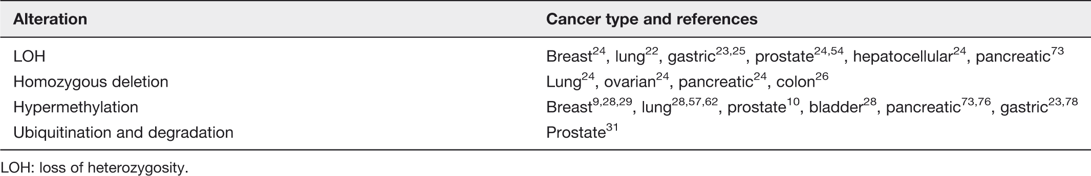
LOH: loss of heterozygosity.
Loss of heterozygosity
LOH at the WWOX locus has been described in numerous malignancies (Table 1), including breast, lung and gastric cancer.22,23 LOH was demarcated in the 16q23 region in 67%, 53% and 52% of breast, prostate and hepatocellular carcinomas, respectively. 24 LOH at the WWOX locus was found in 45.6% of gastric cardia adenocarcinoma samples. 25
Homozygous deletion
Finnis et al. detected homozygous deletions within WWOX in three colon cancer derived cell lines (Co115, KM12C and KM12SM). 26 Homozygous deletion of WWOX exons from an ovarian cancer cell line, two small cell lung cancer cell lines and a pancreatic cancer cell line were reported as well. 24
Hypermethylation
Epigenetics has been underscored as an influential factor in tumorigenesis. Hypermethylation in a tumor suppressor gene’s promoter region can silence that gene’s expression and consequently give the cells a growth advantage. 27 Iliopoulos et al. demonstrated WWOX promoter region hypermethylation as a means of WWOX inactivation in breast, lung and bladder neoplasias. A correlation between WWOX CpG methylation and reduced WWOX protein expression in all three types of cancers was described. 28 In a later study, Iliopoulos performed methylation specific PCR of the WWOX promoter region on three breast cancer cell lines, MCF7 which expresses WWOX strongly, and MDA-MB-231 and HCC1937 and found both promoter regions unmethylated in the MCF7 cells and methylated in the MDA-MB-231 cells. 9 Wang et al. found similar results regarding hypermethylation of WWOX promoter CpG islands and decreased WWOX expression in breast carcinoma samples and two cell lines. 29
Ubiquitination
Ubiquitination, an enzymatic cascade whereby proteins are labeled with a ubiquitin tag and thus designated for degradation via the proteasomal system, is another means by which WWOX could be inactivated and thereby subsequently promote tumorigenesis. 30 Mahajan et al. demonstrated that Ack1, an intracellular tyrosine kinase induces WWOX polyubiquitination and subsequent degradation in prostate cancer cells. In that same study human specimens of androgen independent prostate cancers (AICaP) and benign prostate tissue were obtained and immunprecipitation and immunoblotting revealed that AICaP had increased levels of phosphorylated Ack1 and reduced levels of WWOX compared to the benign prostate samples, suggesting WWOX degradation due to tagging for ubiquitination by Ack1. 31 So far no ubiquitin E3 ligase was associated with WWOX under normal or disease states.
WWOX alteration in cancer
Breast cancer
Breast cancer is the most common cancer among women worldwide and its incidence is rising. 32 There have been numerous studies done underscoring the role of WWOX alteration in breast cancer. A locus within WWOX has even been identified as a genetic breast cancer risk variant. 33 WWOX has been shown to be of significant importance in mammary gland physiology as WWOX ablation in mice is associated with increased fibronectin levels and impaired ductal growth. 34 In Wwox (C3H)+/− mice 50% of female mice formed tumors while only 7% of female wild type mice did. Furthermore, it has been reported that among those Wwox (C3H)+/− mammary tumors, WWOX expression was often lost suggesting LOH, a hallmark of tumor suppressors. 14 Guler et al. were the first to notice the correlation between reduced WWOX expression in human breast cancer samples and breast carcinogenesis. Reduced WWOX expression was reported in 63.2% of breast cancer specimens. 35 In a study analyzing WWOX mRNA expression in 9 breast cancer cell lines and 20 human breast tumor samples, reduced WWOX expression in 3 cell lines was found as were WWOX mRNA variants in a number of the tumors. 36 In addition to the initial observation by Bednarek et al.,2,37 these data paved the road for a possible tumor suppressor function of WWOX in breast cancer.
There have been several studies demonstrating WWOX restoration in vitro and in vivo and subsequent inhibition of breast cancer cell growth. Bednarek et al. noted WWOX’s ability to inhibit tumorigenicity both in vitro and in vivo using MDA-MB-435 breast cancer cell lines. 37 Huebner’s lab elegantly displayed this phenomenon as well and found that exogenous WWOX restoration inhibited breast cancer cell growth in vitro and in vivo and endogenous WWOX restoration via de novo methyltransferase inhibitor treatment inhibits breast cancer cell growth and shrinks xenograft tumor growth. 9 A mechanism for WWOX’s tumor suppressor role was proposed by Aqeilan et al. who demonstrated WWOX’s relationship with Ap2gamma transcription factor, an oncogene in breast cancer and suggested WWOX precludes its activity by sequestering it in the cytoplasm. 38
WWOX expression has been significantly associated with a number of clinicopathologic factors in breast cancer. WWOX reduction is significantly correlated with that of fragile histidine triad (FHIT), encompassing the most active common fragile site FRA3B, 39 in breast cancer specimens. 35 Reduced FHIT and WWOX were found to be associated with ductal carcinoma in situ (DCIS). 40 Subsequently, it was established that expression of WWOX and FHIT is significantly reduced in triple negative breast cancer specimens, a more aggressive breast cancer type. 41 An association between WWOX expression, intramembranous ErbB4 expression (in contradistinction to intranuclear expression) and increased disease free survival has been reported. 42 Conversely, loss of WWOX expression was associated with increased metastatic axillary lymph nodes and decreased disease free survival. 42 In agreement to these observations, Guler et al. found significantly reduced WWOX and ErbB4 in metastases compared to their matched primary breast tumor. 41 Nunez et al. found a correlation between estrogen receptor status and WWOX expression. A greater number of estrogen receptor (ER)-breast cancer samples, a subtype associated with a poorer prognosis, 43 had absent or weakly staining WWOX expression. 44 Pluciennik et al. delineated the same phenomenon and found it to be true regarding progesterone receptor status as well. Conversely, higher WWOX expression was found to be associated with greater disease free survival. 45 Wang et al. found WWOX to be associated with a basal like phenotype of breast cancer as well as decreased disease free survival. 46
A significant association between WWOX and tamoxifen resistance has been described. This study suggested that WWOX and Ap2gama may be more telling biomarkers regarding tamoxifen efficiency than progesterone receptor and Her2 status. 47 Gothlin et al. found an association between increased WWOX expression and decreased risk of recurrence in patients treated with tamoxifen. 48
Decreased WWOX expression in breast cancer has been significantly associated with a number of clinicopathologic parameters such as more aggressive tumor phenotypes, increased metastases and decreased disease free survival. Therefore, WWOX might be a promising biomarker to gauge chemotherapy affectivity. This has exciting implications regarding WWOX’s clinical role in the future.
Prostate cancer
Prostate cancer accounts for 6% of cancer deaths among males worldwide. 49 Prostate cancer derived cells were found to have decreased WWOX mRNA and protein expression and 84% of prostate cancer samples were found to have decreased WWOX expression via immunohistochemical staining. WWOX overexpression in vitro induced apoptosis via a caspase-dependent mechanism and halted cell growth. Ectopic WWOX expression was found to suppress tumorigenicity in xenografts. 10 One mechanism by which WWOX exerts its tumor suppressor role in prostate cancer is by binding to Ap2gamma in the cytoplasm, precluding it from binding with ErbB2’s (an effecter of prostate cancer cell growth) promoter region in the nucleus. 50 In prostate cancer cells, WWOX itself might be activated by complement C1q as suggested by Hong et al.’s study. 51
WWOX was identified as an area of genomic loss via array comparative genetic hybridization of 16 prostate tumor samples. 52 A single-nucleotide polymorphism (SNP)-based genome wide linkage scan of 161 families with prostate cancer detected a strong signal on 16q23, which could very well be WWOX. 53 WWOX promoter hypermethylation was noted in prostate cancer derived cells. Increased WWOX expression in prostate cancer derived cells was delineated after treatment with AZA, a DNA methyltransferase inhibitor and trichostatin A, a histone deacetylase inhibitor. Intratumoral AZA treatment suppressed tumor growth in nude mice. 10 As mentioned above, another means of WWOX inactivation in prostate cancer is its phosphorylation by Ack1, tagging it for polyubiquitination and subsequent degradation. 31 In a CNA study of prostate tumors, 17.7% were found to have LOH of WWOX which was significantly associated with a more advanced disease staging. 54 Further associations between reduced or lost WWOX and other clinicopathologic parameters including disease free survival must be explored.
Lung cancer
Lung cancer is the most common cause of cancer death among males worldwide. 49 A number of studies have delineated WWOX alteration in lung cancer. Yendamuri and colleagues initially analyzed 8 non-small cell lung cancer (NSCLC) cell lines and 27 normal lung and lung tumor tissues for WWOX alterations. They found missing WWOX transcripts in 25.9% of primary tumors and five out of eight cell lines. Thirty-seven percent of the primary tumors displayed LOH at the WWOX locus. 22 WWOX expression was absent or reduced in 84.9% of NSCLCs in a study of 170 samples and present in 80.5% of adjacent normal lung tissue. 55 A study of the tissue of 44 Chinese patients who had undergone resections for NSCLC found 63.6% of samples to have loss of WWOX exons 6–8 transcript. 56 In a different study of 50 lung cancer patients, 2 germline mutations and 2 polymorphic mutations were isolated in WWOX upon DNA sequencing. 57 WWOX restoration in WWOX protein negative lung cancer cell lines in vitro and in nude mice xenografts in vivo induced apoptosis and a striking suppression of tumorigenicity, respectively.8,11,58
The degree of WWOX expression was found to correlate with histopathologic features of NSCLC including histiotype and increased tumor aggressiveness. Weak WWOX staining was associated with high cell proliferation and poor differentiation. 55 WWOX deletion was significantly higher in the tumors of those patients who smoked. 56 WWOX preservation in resected NSCLC tumors of 85 patients was associated with better outcomes. 11 Certain SNPs within WWOX were identified as being significantly associated with increased cancer risk. 59 A common WWOX copy number variation isolated among the Chinese was found to significantly increase the carrier’s likelihood of developing lung cancer. 60
WWOX promoter hypermethylation was identified in 76% of 50 lung cancer patients. 57 Epigenetic modulation of nude mice with H1299 lung cancer tumors (which lack FHIT and WWOX) via AZA (a demethylating agent) injection restored WWOX (among other tumor suppressor genes) expression and decreased tumorigenicity, as well as increased the apoptosis fraction, increased caspase 3 activation and decreased mitosis in those tumors that responded. 61 Another study identified the downregulation of the microRNA 29 family as causing epigenetic WWOX attenuation by allowing DNMTA upregulation which hypermethylates WWOX’s promoter region and silences WWOX. Upon forced miR-29 expression in NSCLC, WWOX and other tumors suppressor genes affected by hypermethylation were found to be reexpressed and tumorigenicity was found to be inhibited in vitro and in vivo. 62 Another means of epigenetic WWOX modulation in lung cancer was proposed by Kimura et al. who found that Bmi1, an epigenetic regulator protein which is highly expressed in SCLC, targets WWOX thereby attenuating it. Bmi1 deletion caused increased apoptosis presumably via restored WWOX functioning. 63
Osteosarcoma
Osteosarcoma comprises 35% of all primary bone tumors. It is the most common primary malignant bone tumor in children and adolescents. While long-term survival for nonmetastatic ostosarcoma is 60–70%, it is only around 30% for patients with metastases. 64 WWOX has been found to play a role in osteosarcoma. As referred to above Wwox knockout mice develop focal lesions resembling osteosarcomas within their life of one month.13,65,66 WWOX was found to be deleted in 30% of human osteosarcoma samples 67 and its protein is lost or reduced in 58% of human osteosarcoma samples. 65 Decreased WWOX mRNA was detected by RT-PCR and associated with malignancy in a number of osteosarcoma samples. 68 Furthermore, WWOX levels were increased in tumors resected post chemotherapy in comparison to primary tumors. 65 WWOX levels were decreased in lung metastases in comparison to levels in the primary tumor. WWOX was reduced in human osteosarcoma cell lines as well. WWOX restoration inhibited proliferation and modified invasion capability in vitro and suppressed tumorigenesis in vivo. RUNX2, a transcription factor responsible for bone development and regulation and a WWOX binding partner, 69 was found to be elevated in the femurs of Wwox knockout mice. RUNX2, which displays elevated levels in osteosarcoma, 70 expression was found to be elevated in osteosarcoma cell lines with reduced WWOX expression. RUNX2 expression was positive in most clinical osteosarcoma samples and negative in most post chemotherapy samples. Additionally, WWOX restoration in osteosarcoma cell lines was associated with RUNX2 downregulation. These findings support the notion that WWOX’s binding with RUNX2 inhibits RUNX2’s transactivation function. 65 Del Mare et al. further explicates the WWOX-RUNX2 functional cross talk and its implications for understanding the pathogenesis of osteosarcoma. 71
Pancreatic cancer
Pancreatic cancer, one of the most lethal cancers, has been deemed the fourth leading cause of cancer death. Pancreatic resection is the only curative option, yet most patients present too late to be candidates for that procedure. Those patients who do undergo pacreaticoduodenectomies only have a dismal five year survival rate of anywhere from 10% to 25%. 72 Kuroki et al. studied WWOX expression and promoter methylation in paired pancreatic cancer samples as well as in pancreatic cancer cell lines. The study found LOH in the WWOX locus in 27% of primary tumors and WWOX promoter hypermethylation in 13% of primary tumor samples and in 22% of pancreatic cancer cell lines. Significant reduction in WWOX mRNA expression and WWOX protein levels was found in all of the cell lines. Forty percent of primary tumors were found to have a significant reduction in WWOX expression as well. Pancreatic cell colony formation was inhibited upon WWOX transfection via apoptosis. 73 WWOX restoration inhibited further cell growth in vitro and in vivo. Not only was cancerous tissue found to have reduced WWOX, but precancerous pancreatic cancer tissue samples were found to have decreasing levels of WWOX expression in accordance with their pancreatic intraepithelial neoplasia grade and with eventual less favorable patient outcomes. Normal pancreatic tissue from the surrounding tissue of the samples was found to have a robust WWOX expression. 74 Bloomston et al. found reduced WWOX and FHIT expression in pancreatic, gall bladder and ampullary cancers and robust WWOX expression in physiologic tissue as well. They did not, however, find any correlation between the gradation of WWOX expression and tumor progression or patient survival. 75 Nakayama et al. studied 41 intrapapillary mucinous neoplasms of the pancreas to further delineate WWOX’s role in pancreatic tumorigenesis and found WWOX loss or reduction in 15% of intrapapillary mucinous adenomas (IPMAs) and 81% of intrapapillary mucinous carcinomas (IPMCs). Furthermore, hypermethylation of the WWOX promoter region was found in 33% of WWOX negative IPMAs and 53% of WWOX negative IPMCs. 76
Gastric adenocarcinoma
Gastric cancer is responsible for 10% of cancer deaths globally. 49 Aqeilan et al. were the first to note WWOX alteration in gastric adenocarcinoma. LOH in 31% of primary tumors and loss of WWOX protein expression in 65% of primary tumors and 33% of gastric adenocarcinoma cell lines was described. 23 Maeda et al. also found loss of WWOX expression in gastric cancer specimens and cell lines. Hypermethylation at the WWOX regulatory site was appreciated in two of the cell lines. 77 Furthermore, WWOX promoter and exon 1 methylation was noted in gastric cardia adenocarcinoma specimens. 25 Additionally, WWOX hypermethylation has been correlated with Helicobacter pylori infection. Yan et al. suggested based on this and other findings that H. pylori induces WWOX hypermethylation and subsequent gastric carcinogenesis. 78 Takeuchi et al. suggested another means by which WWOX function might be attenuated in gastric signet cell carcinoma. They isolated a gene, transmembrane protein 207 (TMEM 207), which they found to be expressed in a gastric signet ring cell carcinoma cell line as well as in a number of tissue samples. They found it to bind with WWOX upon coimmunoprecipitation. They also noted WWOX inhibition of invasion in a different signet ring cell carcinoma cell line to be attenuated upon TMEM 207 overexpression. 79 A correlation between WWOX and FHIT expression has been discovered in gastric adenocarcinoma as well. 23 Upon intragastric administration of N-nitrosomethylbenzylamine to Wwox-heterozygous (WWOX+/−) and Wwox-wild type (WWOX+/+) mice and subsequent analysis, 96% of WWOX+/− mice had developed forestomach tumors whereas only 29% of WWOX+/+ mice did. Furthermore, 29% of WWOX+/− mice developed invasive squamous cell carcinoma of the forestomach whereas none of the WWOX+/+ mice did. This study suggests that loss of one WWOX allele enhances tumorigenicity. 80 WWOX restoration into a gastric adenocarcinoma cell line effectively suppressed cell growth and increased the number of cells in the subG1 stage of the cell cycle. The degree of WWOX expression was found to be significantly correlated with clinical staging, depth of invasion, lymph node status, metastases and venous infiltration. 77 These data indicate that WWOX may play a role in gastric carcinogenesis and furthermore may be a valuable biomarker in terms of gauging clinical features of tumor aggressiveness.
Ovarian cancer
Ovarian cancer is the seventh most common cancer in women globally. 49 In a comparison between normal and malignant ovarian tissue, normal tissue was found to have strong WWOX expression whereas ovarian carcinoma tissue was found to have reduced expression.81,82 A homozygous deletion within WWOX was detected in an ovarian cancer derived cell line with the corresponding aberrant transcript and lack of full WWOX product. 24 WWOX restoration or overexpression, depending on the cell line, inhibited tumorigenicity in vivo. Interestingly, WWOX restoration or overexpression in vitro, caused decreased attachment and migration onto fibronectin, an extracellular membrane component integral to peritoneal metastasis. WWOX knockdown in ovarian cancer cell lines with endogenous WWOX expression caused increased fibronectin adhesion. Based on these in vitro findings, Gourley et al. proposed loss of WWOX as an important factor in peritoneal metastasis in ovarian cancer. 12 Xiong et al. found WWOX overexpression in vitro to induce apoptosis and inhibit further ovarian cancer cell growth. 83
A significant association between reduced WWOX expression and overall survival as well as clinical staging (FIGO stage IV), estrogen and progesterone receptor negative status and lymph node metastasis has been reported.81,84 Eight previously identified SNPs within WWOX were genotyped in 554 ovarian cancer samples and analyzed for association with clinicopathologic factors such as tumor grade and survival. Three SNPs were identified as being significantly associated with one or more clinicopathologic features including tumor grade, histology and progression free survival. These associations, however, were not confirmed in a different ovarian cancer population. 85 In a study evaluating sodium valproate as a treatment for ovarian cancer in ovarian cancer cell lines and xenografts, WWOX was used as a marker to determine treatment efficacy, as WWOX protein expression increased post treatment in ovarian cancer cell lines and WWOX was upregulated in xenografts post treatment as well. 86
Squamous cell carcinoma
Cancers of the skin, including melanoma and basal and squamous cell skin cancers, are by far the most common malignancies in humans. 87 The incidence of these types of skin cancer has increased greatly over the past decade. However, the etiology is largely unknown. Unlike basal cell carcinoma, cutaneous squamous cell carcinoma (SCC) is malignant and tends to metastasize. Lai et al. noted that WWOX expression is induced during normal keratinocyte differentiation suggesting a physiological role in skin biology. 88 However, in the vast majority of SCC cases, WWOX protein levels are reduced, while WWOX mRNA was not reduced at all and thus suggested that cancerous cells inhibit WWOX mRNA’s translational capacity. 88 Interestingly, exposure of SCC cells to UVB or methotrexate enhanced WWOX expression, sensitized the cells to undergo apoptosis and inhibited autophagy. 89 Conversely, WWOX depletion in these cells rendered them more resistant to apoptosis, a hallmark of cancer cells. In vivo, acute exposure of hairless mice to UVB was associated with upregulation of WWOX in epidermal cells in 24 h. In accordance with WWOX loss in human SCC, chronic UVB-treated mice developed cutaneous SCCs in three months, with significant reduction of WWOX protein levels and no effect on WWOX mRNA. 88 This notion that WWOX levels are increased upon UVB radiation may suggest that WWOX might be activated upon DNA damage and/or play a role in DNA damage response.
Clinical significance
WWOX alteration in different cancers and its clinical significance

LOH: loss of heterozygosity; NSCLC: non-small cell lung carcinoma; HCC: hepatocellular carcinoma; ACC: adenoid cystic carcinoma; SCC: squamous cell carcinoma; GBM: glioblastoma multiforme.
Concluding remarks
WWOX research is burgeoning and WWOX’s activity in disparate types of neoplasias is ever being discovered. WWOX was found to have a significant role in hepatocellular carcinoma as well, activating the Wnt/beta-catenin pathway when it is downregulated and inducing apoptosis when it is overexpressed.93–96 Pluciennik et al. found WWOX to be altered in Wilms’ tumor. 97 Zelazowski et al. found a significant correlation between increased WWOX expression in colorectal cancer samples and disease free survival. 98 Dincer et al. demonstrated reduced WWOX and FHIT expression in adenoid cystic carcinoma, a basaloid salivary gland neoplasm. 99 WWOX has been found to play a role in glioblastoma multiforme (GBM) as well. Kosla et al. found WWOX LOH and WWOX promoter methylation in GBM. They also found WWOX to be correlated with antiapoptotic marker BCl2 and marker of cell proliferation Ki67 in GBM. 100 Chiang et al. overexpressed WWOX in GBM cell lines with a mutant p53 and reported that WWOX overexpression induces apoptosis. 101 Absent or abnormal WWOX transcripts were detected in 51% of primary hematopoetic neoplasias. 102 Cui et al. recently further elucidated the role of WWOX in leukemia. 103 We have had promising preliminary results demonstrating WWOX’s role in Ewing’s sarcoma as well (unpublished data). WWOX’s role in these cancers must be further explored. The fact that WWOX lies in a common fragile site could render it more sensitive to genomic alteration and deletion. One allele could be lost due to genomic alteration (i.e. as a result of replication stress) while the other is selected against and lost during transformation likely due to silencing by hypermethylation or due to LOH. Further characterization of WWOX function in pre-malignant stages is of great interest and would shed light on its role(s) in early stages of transformation. Though we are cognizant of the argument that WWOX loss is secondary due to its localization within a fragile site and not a primary event in cancer pathogenesis, we are convinced that the fact that WWOX restoration in WWOX-negative cells suppresses tumorigenicity in vitro and in vivo and that Wwox knockout mice are more prone to form tumors suggests otherwise. Further research will further our understanding of WWOX biology and its role in cancer pathogenesis. The delineation of WWOX’s role(s) in pre-malignancies, where WWOX levels have been shown to be elevated in some tissues, is of great interest and requires further investigation. WWOX’s use as a biomarker to gauge tumor aggressiveness, clinical outcomes and chemotherapy effectiveness could be around the corner. Future translational work involving WWOX restoration in clinical trials in these disparate cancers is an exciting prospect we would like to see in the not too distant future.
Author contribution
Both authors AG and RIA wrote and designed this review article.
Footnotes
Funding
The Aqeilan lab is funded, in part, by ICRF-Research Career Development Awards.
Declaration of interest
The authors have no relevant affiliations or financial involvement with any organization or entity with a financial interest in or financial conflict with the subject matter or materials discussed in the manuscript. This includes employment, consultancies, honoraria, stock ownership or options, expert testimony, grants or patents received or pending, or royalties.