
Abstract
Enzymes play a central role in a spectrum of fundamental physiological processes and their altered expression level has been associated with many diseases and pathological disorders. Enzymes therefore can be exploited as a pristine biological trigger to tune material responses and to achieve controlled release of biomolecules at desired sites. This mini-review highlights enzyme-responsive polymer hydrogels for therapeutic delivery applications developed within the last five years, focusing on protease- and glycosidase-based catalyzed reactions. Strategies employed to produce responsive materials are described. Successful applications for controlled drug delivery are highlighted, and finally, future opportunities and challenges are presented.
Introduction
Stimuli-responsive polymeric materials have emerged as powerful tools and play an increasingly important role in a wide range of biomedical applications, including drug delivery,1,2 tissue engineering,3,4 bioimaging,5,6 and biosensing.7,8 These materials can be finely tuned to exhibit (ir)reversible changes in their physical or chemical properties in response to a number of different stimuli, for example pH,9,10 temperature,11,12 light,13,14 redox potential,15,16 electric field,17,18 ultrasound,19,20 and enzyme activity.21,22 Most notably, the use of enzymes as a stimulus has attracted significant attention and there has been a dramatic escalation in the development of enzyme-responsive materials over recent years, driven by the motivation to mimic biomolecular recognition processes.
Enzymes are key biological catalysts that play a vital role in many chemical and biological processes within cells. Enzyme-catalyzed reactions are highly specific and selective toward their substrates. They generally require milder conditions compared to other conventional chemical reactions, i.e. enzyme reactions can occur in aqueous environments, at relatively low temperatures, and typically at neutral or slightly acidic or alkaline pH. These defining advantages make enzyme-catalyzed reactions suitable for biomedical applications. As their presence and activities are essential in maintaining physiological homeostasis, enzyme dysregulation is associated with many diseases and pathological disorders, such as cancer, cardiovascular disease, inflammation, osteoarthritis, and Alzheimer’s disease.23–27 Altered expression level of specific enzymes therefore can be exploited as a pristine biological trigger to achieve enzyme-mediated biomaterial responses and controlled release of biomolecules at desired sites and this aspect is the focus of this mini-review.
To achieve functional enzyme-responsive biomaterials, three general requirements need to be met (Figure 1). First, the materials have to incorporate recognition elements or substrate mimics that can be specifically recognized by enzymes. For proteolytic enzymes, amino acid sequences generally determine enzyme-substrate specificity, therefore common recognition elements include peptide chains/linkers or polymer–peptide conjugates with specific amino acid sequences toward enzymes of interest. Second, the anchored substrates have to be accessible by enzymes and this can greatly affect the kinetics of the enzyme-catalyzed reactions. Third, the enzyme–substrate reactions have to be translated to changes in material properties. These can include degradation or morphological transformation of biomaterials. Therapeutic molecules can be encapsulated through physical encapsulation or covalent attachment into the polymeric materials and are locally delivered through diffusion or degradation of biomaterials upon enzymatic actions.
Enzyme-responsive polymer hydrogels for therapeutic delivery. To achieve functional enzyme-responsive biomaterials, three general requirements need to be met. (a) The materials have to incorporate recognition elements, e.g. enzyme-cleavable linkers. (b) The linkers have to be accessible by enzymes and this can greatly affect the kinetics of the enzyme-catalyzed reactions. (c) The enzyme–substrate reactions have to be translated to changes in material properties, for example degradation or morphological transformation of hydrogels. Therapeutic molecules can be encapsulated through physical encapsulation or covalent attachment into the polymeric materials and are locally delivered through diffusion or degradation of biomaterials upon enzymatic actions. (A color version of this figure is available in the online journal.)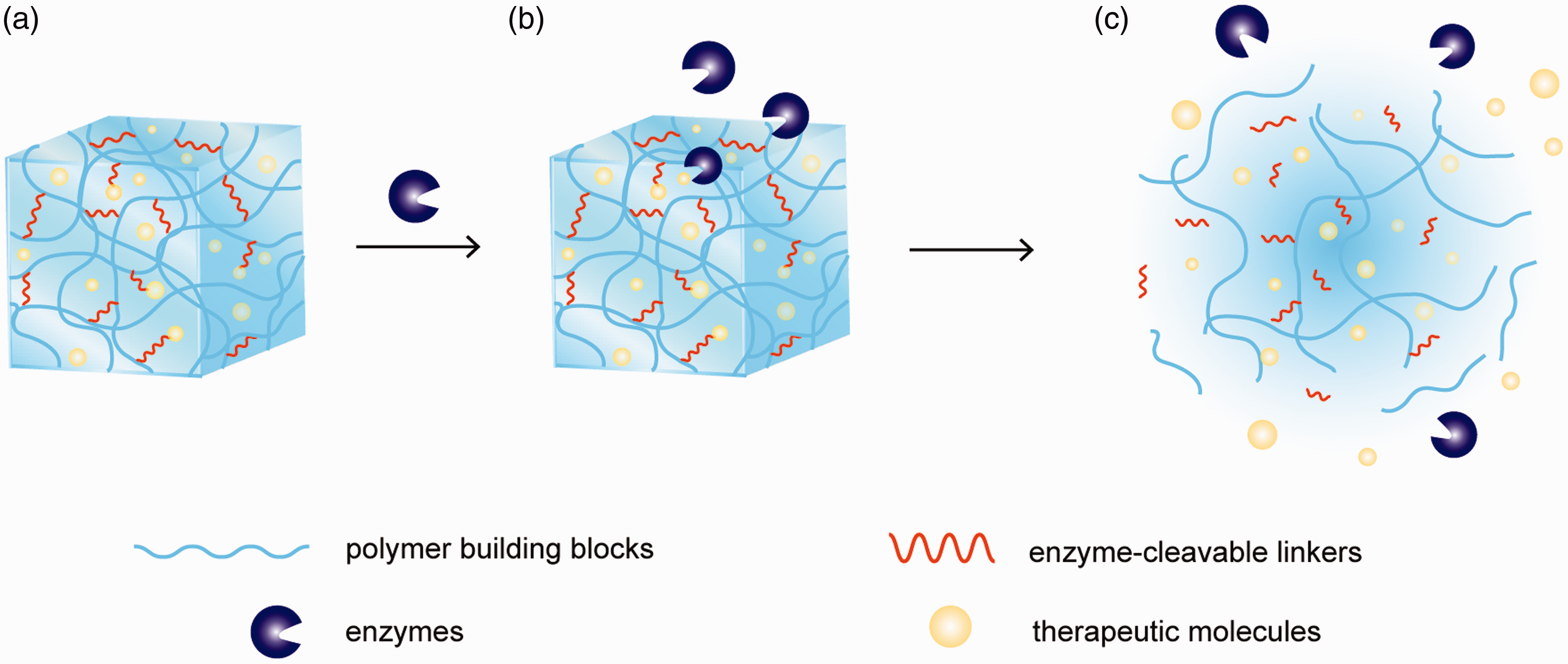
This mini-review highlights and discusses the most recent advances of enzyme-responsive materials developed within the last five years, focusing on polymer hydrogels programmed to release therapeutics in response to specific enzymatic activities. Polymer hydrogels are crosslinked three-dimensional polymer networks that serve as a prime candidate for therapeutic delivery.28–33 Other classes of enzyme-responsive polymeric materials, such as polymer nanoparticles, polymer conjugates, and supramolecular polymer assemblies, have been described in detail in previous reviews.7,21,34–37 Medically relevant classes of enzymes that serve as biological stimuli are outlined and assembly approaches employed to construct enzyme-responsive polymer hydrogels are described. Successful applications of these materials for enzyme-controlled drug delivery are highlighted. Finally, areas for future developments of enzyme-responsive assemblies are discussed.
Protease-responsive polymer hydrogels
Proteases are enzymes that catalyze the hydrolysis of peptide bonds. Overexpression of protease level underlies many diseases, including cancer, cardiovascular, inflammatory, neurodegenerative, bacterial, viral, and parasitic diseases. 38 They are therefore considered to be major targets exploited as physiological stimuli to allow for selective activation of drug release from biomaterials. In their pioneering work in 1999, West and Hubbell were the first to design and develop protease-responsive polymer hydrogels. 39 These hydrogels were prepared by chain-growth photoinitiated polymerization of peptide-containing poly(ethylene glycol) (PEG) diacrylate macromers. The reactive peptide domains that were incorporated, APGL or VRN, were designed to be specifically recognized and cleaved by matrix metalloproteinase-1 (MMP-1) or plasmin, respectively, two enzymes that play key roles in cell migration and extracellular matrix remodeling. Through enzyme–substrate recognition, selective degradation of hydrogels by targeted proteases was reported.
PEG is a bioinert and biocompatible synthetic polymer, and a range of crosslinking chemistries have been reported to present PEG with attractive functional cues.40,41 Furthermore, with a high degree of tunability of material properties, it is not surprising that a significant amount of work on enzyme-responsive synthetic polymer hydrogels focuses on PEG-based hydrogels. Anseth et al. created MMP-responsive PEG hydrogels via step-growth thiol-norbornene photopolymerization scheme.42,43 In their work, four-arm norbornene-functionalized PEG macromers were mixed with peptide crosslinkers containing MMP-cleavable sequences with dual thiol groups ( Peptide-crosslinked poly(ethylene glycol) (PEG) hydrogels. PEG macromers can be modified to yield functional terminal groups, including norbornenes and maleimides. Enzyme-cleavable peptides with thiol groups serve as crosslinkers. Examples of crosslinking chemistries used to form PEG hydrogels include: (a) thiol-norbornene and (b) thiol-maleimide reactions. (A color version of this figure is available in the online journal.)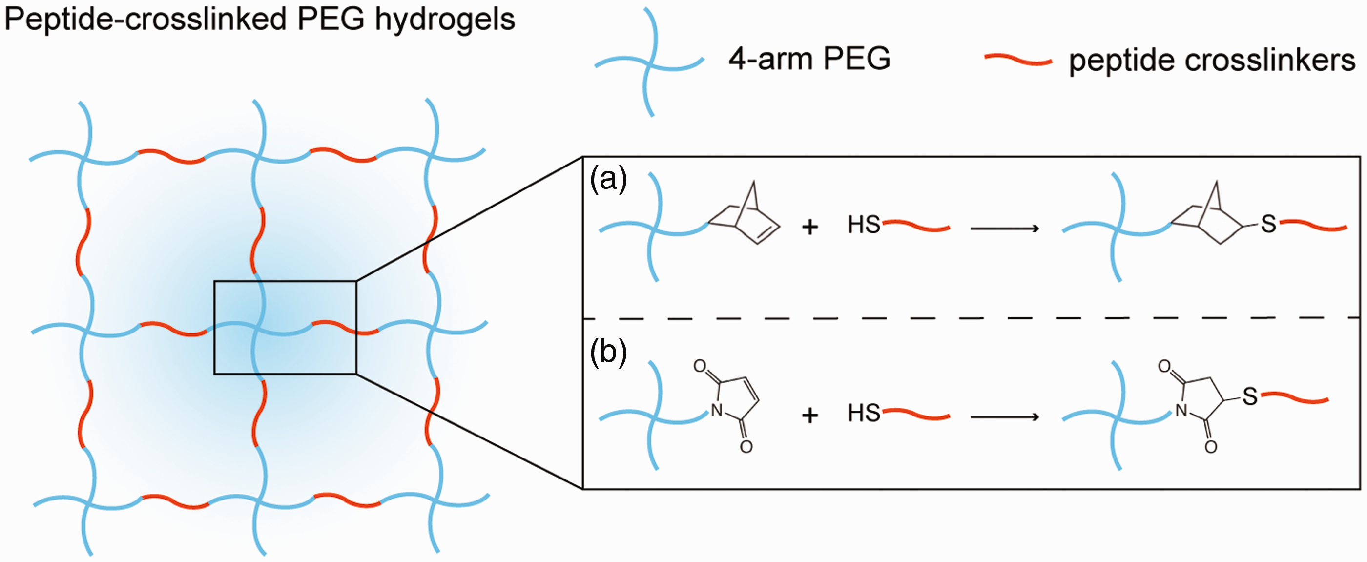
Another reaction mechanism used to create enzyme-responsive PEG hydrogels is Michael-type addition of thiols to maleimides (Figure 2(b)). García et al. developed protease-responsive PEG hydrogels via thiol-maleimide crosslinking chemistry.45,46 In their approach, four-arm maleimide-functionalized PEG macromers were first conjugated with thiol-containing RGD adhesive peptides, followed by crosslinking reaction in the presence of dithiol MMP-cleavable peptide linkers (
Enzyme-responsive polymer hydrogels can also be prepared from natural materials. In general, natural polymers offer advantages over synthetic polymers as they can be metabolically processed and they often possess pristine biologically relevant functionalities. Hyaluronic acid (HA) is a naturally occurring polysaccharide, inherently immunogenic, and is an essential component of extracellular matrix.47,48 Burdick et al. developed MMP-responsive HA hydrogels formed via unique sequential crosslinking process, i.e. a thiol-maleimide Michael-type addition followed by photoinitiated free-radical polymerization.
49
HA functionalized with both methacrylate and maleimide moieties was reacted with RGD peptides and bifunctional MMP-cleavable peptides (
The first proof-of-concept application of MMP-responsive HA hydrogels for on-demand drug delivery was demonstrated in a porcine model of myocardial infarction.
50
In this system, HA hydrogels serve as a depot for the therapeutic agent rTIMP-3, which is a recombinant tissue inhibitor for MMPs (Figure 3). MMP inhibition has emerged as a potential therapeutic approach for treatment of inflammatory and cardiovascular diseases.
51
MMP-responsive HA hydrogels containing rTIMP-3 were injected 14 days following myocardial infarction. At targeted sites where MMP expression was elevated, the active enzymes led to hydrogel erosion and locally released rTIMP-3, which then inhibited interstitial MMP activity and eventually attenuated cardiac remodeling. To ensure minimized passive release of encapsulated rTIMP-3, negatively charged dextran sulfate was incorporated within the hydrogels and positively charged rTIMP-3 could be sequestered within the hydrogels via electrostatic interaction. This study presents a highly promising development of MMP inhibitor delivery and potentially overcomes the major limitation of off-target effects. Furthermore, this technology may be broadly applicable to other diseases caused by imbalance of MMPs and their inhibitors.
Matrix metalloproteinase (MMP)-responsive hyaluronic acid (HA) hydrogels for controlled delivery of MMP inhibitors (rTIMP-3). (a) Chemical modification of HA and dextran sulfate (DS), which are building blocks for hydrogel formation. (b) Hydrogel is formed with MMP-cleavable crosslinks. Negatively charged DS is incorporated into the hydrogels to immobilize positively charged rTIMP-3 via electrostatic interaction and to minimize their passive diffusion. In the presence of MMP, hydrogel degrades and releases encapsulated rTIMP-3, which inhibits local MMP activity and attenuates further hydrogel degradation. (Reproduced from Purcell et al.,
50
with permission from Nature Publishing Group.) (A color version of this figure is available in the online journal.)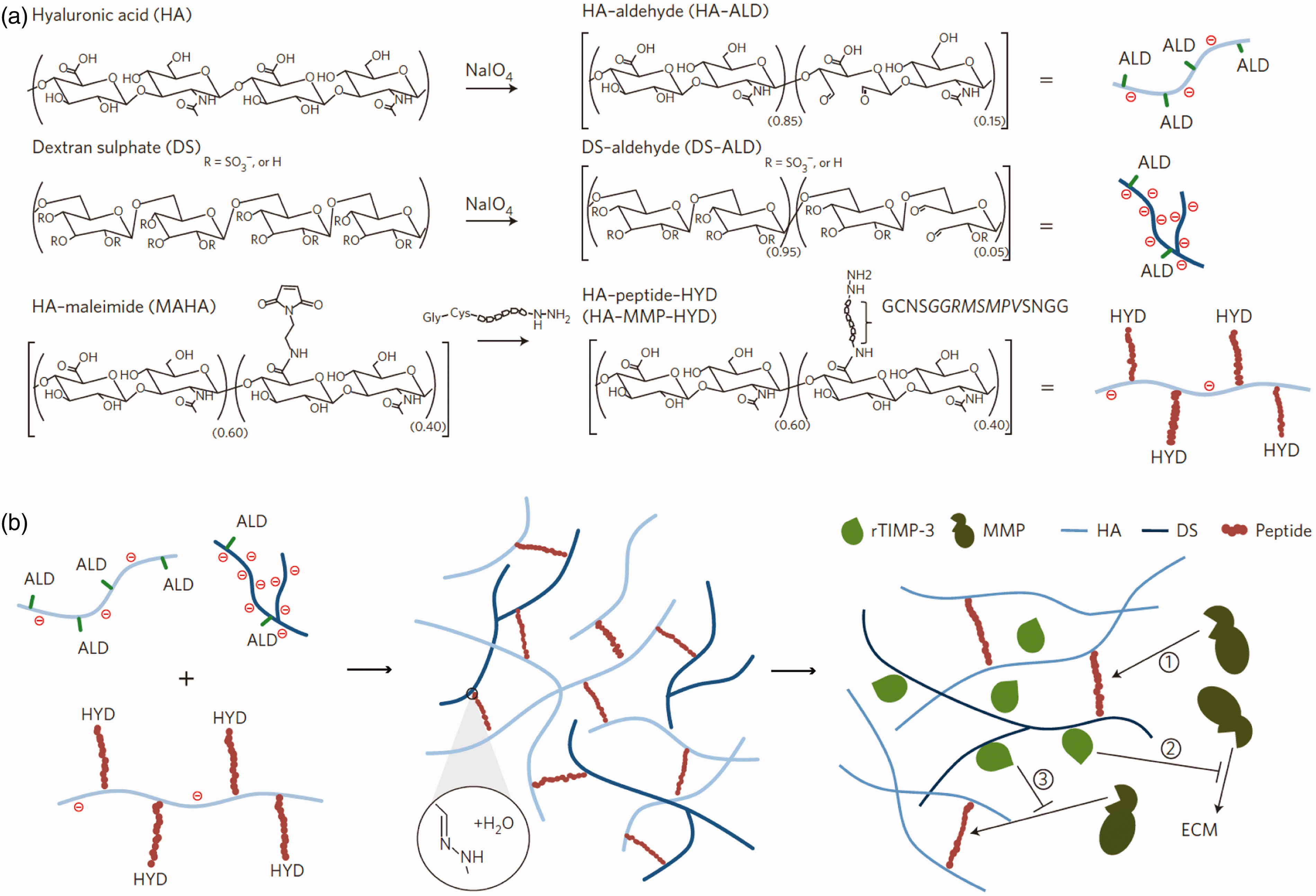
Other examples of peptide-crosslinked MMP-cleavable hydrogel systems include those prepared from Pluronic® triblock copolymer,
52
silk and elastin,
53
collagen,
54
alginate,
55
or heparin.
56
Werner et al. developed starPEG-heparin hydrogel networks crosslinked via Michael-type addition with MMP-cleavable peptides for controlled delivery of a range of therapeutic molecules, including VEGF,
56
stromal cell-derived factor-1α (SDF-1α),
57
transforming growth factor beta,
58
and growth factor cocktail consisting of VEGF, SDF-1α, and basic fibroblast growth factors that have synergistic effect for angiogenesis.
59
With the diverse roles of MMPs in pathological processes and disease progression, increasing investigations of amino acid sequences designed to be highly specific to certain MMPs, as well as development of broad spectrum of MMP inhibitors, are anticipated to enable more precise targeting to specific diseases.51,60–62 Figure 4 highlights some examples of peptide sequences derived from peptide libraries or phage display libraries that were screened for degradation by MMPs. In addition to MMPs, polymer hydrogels responsive to other classes of proteases, such as chymotrypsin,
63
cathepsin,
64
and elastase,
65
have also been reported.
Examples of peptide sequences derived from peptide libraries or phage display libraries that are screened for degradation by matrix metalloproteinases (MMPs): (a) MMP-1 and (b) MMP-2. (Reproduced from Patterson and Hubbell,
61
with permission from Elsevier.)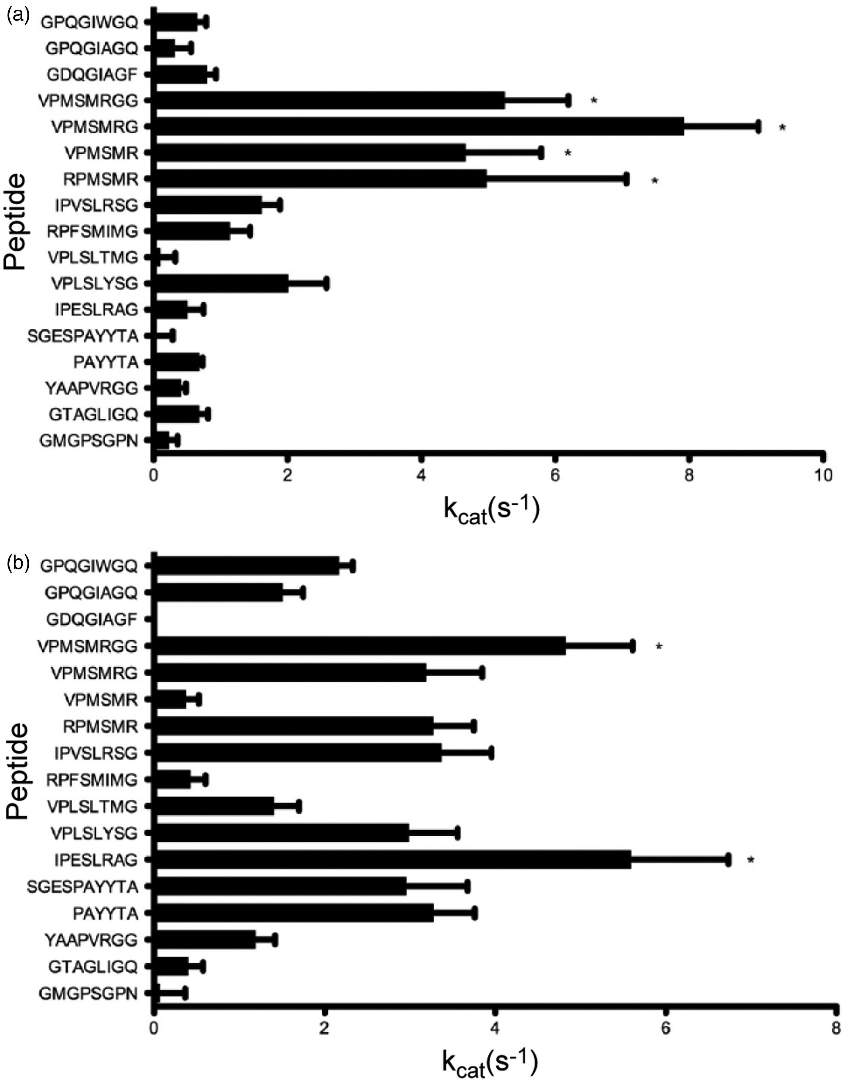
Glycosidase-responsive polymer hydrogels
Glycosidases play a central role in a spectrum of fundamental biological events, and with rate enhancement of 10 17 -fold, they are considered to be the most proficient catalyst. 66 Glycosidases catalyze the hydrolysis of glycosidic bonds in polysaccharides, hence this class of enzyme-responsive hydrogels is typically based on biodegradable carbohydrate polymers. β-mannanase is of particular interest as it is secreted by colon microflora and is largely localized in gastrointestinal tract, thereby allowing for colon-targeted drug delivery. A large number of polysaccharides have been studied for their potential applications as colon-specific drug carriers and are detailed in previous reviews.67–69 Qi and coworkers developed peptide–polysaccharide hybrid hydrogels made of Fmoc-diphenylalanine peptide and konjac glucomannan. 70 Docetaxel (anticancer drugs) were physically encapsulated within the hydrogels and β-mannanase-catalyzed reactions mediated controlled drug release. By varying the concentration of the glucomannan composition, the rate of therapeutic release can be modulated. In another example, Kono et al. reported β-mannanase-responsive hydrogels based on guar gum. 71 One-pot synthesis of guar gum hydrogels was achieved with 1,2,3,4-butanetetracarboxylic dianhydride crosslinker. β-mannanase-triggered hydrogel degradation and tunable release of encapsulated model proteins, lysozyme and bovine serum albumin, were demonstrated.
In a different approach, a number of recent studies demonstrated β-galactosidase-mediated release of nitric oxide (NO) from hydrogels.72–74 NO is an important cellular signaling molecule and biological regulator in many physiological processes, including vascular functions, neurological functions, immune responses, wound healing, and tumor progression.75–77 These molecules have short half-life (seconds), passively diffuse in short distances (∼100 µm), and their therapeutic effects are dose-, duration-, and location-dependent.
78
Hence, to achieve effective NO-based therapies, on-demand local delivery of biologically active NO is critical. Zhao et al. developed injectable chitosan hydrogels with β-galactosidase-sensitive NO donors (β-gal-NONOate) covalently bound to the polymer backbones via click chemistry.
73
These materials showed remarkable stability with no sign of decomposition of NO donors for over six months upon storage at room temperature. Release of NO from the hydrogels was controlled by the enzymatic activity of β-galactosidase, which hydrolyzed the glycosidic bonds and enabled release of active therapeutic molecules in micromolar concentrations (Figure 5). By exploiting endogenous β-galactosidase activity, local delivery of NO leading to enhanced proliferation of endothelial cells and angiogenesis stimulation in vivo was reported. While a number of enzyme-mediated NO releasing systems have demonstrated their ability to deliver NO on targeted sites, the fine control of dose and duration is still lacking. For example, angiogenic effects can be achieved with picomolar concentrations of NO, but micromolar NO concentrations are required to inhibit tumor growth.
76
Therefore, control over dose and rate of delivered NO remain as key challenges to develop effective NO-based therapies.
β-galactosidase-activated delivery of nitric oxide (NO) from chitosan hydrogels. β-galactosidase-sensitive NO donors (β-gal-NONOate) are covalently conjugated to the chitosan backbones via click chemistry. NO release is mediated and controlled by enzymatic activity of β-galactosidase, which hydrolyzes the glycosidic bonds and enables release of the active therapeutic molecules. (Reproduced from Zhao et al.,
73
with permission from Elsevier.) (A color version of this figure is available in the online journal.)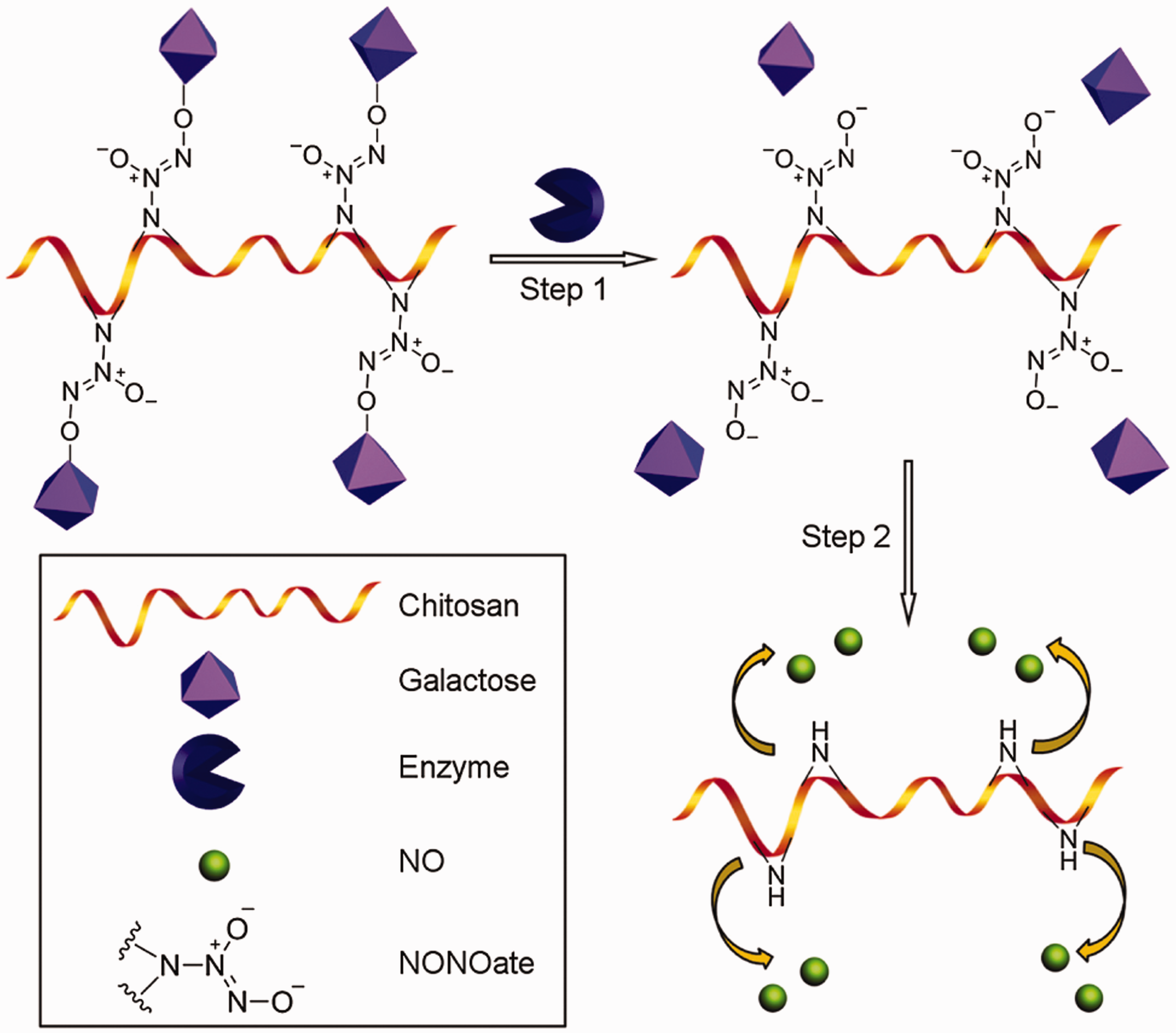
Summary and future perspectives
This mini-review provides an overview of the state of the art of enzyme-responsive polymer hydrogels and their applications in controlled drug delivery. Material responses translated from the activities of biologically relevant enzymes, such as proteases and glycosidases, are discussed. Enzymatic approaches offer a number of key advantages over traditional chemical reactions. Enzymes are inherently biocompatible and the catalyzed reactions are highly specific and selective, and can be performed under mild conditions. Polymer hydrogels programmed to release therapeutics in response to specific enzymatic activities hold enormous promise for biomedical applications and it is anticipated that these unique biomaterials will lead to significant impact in both research and clinical practices.
There has been a remarkable progress and great success in the design and development of enzyme-responsive polymer hydrogels with excellent tunable physical and chemical properties. While the abundance of proof-of-concept studies proves that such unique materials can deliver, there are many interesting challenges ahead before their full potential can be realized. Many enzymes have been characterized with a high degree of specificity for their substrates; however, their activities may alter with synthetic substrate mimics. Furthermore, substrates are generally bound to the materials and enzyme-catalyzed reactions in insoluble hydrogel networks may differ substantially from reactions involving solubilized substrates. Further investigation of enzyme kinetics in the crosslinked networks can provide more precise control of drug delivery. In particular, a fine control over spatiotemporal drug release from enzyme-responsive biomaterials will offer unprecedented opportunities in the biomedical field.
The expression level of enzymes varies in different diseases as well as at different stages of one disease. Hence, it will be desirable to develop smart hydrogels that not only respond to a specific enzyme but can also exhibit programmable responses toward varying enzyme concentrations. Furthermore, current studies demonstrate irreversible responses of the polymeric materials after enzymatic actions, which means, once initiated, encapsulated therapeutics will be continuously released. Incorporating functionalities that allow reversible changes of the hydrogel platforms will facilitate on-demand release of therapeutic payload, in particular it provides opportunities to achieve interactive adjustment of dose and rate of drugs tailored by enzyme expression levels and this unique feature will be highly valuable for long-term therapy. Inspired by complex biological environments, the ability of polymeric materials to respond to a number of enzyme stimuli will offer a new dimension and equip them with self-regulating systems to perform multienzymatic cascade reactions. Lutolf and coworkers developed dual enzyme-responsive PEG hydrogels able to undergo formation and degradation in response to transglutaminases Factor XIIIa and MMPs, respectively.79,80 We can envision smart hydrogel systems that can dynamically switch between crosslinked and non-crosslinked state repeatedly.
As the aberrant expression of enzymes serves as a disease biomarker, integration of enzyme activity detection into enzyme-responsive hydrogel drug depot will be a highly attractive tool in theranostics (combining therapeutic delivery and diagnostics). This emerging field provides a unique and powerful approach to allow real-time monitoring of enzyme expression levels and the effects of given therapy, paving the way for personalized medicine. The ongoing intensive research to discover novel enzyme biomarkers will drive further exciting development of enzyme-responsive biomaterials. Finally, assessment of these materials in in vitro and in vivo settings will push the technology advances closer toward bench-to-bedside translation.
Footnotes
Acknowledgements
RC acknowledges the support of The University of Sydney Early Career Researcher Grant and thanks Dr Philip Howes for critical reading of the manuscript.
Declaration of Conflicting Interests
The author(s) declared no potential conflicts of interest with respect to the research, authorship, and/or publication of this article.