
Abstract
The clinical data of 183 patients with hepatitic cirrhosis and portal hypertensive splenomegaly complicated by peripheral cytopenia were retrospectively analyzed to investigate the causes of peripheral cytopenia, as well as the proportion of the causes in these patients. All patients underwent splenectomy. Before operation, these patients had one or more types of peripheral cytopenia (cumulative cytopenia: 390 patient-times). After splenectomy, blood counts in 79.2% (309/390) returned to normal, while in 15.9% (62/390) they increased but failed to reach to normal levels, and in 4.9% (19/390) they became lower than before the operations. For the last group of patients (n = 19), long-term follow-up showed that blood counts returned to normal in five patients. In other words, in 80.5% [(309 + 5)/390 or 314/390] of patient-times, the peripheral cytopenia was due to hypersplenism, in 15.9% it was due to a combination of factors, and in 3.6% [14/390] it had nothing to do with the hypersplenism. Thus, hypersplenism is a major cause, but not the only cause, of peripheral cytopenia in patients with hepatic cirrhosis and portal hypertensive splenomegaly, and splenectormy is an effective treatment for these patients.
Impact statement
For a long time, the development of peripheral cytopenias as a complication to cirrhotic portal hypertension has been attributed to hypersplenism; however, this has never been fully demonstrated. Dameshek summarized that hypersplenism should be diagnosed by the presence of four conditions: (a) mono- or multi-lineage peripheral cytopenias; (b) compensatory hyperplasia of bone marrow; (c) splenomegaly; and (d) correction of cytopenias after splenectomy. We retrospectively analyzed the clinical data from 183 surgical patients, and found that 80.5% of peripheral cytopenias was caused by hypersplenism, 16% by a combination of factors, and 3.5% by other factors unrelated to hypersplenism. As the first quantitative findings in this field, our results verify that hypersplenism is a major, but not exclusive, cause of peripheral cytopenias, and provides important clinical evidence for investigating the cause of peripheral cytopenias.
Introduction
Globally there are approximately 360 million hepatitis B virus (HBV) carriers, and more than half are in the Asia-Pacific region. China is a high prevalence area of HBV infection where the positive HBV carrier rate is 9.8%. On average, 20% of these carriers will develop into chronic hepatitis, 1 and 50% of those with chronic hepatitis will develop into cirrhosis and portal hypertension. Peripheral cytopenia, defined as a peripheral white blood cell (WBC) count <4.0 × 10⁹/L, red blood cell (RBC) count <3.5 × 10¹²/L, or platelet (PLT) count <100 × 109/L, is very common in patients with hepatitic cirrhosis and portal hypertensive splenomegaly.2,3 Ninety percent of these patients have one or more types of peripheral cytopenia,4,5 which affects the prognosis.
In the past, peripheral cytopenia was considered to be caused solely by hypersplenism.6,7 However, according to Kalambokis and Tsianos, this etiology has not been well documented. 8 Many scholars have now reported that toxicity of hepatitis virus to bone marrow,9–11 liver hypofunction,12,13 gastrointestinal bleeding, 14 immune dysfunction,15,16 drug toxicity,17–19 peripheral platelet destruction, 20 hematopoietic disorders caused by vitamin and nutritional deficiency21–23 and at the same time with the blood system diseases can also lead to peripheral cytopenia. Karasu and Tekin 24 stated that hypersplenism cannot be the only cause for patients with peripheral thrombopenia. Dameshek 25 proposed four criteria for diagnosis of hypersplenism: (1) splenomegaly; (2) one or several types of cytopenia; (3) bone marrow normal or in hyperplastic state; (4) disappearance of pathological changes of blood cells after splenectomy. The clinical manifestations of portal hypertension include splenomegaly, and for patients with hypersplenism, peripheral cytopenia should be present and blood counts should become normal after splenectomy 26 and state the hypothesis addressed in this study: for example, this study addressed the hypothesis that hypersplenism is a major cause, but there are other causes, of peripheral cytopenia in patients with hepatic cirrhosis and portal hypertensive splenomegaly. We studied the clinical data of 183 patients with posthepatitic cirrhosis and portal hypertensive splenomegaly complicated by mono- or multiple-lineage peripheral cytopenia treated in our hospital from January 1996 to December 2013, and investigated the causes of peripheral cytopenia, as well as the proportion of the causes, in these patients to facilitate the choice of treatment.
Materials and methods
Patient cohort
The inclusion criteria were patients with posthepatitic cirrhosis and portal hypertensive splenomegaly who were complicated by peripheral mono- or multiple-lineage cytopenia treated with splenectomy. Exclusion criteria included (1) incomplete clinical data and (2) cytopenia blood diseases. All patients signed an informed consent form approved by the hospital ethics committee to comply with ethical requirements. There were 141 males and 42 females, making a male to female ratio of 3.4:1. Their ages ranged from 13 to 79, with an average of 43 years. There were 150 patients with posthepatitic B cirrhosis, and 33 patients with posthepatitic C cirrhosis. The diagnoses were confirmed by histopathological examination of liver tissues. The spleens in these patients were enlarged and could all be felt under the left costal margin. In 80 patients, the spleen was felt <5 cm under the left costal margin (splenomegaly degree I), in 72 patients the spleen was felt from >5 cm under the left costal margin to the umbilicus (splenomegaly degree II), and in 31 patients the spleen was felt at the umbilicus or beyond the abdominal midline (splenomegaly degree III). Ultrasound or computed tomography (CT) showed the average size of the spleen to be 226 mm × 162 mm × 96 mm. Radiological imaging of the upper digestive tract or gastroscopy showed moderate to severe varices in the lower esophagus and gastric fundus of these patients (Figure 1). Bone marrow biopsy was carried out in 142 patients (77.6%). In 82 patients, the bone marrow was normal and in 60 patients it showed hyperplasia. Splenectomy was carried out in 97 patients for massive digestive tract bleeding (blood loss ≥ 500 ml), in 55 patients for a PLT ≤ 5 × 10⁹/L, and in 31 patients for splenomegaly. Concomitant pericardial devascularization was carried out in 163 patients.
Severe varicose veins existed in lower esophagus and gastric fundus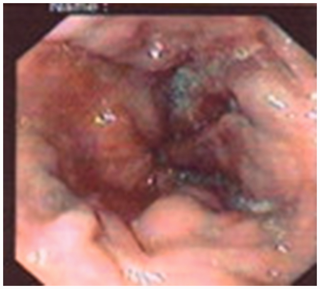
Statistical analysis
The SPSS 18.0 (SPSS, Chicago, IL, USA) statistical software was used. The Student’s t test was used to process numerical variables, which were expressed as mean ± standard deviation (S.D.); a P < 0.05 was considered statistically significant.
Results
Changes before and after operation in 183 patients with peripheral cytopenia (n)
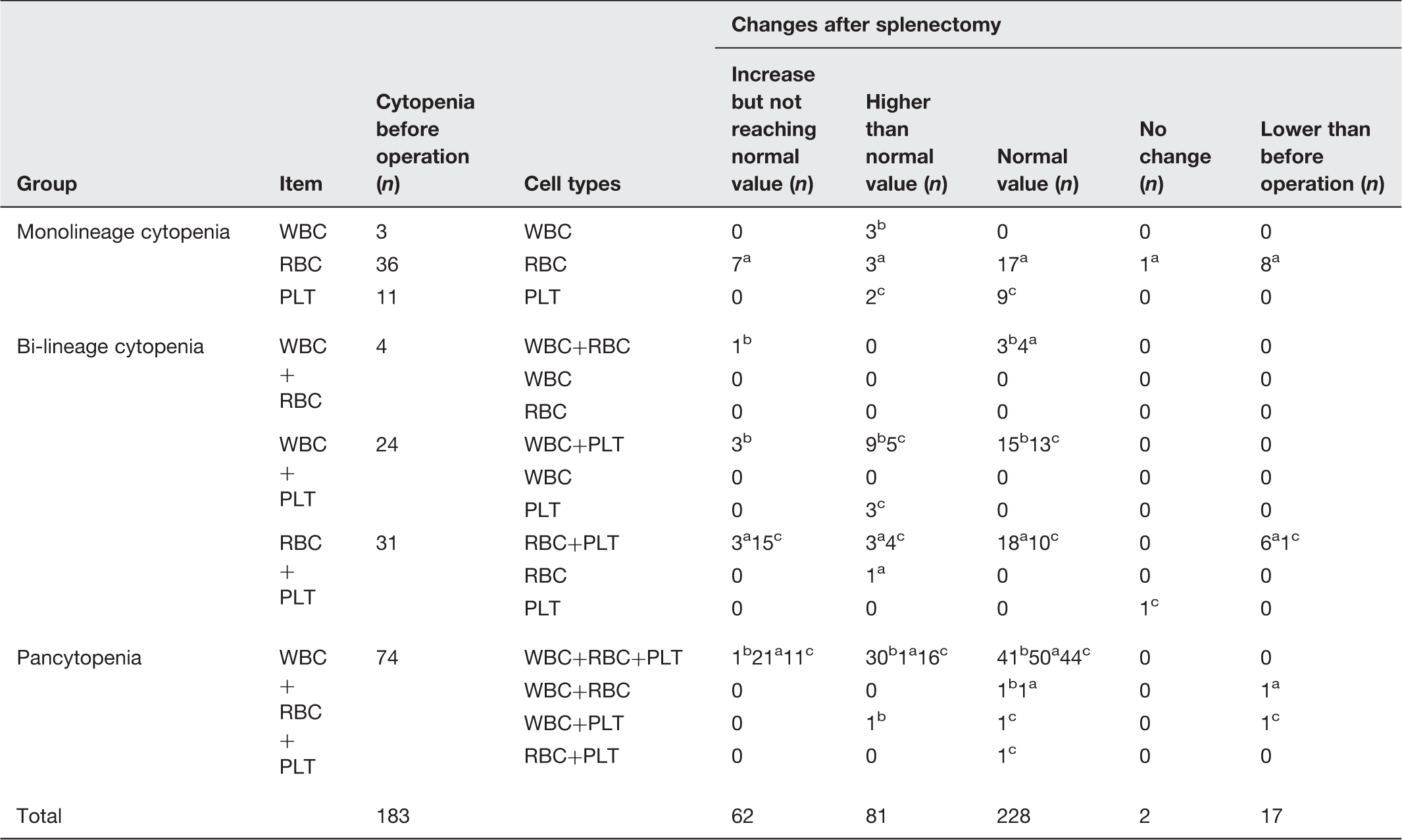
RBC.
WBC.
PLT.
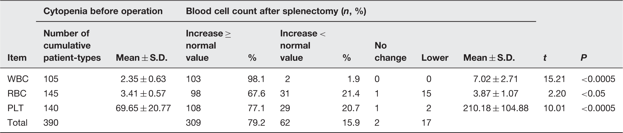
Comparison between postoperative state and preoperative state in the cumulative 390 patient-times of cytopenia
Comparison between postoperative follow-up and preoperative state in 239 patient-times of blood cell change (mean ± SD)
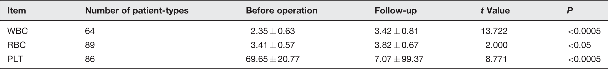
In 17 patients whose postoperative blood cells decreased rather than increased and in two patients whose postoperative blood cells did not change, isotopic scans did not show any accessory spleen. These patients were followed for 1 to 23 years (average seven years). In 15 patients with erythropenia, three died (two patients died of massive bleeding from the digestive tract, and one patient died of a stroke). In five patients, the RBC counts returned to normal (33.3%). In the remaining seven patients, the RBC counts were still low. In addition, two patients with thrombocytopenia died. For the two patients whose postoperative blood cells did not change, one patient’s RBC count returned to normal on follow-up, and one patient with postoperative unchanged PLT count died five years ago after surviving the operation for 20 years. Thus, the cytopenia was due to hypersplenism and two-thirds were related to other factors. A decrease or no change in postoperative PLT count predicted poor prognosis. The proportion of causes of peripheral cytopenias among the 183 patients is shown in Figure 2.
The proportion of causes of peripheral cytopenias among the 183 patients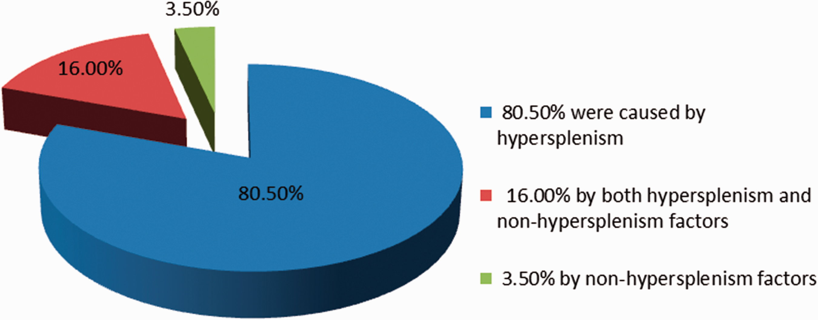
In about 4.9% (19/390) of patient-types, the peripheral blood cells became lower than before operation. In most of these patients, the decrease was in RBC. In 15 patients with a decrease in postoperative RBC, the decrease occurred mainly in the mono-lineage cytopenia group (n = 8) and in the bi-lineage cytopenia group (n = 6). However, in the pancytopenia group, there was only one such patient. Besides, all the preoperative RBCs were more than 3.2 × 10¹²/L, the WBC was >3.5 × 10⁹/L, and the PLT > 50 × 109/L. Thus, patients with mildly low blood cells (WBC + PLT) had a decrease in postoperative red blood cells. Two patients had a decrease in postoperative PLT. One was in the bi-lineage cytopenia group and the other was the pancytopenia group. To find out the causes in these two patients, we carefully studied these patients’ spleen size, liver function, and postoperative bleeding volume and compared these patients with other patients during the same period. However, we did not find any explanation. Isotopic scans also found no accessory spleen.
Discussion
In this study, all patients underwent splenectomy carried out for massive bleeding from ruptured lower esophageal and gastric fundic varices, or severe cytopenia from hypersplenism. The vast majority of the operations were performed before 2008. With recent developments in endoscopic hemostasis,27–29 transjugular intrahepatic portosystemic shunt (TIPS), 30 and hemostasis through medication, 31 operation is now only considered when non-surgical treatment is unsuccessful. As less and less patients underwent operation, the clinical data of these 183 patients become valuable to provide important information to study the causes of peripheral cytopenia. Table 1 shows that peripheral cytopenias are usually multiple linage cytopenias, accounting for 72.2% of patients (133/183). The more the types of blood cells are involved, the more severe is the disease and the worse is the prognosis. 32 The spleen is the target organ in hypersplenism. If peripheral cytopenia is caused by hypersplenism, the blood cells should return to normal after splenectomy. 33 Otherwise, causes other than hypersplenism should be considered. In this study, of the 183 patients, the cumulative monolineage cytopenia before operation was 390 patient-times. After splenectomy, approximately 80.5% (314/390) of patient-times with peripheral cytopenia had their blood cells counts returned to normal, indicating that these peripheral cytopenias were caused by hypersplenism. Hypersplenism is related to spleen size and phagocytic capacity of splenic macrophages. 34 A larger spleen size, which indicates more blood cells stored, and a higher phagocytic capacity of splenic macrophages would result in lower peripheral blood cell counts, and thus a more obvious increase in peripheral blood cell counts after splenectomy. In 15.9% (62/390) of patient-times with peripheral cytopenia, the blood cells counts increased but they did not reach normal, indicating that in addition to hypersplenism other factors were involved. 23 Also, thrombocytopenia is related to thrombopoietin (TPO), and the more severe the liver cirrhosis, the worse the liver functional reserve, thus the more obvious the thrombocytopenia, and the poorer the prognosis. 35 TPO is almost specially produced by liver cells. When cirrhosis develops, the number of functional liver cells secreting TPO decreases,24,36 resulting in a reduction of TPO secretion37,38 and imbalance in production and destruction of TPO. 39 Therefore, even if the spleen does not store and does not destroy PLT, the PLT in circulating blood might decrease. As these factors cannot be eliminated through splenectomy, the blood cell counts in these patients with cytopenia cannot fully recover.
Kalambokis and his associates 8 found an increase in incidence of peripheral cytopenia in cirrhotic patients being related to activated monocytes and promoted proinflammatory cytokines, such as serum interleukin-1, interleukin (IL)-6, tumor necrosis factor (TNF)-α, and interferon-γ, caused by hypersplenism in cirrhosis. The endotoxin produced by gut bacteria and antibiotic therapy for endotoxemia could also increase the number of blood cells in cirrhotic patients. Of the 105 patient-times of leukopenia before operation, the white cell counts returned to normal in 103 patient-types after operation. In the remaining two patient-types (1.9% or 2/105), the WBC count increased above normal. Thus, there was no patient who had a long-lasting decrease in WBC on follow-up. In 1979, Spigos et al. 40 first successfully used partial splenic arterial embolization (PSE) to treat hypersplenism. Subsequently, more clinicians41–43 used PSE to treat portal hypertension, hypersplenism, and hemorrhage from esophageal and gastric fundal varices. PSE not only enhances PLT and WBC counts,44,45 but also decreases splenic size, improves pancytopenia, 46 and induces immune function. 44 Zhengran et al. 47 reported that when the splenic artery embolization region was controlled at 60–80%, peripheral cytopenia caused by cirrhotic hypersplenism improved, portal venous blood flow, portal pressure, and degree of esophageal-gastric fundic varices reduced. Kontchou and his associates, 48 on the other hand, proposed that although PSE can treat splenomegaly and hypersplenism, serious complications such as splenic infarction or splenic abscess resulted in a high risk of mortality and the indications of PSE were limited. As splenectomy, especially laparoscopic splenectomy, has few serious complications,49–51 splenectomy is still a common and effective method to treat splenomegaly and hypersplenism.52,53
For a long time, the causes of peripheral cytopenias in patients with cirrhosis and portal hypertension have been controversial. The present clinical study of 183 patients showed that hypersplenism was still a major cause of peripheral cytopenias (80.5%). However, 16% of the cases were caused by a combination of factors, and 3.5% were not related to hypersplenism at all, i.e. caused by out-of-spleen factors. This conclusion not only reveals the proportion of causes of peripheral cytopenias, but more importantly, can be used to guide the treatment and determine the treatment effect. Non-surgical treatment is recommended for patients with mild to moderate cytopenias or hypersplenism, while splenectomy may be ideal for patients with severe cytopenias or hypersplenism. 54
Footnotes
The research statement
This research has been approved by the Ethics Committee of the Hainan Province People’s Hospital. All patients gave informed consent for their data to be used for research. All the authors have read and approved this article.
Funding
This research is supported by the China's Hainan Province Science and Technology special funds for international cooperation projects (grant no. KJHZ2015-28).
Authors’ contribution
YL conceived and designed the research; WYL took part in the design and writing of the paper; HWu, XYH, XG contributed to data collection and proofreading; NL, JY, YJL, JD, and QL took part in data statistical analysis and preparation of figures and tables.