
Abstract
Presented are the results of a study of the expression pattern of different proteins in the course of bovine leukemia virus-induced leukemia in experimental sheep and I discuss how the obtained data may be useful in gaining a better understanding of the pathogenesis of the disease, diagnosis, and for the selection of possible therapeutic targets. In cattle, the disease is characterized by life-long persistent lymphocytosis leading to leukemia/lymphoma in about 5% of infected animals. In sheep, as opposed to cattle, the course of the disease is always fatal and clinical symptoms usually occur within a three-year period after infection. For this reason, sheep are an excellent experimental model of retrovirus-induced leukemia. This model can be useful for human pathology, as bovine leukemia virus is closely related to human T-lymphotropic virus type 1. The data presented here provide novel insights into the molecular mechanisms of the bovine leukemia virus-induced tumorigenic process and indicate the potential marker proteins both for monitoring progression of the disease and as possible targets of pharmacological intervention. A study of the proteome of B lymphocytes from four leukemic sheep revealed 11 proteins with altered expression. Among them, cytoskeleton and intermediate filament proteins were the most abundant, although proteins belonging to the other functional groups, i.e. enzymes, regulatory proteins, and transcription factors, were also present. It was found that trypsin inhibitor, platelet factor 4, thrombospondin 1, vasodilator-stimulated phosphoprotein, fibrinogen alpha chain, zyxin, filamin-A, and vitamin D-binding protein were downregulated, whereas cleavage and polyadenylation specificity factor subunit 5, non-POU domain-containing octamer-binding protein and small glutamine-rich tetratricopeptide repeat-containing protein alpha were upregulated. Discussed are the possible mechanisms of their altered expression and its significance in the bovine leukemia virus-induced leukemogenic process.
Impact statement
The submitted manuscript provides new data on the molecular mechanisms of BLV-induced tumorigenic process indicating the potential marker proteins both for monitoring the progression of the disease and as possible targets of pharmacological intervention. This is to my knowledge the first study of the proteome of the transformed lymphocytes in the course of bovine leukemia virus-induced leukemia in susceptible animals. BLV can be considered as useful model for related human pathogen – HTLV-1, another member of the deltaretrovirus genus evolutionary closely related to BLV. Information gathered in this study can be useful to speculate on possible shared mechanisms of deltaretrovirus-induced carcinogenesis.
Introduction
Enzootic bovine leukosis (EBL), albeit eradicated in most European countries, continues to be a problem in many regions around the world. EBL naturally occurs in cattle, but the disease can be experimentally induced in sheep. The etiological agent, bovine leukemia virus (BLV), was classified as a member of the deltaretrovirus genus which also includes the related human T-lymphotropic virus type 1 (HTLV-1).
Once the virus is integrated into the genome of the host cells, it has a life-long association with the host in the form of an asymptomatic, persistent infection and can induce lethal lymphoma or leukemia only in a small proportion of infected animals.
This virus–host interaction is a kind of equilibrium between the latent and transcriptionally active phase, and probably a somatic mutation associated with genetic instability pushes the transformed cells to clonal expansion which results in leukemia. 1 BLV’s natural host is cattle, but experimentally infected sheep develop tumors at a much higher frequency than cattle and the latent period is shorter. 2
The tumorigenic process resulting from BLV infection is, just as many other malignancies, very complex and, besides the virus, which can be considered the main player, several other genetic determinants have to be taken into consideration. Among them, mutations of the p53 gene appear to be an essential leukemogenesis-predisposing factor, as roughly 50% of BLV-induced solid tumors in cattle exhibit the presence of a mutated p53 gene. Apart from the p53 gene, other abnormalities typical for tumor cells are found, e.g. translocations and rearrangements of the isochromosomes as well as acquisition of additional small chromosomes. 3 It is currently unclear whether these abnormalities are indispensable factors of cellular transformation or just byproducts of the process. There is also evidence supporting the role of the host genome in predisposition to tumor development. In particular, polymorphism of the bovine lymphocyte Ag (BoLA)-DRB3 was studied and the presence of the amino acids Glu-Arg (ER) at putative Ag binding residues 70 and 71 was found only in BoLA haplotypes associated with resistance to persistent lymphocytosis caused by BLV. 4 Analogously, susceptibility to the polyclonal expansion of B lymphocytes was associated with the W12.1 allele in BLV-infected Holstein-Friesian cattle. 5
Although the causative role of BLV is indisputable, the curiosity of BLV-induced leukemogenesis also lies in the modulation of viral expression. Roughly, only one per ten thousand B-lymphocytes was shown to express tax/rex mRNA during persistent lymphocytosis.6,7
It seems clear from the above examples that the mechanism of leukemogenesis in the case of BLV infection is largely unknown. It is, however, accepted that the transition from the asymptomatic, latent phase to the leukemia/lymphoma phase is a multi-step process during which genetic alterations, the microenvironment and/or immune evasion are involved to varying degrees.
Therefore, a study of the proteomics of transformed lymphocytes during clonal expansion can help to dissect the molecular pathways of the leukemogenic process. Using an experimental model of sheep instead of cattle is much more reasonable, not only for economic reasons but mostly because the process of tumor induction in sheep is faster and therefore more convenient for research purposes.
Material and methods
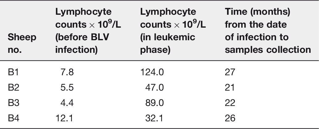
Lymphocyte counts of sheep used in the experiment (at the time of samples collection)
Data analysis
The acquired MS/MS data were pre-processed using Mascot Distiller software (version 2.4.2.0, MatrixScience) and the search was performed with the Mascot Search Engine (Mascot Daemon v. 2.4.0, Mascot Server v. 2.4.1, MatrixScience) against the merged database: the Uniprot-derived, complete bovine proteome database and the Swiss-Prot database (547,357 sequences; 194,874,700 residues) with a Mammalia filter (66,416 sequences). To reduce mass errors, the peptide and fragment mass tolerance settings were established separately for individual LC-MS/MS runs after measured mass recalibration, as described previously. 9 The parameters were as follows: enzyme—semiTrypsin, missed cleavages—1, variable modifications —Methylthionine (C), Oxidation (M), quantitation—iTRAQ 8plex, taxonomy—Mammalia, instrument—HCD, Decoy option—active. The Mascot Percolator was used to re-rank peptide matches. A statistical assessment of the confidence of peptide assignments was based on the target/decoy database search strategy. 10 This procedure provided q-value estimates for each peptide spectrum match in the dataset. All queries matched with q-values>0.01 were removed from further analysis. A protein was regarded as identified confidently when at least two peptides of this protein were found. Proteins identified by a subset of peptides from another protein were excluded from the analysis. Proteins that exactly matched the same set of peptides were combined into a single group (family). Mass calibration and data filtering as described above were carried out with MScan software, developed in-house (http://proteom.ibb.waw.pl/mscan/).
During initial statistical analysis, one of the controls was identified as an outlier and removed from further computations. Further statistical analysis of the quantitative results was performed using in-house software Diffprot with the following parameters: data normalization—LOWESS, a clustering of peptide sets with over 80% similarity. 9
All animal experiments were performed in compliance with the Guide for the Care and Use of Laboratory Animals published by AAALAC and approved by the Local Ethical Committee at the University of Life Sciences in Lublin (decision no 75/2012).
Results
A total of 1816 protein families were identified in the source material. After quantitative analysis, statistically significant (q-value equal or below 0.05) expression change was confirmed for 11 proteins. Eight of them, i.e. trypsin inhibitor (TI), platelet factor 4 (CXCL4/PF-4), thrombospondin-1 (TSP-1), vasodilator-stimulated phosphoprotein (VASP), fibrinogen alpha chain, zyxin, filamin-A (FLNa) and vitamin D-binding protein (DBP) showed decreased expression while the remaining three proteins, i.e. cleavage and polyadenylation specificity factor subunit 5 (CPSF5), non-POU domain-containing octamer-binding protein (Non-POU) and small glutamine-rich tetratricopeptide repeat-containing protein alpha (SGTA) were upregulated. Most of the downregulated proteins are engaged in cell–cell and cell–matrix signaling pathways, cytoskeleton organization and inflammatory response, while upregulated proteins play mainly regulatory functions in numerous nuclear processes.
Identification of proteins showing statistically significant altered expression (cumulative results for all four lymphocytotic animals)
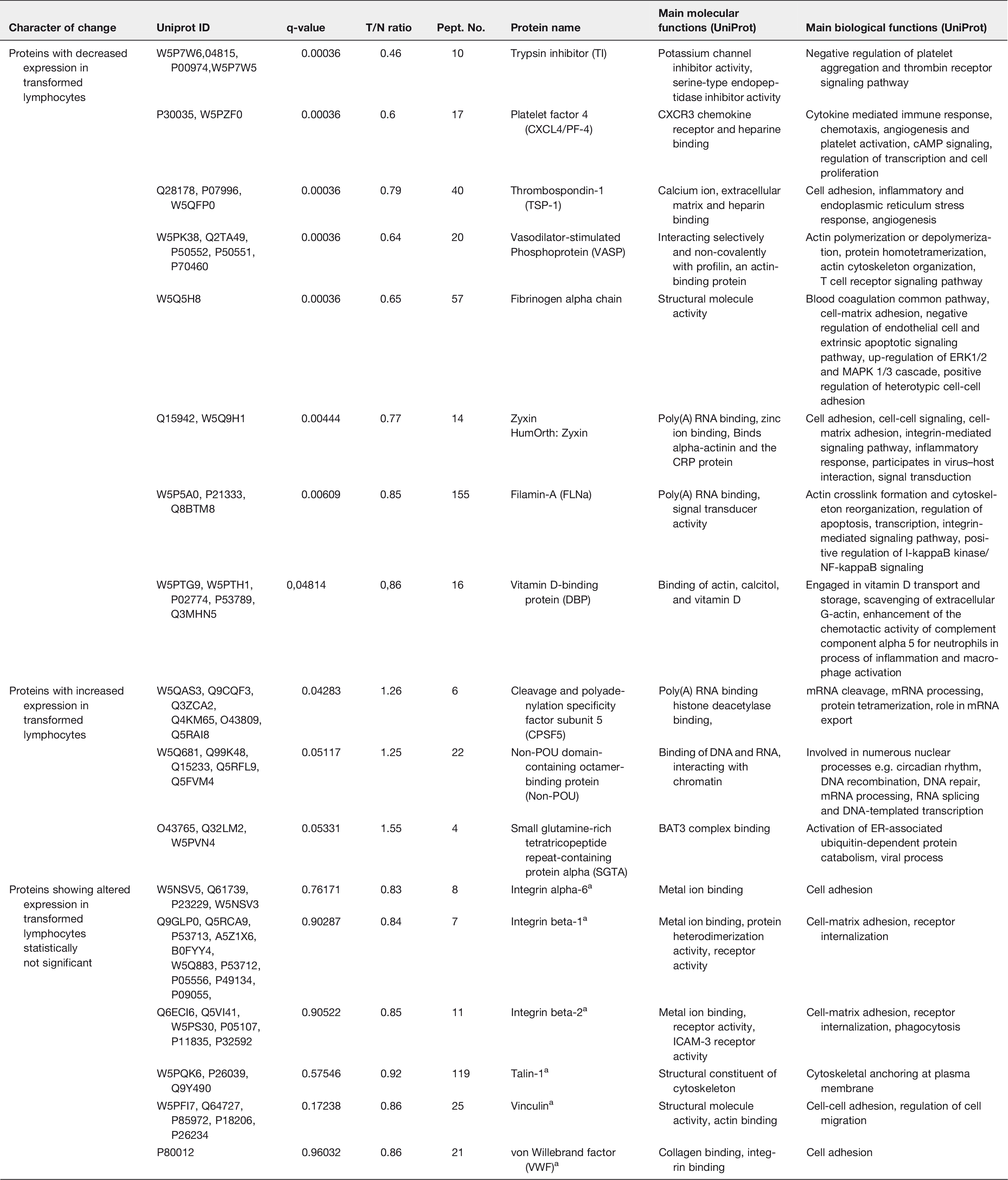
Note: Proteins with altered expression are ranked in descending order of statistical importance (T/N ratio and q-value).
T/N ratio: transformed (T) versus normal (N), healthy lymphocytes protein expression; Pept. no.: number of identified peptides.
Proteins showing altered expression statistically not significant.
There were few other proteins that showed changed expression in our study, but due to the high diversity of the obtained results, their altered expression was not statistically significant. These proteins include integrin, talin-1, vinculin, tubulin, alpha actinin-1, and the von Willebrand factor (Table 2). We believe that these results should be taken into account despite this shortcoming in order to better understand the entire proteome changes in the course of BLV-induced transformation of sheep lymphocytes.
Discussion
Downregulated proteins
Trypsin inhibitor (TI) is the protein that demonstrated the most profound changes in the level of expression between BLV-transformed and healthy lymphocytes in our study. It was also the most significantly downregulated protein in the transformed lymphocytes (T/N ratio = 0.46, q-value 0.00036) (Table 2). Trypsin inhibitor is one of many known serine protease inhibitors (also known as serpins). Until now, more than 1000 serpins have been described in all main taxons of plants and animals. 11 Most known serpins play a role in controlling proteolytic cascades, but some of them function as molecular chaperones, carriage proteins, chromatin remodeling molecules and others. The tumor-associated trypsin inhibitor (TATI), which is related to the protein identified in our study but not an identical member of the serpin family, was found to be upregulated in many clinical studies, and usually its increased expression was associated with poor prognosis and metastatic potential of the tumor.12–14 According to the UniProt Database, the protein identified in our study is involved in negative regulation of the thrombin receptor signaling pathway and it downregulates platelet aggregation. The latter was also reported by others in studies on CLL and CLL-related disorders. 15 Also, downregulation of related inter-alpha-trypsin inhibitors (ITI) was reported for a variety of solid tumors. 16
In our study, expression of TI in transformed lymphocytes from leukemic sheep was over two times lower as compared to lymphocytes from healthy, control sheep. Decreased expression of the trypsin inhibitor was also found in the sera of acute leukemia patients by Zheng and Ma. 17 There are many possible explanations for TI downregulation in BLV-transformed cells. The frequency and extent of downregulation of ITIs in multiple human tumors suggest that their tumor-suppressive role can be exerted at different levels of tumor development and metastasis involving complex pathways of cellular regulatory mechanisms.
One of these might be the interaction of heavy chains of ITI polypeptides with hyaluronic acid involved in the stabilization of the pericellular matrix, thereby hampering tumor metastasis. Inter-α-trypsin inhibitor is the precursor of urinary trypsin inhibitor—UTI was shown to inhibit the activity of plasmin, chymotrypsin and other proteinases, thereby reducing invasion and metastasis of tumor cells in vitro and in vivo, including human promyeloid leukemia U937 cells
18
(Figure 1). A similar mechanism may function in the case of BLV-induced leukemia in sheep.
Schematic drawing of the cellular pathways contributing to the pathogenesis of BLV-induced leukemia. Known signaling pathways (red), present study based hypothetical signaling pathways (blue), (references marked in parentheses). MA-SP: membrane anchored serine proteases. (A color version of this figure is available in the online journal.)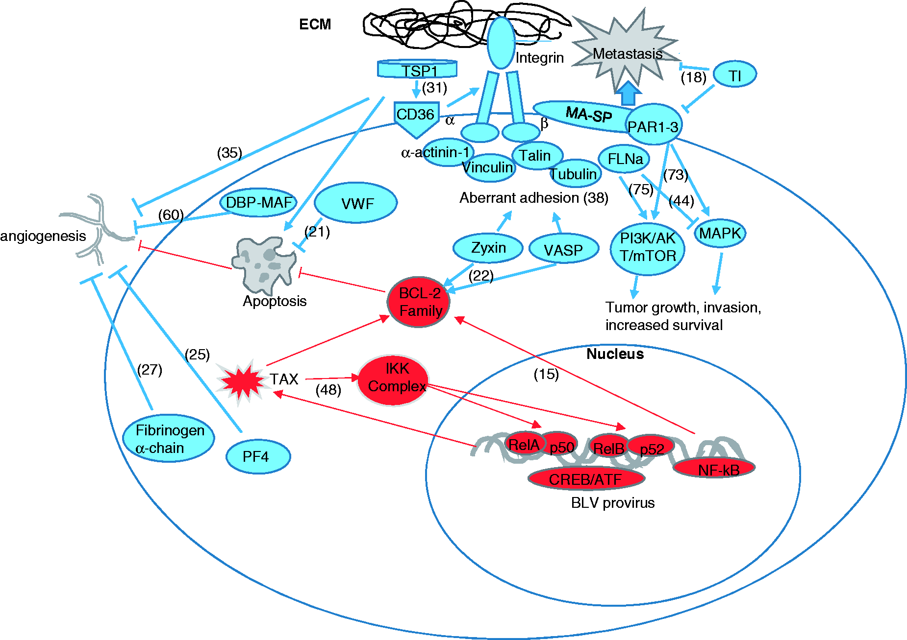
Platelet factor 4 (also known as C-X-C Motif Chemokine 4) belongs to a large group of cytokines exhibiting chemotactic properties and therefore named chemokines. CXCL4/PF-4 plays a crucial role in platelet physiology and can be detected in the storage compartments of megakaryocytes. 19 Other physiological functions of CXCL4/PF-4, e.g. granulocyte activation, platelet coagulation, stimulation of activation, differentiation and migration of monocytes, macrophages as well as NK and T cells have also been well documented20,21 (Table 2). CXCL4/PF-4. The role of CXCL4/PF-4 in pathology is less known; however, a growing body of evidence places this molecule as an important factor in several pathological processes. Probably, the best documented is the role of CXCL4/PF-4 in angiogenesis and tumor formation. It was shown that both CXCL4/PF-4 as well as its variant, CXCL4/PF-4 var, exert angiostatic and chemotactic activities through the CXCR3 ligand expressed on activated T, NK and some epithelial cells. Upregulation of this protein was shown in the course of several solid tumors 22 and decreased levels in others with high metastatic potential. 23 Decreased levels of CXCL4/PF-4 were observed in some hematological malignancies, e.g. in myelodysplastic syndrome, which is a very serious pathological condition frequently leading to acute myeloid leukemia. 24 The ability to stimulate NK and T cells suggests the important role of CXCL4/PF-4 in the immune response to cancer and therefore decreased expression of this factor as observed in the course of leukemia in sheep enables the proliferation of transformed lymphocytes and promotes carcinogenesis and progress of the disease. A similar decrease in the serum concentration of platelet factor 4 was recently found in the sera of adult patients suffering from acute lymphocytic leukemia (ALL). 25 The same authors found that the fibrinogen alpha chain was also downregulated. It is worth noting that both proteins show antiangiogenic activity, and therefore their decreased expression constitutes one of the tumor-promoting factors25–27 (Figure 1). The authors concluded that both proteins might be considered as potential biomarkers for prognosis and assessment of the therapeutic response of ALL and for monitoring of minimal residual disease. 25 Similar conclusions were drawn by Shi et al., 28 who studied the pediatric cases of acute lymphoblastic leukemia (ALL) and found the reduced expression of PF4. They proposed PF4 as a potential marker to differentiate ALL, AML and healthy individuals. As both proteins, i.e. platelet factor 4 and fibrinogen alpha chain, showed a similar proteomic profile in our experiment, i.e. a statistically significant decrease in the course of BLV-induced sheep leukemia, it might be interesting to study the signaling pathways leading to the observed changes in more detail. The mechanism of altered expression of PF4 in the course of hematopoietic system malignancies is not clear; however, it is strictly connected with the conformational changes in platelet membrane glycoproteins (GPs), resulting in increased adherence and aggregation capacity. GPs operate in the form of complex proteins. Among them, the von Willebrand factor (VWF) (an important ligand-binding site) and actin-binding proteins (both found to show altered expression in BLV-transformed sheep lymphocytes) are essential. According to Qian and Wen-Jun, 29 the mechanism of platelet activation is associated with the proliferation of leukemia cells leading to damage of the vascular endothelial cells followed by platelet adhesion and stimulation of intracellular signaling, which results in the secretion of platelet granule contents, including PF4. The role of platelet-associated PF4 as a biomarker of tumor growth was confirmed by other authors, with upregulation of its expression in the early stages of different tumors and decrease as the tumor progressed. 22 Pilatova et al. 30 proposed that increased platelet levels in experimental tumor-bearing mice reflect the feedback loop mechanism in response to the induction of pro-angiogenic factors by the growing tumor. Therefore, it seems reasonable to assume that the reduced level of PF4 in our study was also the effect of an advanced stage of the leukemogenic process, as we performed the proteomic study in advanced clinical stages of BLV-induced leukemia (lymphocyte counts from 32,000 to 124,000 per ul) (Table 1). Statistical analysis showed that there was no correlation between platelet counts and PF4 expression. PF4 is an important antiangiogenic factor, and therefore its reduced level was consistent with its reduced antiangiogenic activity. Similar results were found by others studying cases of patients affected by acute lymphocytic leukemia (ALL). 25
Thrombospondin-1 (TSP-1) is a known apoptosis-inducing compound of endothelial and tumor cells and acts through receptor CD36 (Figure 1). Li et al. 31 have shown that TSP-1 induces apoptosis in primary leukemia cells as well as in leukemia cell lines with caspase-3 being an indispensable component of the signaling pathway. Protection of leukemic cells from apoptosis can be considered a significant component of the process of neoplastic transformation, therefore downregulation of this proapoptotic and anti-angiogenic protein in the mechanism of tumor formation was frequently reported.32,33 We previously showed that the caspase-dependent pathway is also engaged in a glutathione-involved mechanism of protection of BLV-infected cells from apoptosis. 34 Therefore, downregulation of TSP-1 can play a similar role as experimental depletion of glutathione in experiments with BLV-infected sheep cells. 34 To dissect the role of thrombospondin in the leukemogenic process, the example of the CLL model can be considered, as BLV-induced leukemia, due to its phenotypic similarities, is frequently compared to CLL. Edelmann et al. 35 demonstrated that CLL cel viability was greatly enhanced when CLL cells were co-cultured with components of the stroma microenvironment, i.e. the bone marrow fibroblast cell line. The mechanism of the stroma-induced prolonged survival of CLL cells was dual and relied on upregulation of the genes of the phosphoinositol-3 kinase signaling pathway conferring chemoresistance and pro-angiogenic properties and downregulation of the anti-angiogenic thrombospondin-1 (Figure 1). Such a mechanism can also play an important role in the pathogenesis of BLV-induced leukemia, as a part of downregulation of TSP-1 and changed expression of some proteins of the phosphoinositol-3 kinase signaling pathway, e.g. tyrosine kinases, was also noted.
Vasodilator-stimulated phosphoprotein (VASP) was downregulated together with zyxin in our study. Both proteins are involved in cellular adhesion and are known to interact with each other. VASP is also a substrate of the BCR-ABL oncoprotein, which is a known inducer of chronic myeloid leukemia (CML). Several motifs essential for malignant transformation were identified in the BCR-ABL chimeric protein. Among them, SH1 (kinase) and the binding site for actin are the most essential. 36 Normally, only a small fraction of tyrosine residues in the cell is phosphorylated and the tyrosine phosphatases counteract the activity of tyrosine kinases. In the case of permanently activated tyrosine kinase activity in cells from CML patients (just as in the case of BCR-ABL), this subtle balance is compromised, thus leading to chronic activation of multiple signaling pathways targeting major cellular functions and resulting in reduced apoptosis, increased proliferation, and distorted interaction with ECM. 37 The latter results in impaired adhesion to integrins and other components of ECM. Such abnormalities were also observed in our study. Bernusso et al. 38 showed that VASP plays an important role in the pathogenesis of CML, either through the regulation of BCR-ABL effector proteins or the dephosphorylation of Ser157 in VASP. In particular, they demonstrated that VASP or Zyxin depletion resulted in reduced expression of antiapoptotic proteins (BCL2 and BCL-XL) and reduced expression of selected adhesion proteins (phosphorylated FAK), but there was no effect on the proliferation-related proteins 38 (Figure 1). The authors concluded that the absence of VASP and Zyxin interaction may contribute to CML aberrant adhesion. A similar mechanism may have taken place in our study on the advanced clinical stage of leukemia, as we found reduced expression of both proteins. Aberrant adhesion may substantially contribute to the development of the leukemogenic process in BLV-infected sheep.
Filamin A (FLNa) is a widely expressed protein that crosslinks actin filaments and is involved in anchoring the membrane proteins at the actin cytoskeleton and forming a stable 3D structure. FLNa regulates reorganization of the actin cytoskeleton by interacting with more than 60 different proteins engaged in various signaling pathways, ion channel and transcription regulation, cell migration, adhesion, and other crucial functions. FLNa was originally recognized as a cancer-promoting factor; however, there is evidence of FLNa engagement in tumor prevention as well. Therefore, the precise role of filamin A in cancer development is unclear. This dual role of filamin A partly depends on the intracellular localization of the protein. Full-length filamin A is mainly localized in the cytoplasm and promotes the development of metastasis (reviewed in 39), while the 90-kDa product of cleavage can localize to the nucleus and its expression seems to be necessary for androgen dependence of prostate tumor cells and sensitivity to treatment.39,40
There is no clear trend regarding expression of FLNa in different malignancies. Usually, its expression was correlated with clinical stage, lymph node metastasis, histological grade of the tumor and overall survival, and FLNa’s role can be defined as that of a tumor suppressor. 41 Frequently, FLNa acts as a tumor suppressor directly or through the other FLNa-interacting proteins. 42 Overexpression of such a protein inhibits, directly or indirectly, through the mediating molecules the invasiveness and metastasis of cancer by blocking various downstream pathways. Frequently, FLNa is overexpressed in many cancers, 43 but in the case of malignancies, where it acts as a tumor suppressor, downregulation of this protein was found. This was the case in our study, as quantitative analysis confirmed a statistically significant 0.85-fold decrease of its expression in malignant lymphocytes. Immunosuppressive action of filamin was also evidenced in a study by Sun et al., 44 in which the authors found that expression of FLNa was significantly decreased in prostate cancer tissues as compared to normal tissues. The authors suggested that the tumor-suppressive mechanism of FLNa involves the reduction of matrix metallopeptidase 9 (MMP-9) expression and, finally, the reduction of the invasiveness of cancer through inhibiting the Ras/MAPK/ERK cascade. The crucial role in anchoring MMP-9 to the cell surface of the transformed cells is played by α4β1 integrin, which together with CD44 forms a docking complex for MMP-9. 45 Therefore, decreased expression of integrin impairs MMP-9 anchoring, thereby reducing the invasiveness of the transformed cells. It is possible that FLNa exerts a similar suppressive effect in the case of BLV-induced leukemia, since in our study, we found decreased expression of both the alfa and beta subunits of integrin as well as integrin-linked protein kinase in lymphocytes from BLV-infected sheep (Table 2). It is known that higher levels of MMP-9 at the surface of transformed lymphocytes are correlated with advanced clinical stages and poor patient survival. 46 Integrins are responsible for the transmission of signals from the external environment to the cell. The process of signal transmission requires prior identification of extracellular matrix proteins, e.g collagen, fibrinogen and vinculin. Integrins attach to the actin filaments upon binding of the appropriate ligand from the extracellular matrix. Integrins do not possess enzymatic activity or the kinase domain, so in order to transmit signals they have to rely on their relationship with other signaling molecules. Therefore, altered expression of the integrin-linked protein kinase in our study is not surprising. There is also evidence regarding the role of integrins in the prevention of apoptosis in chronic lymphocytic leukemia B cells. 47 A crucial role was attributed to the iC3b integrin ligand, which is a proteolytically inactive complement cleavage product. Concentrations of iC3b in CLL patients were on average 14 times higher than in healthy patients. The mechanism of apoptosis inhibition in BLV-infected sheep lymphocytes is not known in detail, but NF-κB-dependent upregulation of the Bcl-2 family seems to play a crucial role 48 (Figure 1). Another possibility might be reduced production of oxygen species. 49 It cannot be excluded that the integrin pathway may also be engaged, as we found altered expression of both integrins and integrin ligands in the lymphocytes of BLV-infected sheep. The role of integrins in the pathogenesis of BLV leukemia is further justified by the altered expression of their ligands, i.e. fibrinogen and the von Willebrand factor. The role of the largest plasma protein, i.e. the von Willebrand factor, in hemostasis is well known, but in recent years, other functions of VWF have been evidenced, of which control of angiogenesis seems to be very significant. 50 VWF can exert its regulatory effect through the αvβ3 endothelial receptor, as the expression of αvβ3 was upregulated in tumor-associated blood vessels. 51 The role of VWF in controlling blood vessel formation may have serious clinical implications for patients with a deficiency or dysfunction of VWF. Angiogenesis is also critical for tumor formation, and therefore the altered expression of VWF found in our study is not surprising. Recently, the direct antitumor effect of VWF by negative modulation of apoptosis was suggested by Franchini et al. 52 (Figure 1).
It is known that the plasma level of VWF increases in malignancy and is the result of adverse changes in the endothelium. Severe endothelial dysfunction is frequently present in the course of acute lymphoblastic leukemia. In our study, we observed the opposite effect, i.e. that the endothelial dysfunction is not the hallmark of BLV-induced leukemia in experimental sheep.
Talin was another protein showing reduced expression in our study (Table 2). Talin together with filamin are key molecules in the linkage of the extracellular matrix (ECM) with the intracellular cytoskeleton. Crosstalk of ECM with the cytoskeleton heavily depends on integrins and is indispensable for the regulation of cell adhesion, cellular shape, and migration. Integrins exert their function by engaging ECM ligands. Among them talin is unique for its affinity to bind and activate integrins. Tumor formation, invasiveness, and migration capability are crucial attributes of any cancer, therefore, talin-1, due to its key role in integrin activation and integrin crosstalk, represents a promising diagnostic and prognostic marker that justifies further study. Altered expression of talin was found in many cancers. At the molecular level, talin was functionally associated with induction of proliferation pathways and protected tumor cells from anoikis, thus enabling metastatic spread of primary tumor cells via activation of the Akt survival pathway. 53
Vinculin also showed reduced expression in our study, which is not surprising as it directly interacts with talin and is a key regulator of the focal-adhesion complex. Vinculin binds to talin or alfa-actin and participates in the stabilization of integrin-mediated cell-ECM junctions. A lack of vinculin may decrease cell adhesion by preventing actin polymerization. Vinculin also plays an important role in the process of metastasis of many tumors.54,55 The process is very complex and, despite decades of research, is still not known in detail. Until now, more than 30 metastasis suppressor genes (MSG) have been identified but the underlying molecular mechanism is poorly understood. Recently, Thakur et al. 56 analyzed the tumor transcriptomes of lung cancer patients and found that NME2-encoded nucleoside diphosphate kinase B is a key factor among the suppressor proteins and involves vinculin to control lung cancer cell dissemination. Thus, selective RNA-induced silencing of vinculin diminished the metastatic potential of NME2-depleted cells. This finding confirms the crucial role of vinculin as a focal adhesion factor to regulate lung cancer metastasis. Dissecting the role of NME2–vinculin signaling in the mechanism of metastasis could be important also from the therapeutic point of view and could have potential clinical significance.
The decreased expression of three adhesion-related proteins, i.e. talin, vinculin, and integrin, as observed in our study confirms their strong relationship in controlling membrane-microfilament interactions in BLV-transformed lymphocytes. Expression of talin and vinculin was shown to play an important role in retrovirus infections, as experiments with the human immunodeficiency virus and the Moloney murine leukemia virus showed that overexpression of these proteins increased resistance of human cells (HeLa) to virus entry. 57 Both proteins negatively affect phosphorylation of paxillin, a major focal adhesion scaffolding protein, thereby hampering retrovirus infection. Downregulation of adhesion complex components can be considered as part of the immune response to infection which, nevertheless, has limited significance at advanced stages of BLV-induced leukemia, considered as clonal expansion of transformed lymphocytes. The decreased expression of these proteins as the components of host-virus interplay found in our study can be much more pronounced at early stages of retrovirus infection before clonal expansion has taken place.
The vitamin D-binding protein (DBP) is a key molecule in the metabolism of vitamin D as it transports vitamin D metabolites to different internal organs. Their role in cancer development is not unequivocal, although some authors found a strong protective association between elevated concentrations of DBP and kidney cancer. 58 Another study of a cohort of 148 lung cancer patients revealed the prognostic significance of the DBP level in the sera of the patients. 59 Low DBP levels were unequivocally associated with poor prognosis, and vice versa, higher levels predicted longer survival. According to the authors, a better cancer outcome is associated with the role of DBP in macrophage activation, as these are known scavengers of abnormal cells. The active component is DBP-MAF (the deglycosylated form of vitamin D-binding protein) which exerts its anticancer activity through both stimulation of macrophages to attack tumor cells and by inhibition of angiogenesis. 60 DBP-MAF is known to be secreted in the course of the inflammatory response accompanying tumor development. The lower expression of DBP found in our study is consistent with the above findings as we studied the terminal stages of the disease ending with death in a short time period. Because DBP-MAF can be considered as a potent tumor suppressor, its downregulation in our study is not surprising, as switching off tumor suppressors is a common mechanism of carcinogenesis. However, the detailed mechanism of DBP downregulation in the course of BLV-induced leukemia needs further clarification.
Overexpressed proteins
We also found several upregulated proteins in the transformed sheep lymphocytes. One of them was the non-POU domain-containing octamer-binding protein (non-POU). There is evidence that this RNA- and DNA-binding protein belongs to the family of transcription regulators and can play an important role in the signaling pathways of lymphoid cells. 61 Non-POUs bind intracisternal A-particles (IAPs) —the retroviral elements in the mouse genome that are highly expressed in lymphoid tissues. IAPs can act as transposons and can stimulate neoplastic transformation by augmenting the autonomous growth of host cells. Therefore, the upregulation of non-POUs in the transformed lymphocytes found in our study is not surprising and can be a part of the mechanism of neoplastic transformation.
In our study, overexpressed cleavage and polyadenylation specificity factor subunit 5 (CPSF5) together with subunit 2 and other components of the cleavage factor Im (CFIm) complex play a key role in the processing of pre-mRNA. In particular, both subunits participate in mRNA 3′-end processing, polyadenylation, and splicing via the spliceosome as well as in transcription from the RNA polymerase II promoter and the termination of RNA polymerase II transcription. 62
There are very few reports on the expression of this particular protein in tumor cases. In one study, the authors conducted proteomic analysis of human colon cancer cells expressing Snail1, which is the transcription factor known to induce the transition from the epithelial to mesenchymal phenotype (EMT) that is crucial for the acquisition of tumor invasiveness. It is known from transcriptomic studies that Snail1 is a key player in EMT and that it regulates many proteins implicated in various cellular processes. CPSF6 was among the proteins upregulated in human colon cancer cells by Snail1 which showed two times higher expression as compared with Mock cells. 63 The authors attributed the main role of CPSF to RNA processing and its role in cancer progression. However, colon cancer is a solid tumor, therefore hematological malignancies present a much better model for drawing any conclusions in the context of BLV-induced leukemia. In this respect, one example might be the study of cases of acute lymphoid leukemia and myeloproliferative syndrome as conducted by Hidalgo-Curtiset al., 64 who found CPSF6 as a partner gene fused to fibroblast growth factor receptor 1 (FGFR1), a representative of tyrosine kinase genes together with a group of other functionally related gene products. Tyrosine kinases are mainly involved in signal transduction but their oncogenic role is well known. It is therefore reasonable to assume that a similar mechanism may function in BLV-induced leukemia in sheep.
Small glutamine-rich tetratricopeptide repeat-containing protein alpha (SGTA) was shown to present about 50% higher expression in the transformed lymphocytes as compared to healthy ones. Zhu et al. 65 showed that overexpression of SGTA was correlated with the histological grade and short-term survival of patients affected by breast carcinoma. On the molecular level, the connection with the cell cycle was confirmed and SGTA depletion resulted in downregulation of cyclin A, cyclin B, and CDK2, and with upregulation of p27. Overexpression of SGTA was also positively correlated with expression of Ki-67, a known poor prognosis-related marker in breast cancer and other malignancies. The overexpression of SGTA found in our study, as well as in other reports,65,66 suggests that this molecule is a part of the signaling pathways regulating neoplastic proliferation of BLV-infected sheep lymphocytes. Similar findings were also reported in a study of large group patients suffering from different types of Non-Hodgkin Lymphomas (NHL). 67 Generally, high expression of SGTA was observed in clinical NHL specimens, except for in mucosa-associated lymphoid tissue B cell lymphomas (MALT) which, according to the authors, could be attributed to its indolent character. Based on the above findings, we speculate that the overexpression of SGTA found in our study substantially contributes to the fatal clinical course of BLV-induced leukemia in sheep and that the mechanism of its action involves proteins engaged in the cell cycle, including cyclin-dependent kinase inhibitor (p27Kip1).
Concluding remarks
The proteins identified here can be considered markers indicating which signaling pathways are engaged in the process of cellular transformation. Previous studies on BLV-induced leukemia/lymphoma were frequently presented as an animal model of HTLV-induced pathogenesis in humans.1,68 Author took advantage of the close taxonomic and biological relationship between both retroviruses with the aim of identifying the genetic determinants of the neoplastic process that would be useful in designing potential novel therapies in humans. As a result of those investigations, the crucial role of BLV Tax and G4 genes was indicated and their oncogenic role was confirmed. 68 Tax activates the CREB/ATF signaling pathway through the response element in the 5′ LTR promoter, leading to enhanced transcription of the proviral genome. Also, signaling through the NF-κB-dependent pathway as evidenced by TAX-dependent upregulation of nuclear RelB/p50 and p50/p50 NF-kappaB dimers seems to be essential for the disruption of normal B-cell homeostasis and tumor progression. 48 G4 forms a complex with farnesyl pyrophosphate synthase (FPPS), a component of the mevalonate/squalene pathway leading to prenylation of the Ras oncogene and cellular immortalization. The third essential player in the BLV-induced transformation can be a proviral region located next to the env gene encoding recently discovered microRNAs, 69 although the detailed role of BLV-encoded microRNAs needs further characterization.
The NF-κB complex is involved in a variety of physiological and pathological processes inside the cell, such as transcription of DNA, production of cytokines, cell survival, responses to stimuli as well as in regulating immune response to infection. 70 The latter is particularly important in the context of a BLV-induced transformation. Identified in our study as one of the downregulated proteins, filamin-A (Fln) was found to play a key role in engaging CD28 in the activation of the alternative IKKα-dependent NF-κB pathway in the absence of TCR. 71
As was reviewed in literature,1,68,69 the development of leukemia in BLV-infected animals is a multi-step process starting with the asymptomatic stage and progressing to persistent lymphocytosis (PL), then the leukemia/lymphoma stage. A crucial role in pathogenesis is attributed to the pleiotropic activity of the TAX and G4 genes, leading to immortalization of the infected cells. The result of polyclonal proliferation and accumulation of immortalized cells is a hematological disorder called persistent lymphocytosis (PL). Finally, monoclonal proliferation of a selected clone of the transformed lymphocytes occurs accompanied by a malignant phenotype. Genetic alterations of tumor-associated genes, e.g. TNF-α as well as tumor suppressors, such as p53, constitute the basis for the changed phenotype. Also, the bovine leukocyte antigen (BoLA) is considered a key player in the transition from PL to the leukemic stage of the disease.
All proteins whose changed expression was found in the study here seem to be engaged in this global proteome change at different stages of the transformation process. Particular attention should be given to five proteins, i.e. spleen trypsin inhibitor, CXCL4/PF-4, thrombospondin, vasodilator-stimulated phosphoprotein, and the fibrinogen alpha chain, whose downregulation was noted in our study. These are situated at the top of the list because changes in their expression were much more statistically significant than the remaining ones (q-value 0.00036). In particular, the role of the trypsin inhibitor is worth noting as it possesses the ability to reduce the activity of the thrombin receptor signaling pathway and to efficiently block voltage-dependent K+ channels (Table 2). Thrombin receptor overexpression, associated with increased invasiveness of the tumor,72,73 is frequently noted in highly malignant tumors, as is overexpression of potassium channels which emerge as potential cancer biomarkers. 74 Therefore, downregulation of the trypsin inhibitor as found in our study can be considered a part of the BLV-triggered pleiotropic regulatory mechanism leading to full development of the tumor.
The observed decreased expression of the whole group of cytoskeleton-associated proteins, i.e. filamin-A (statistically significant), integrin, talin-1, tubulin, alpha actinin-1, and vinculin, should also be emphasized. Among them, talin, vinculin, and actinin-1 link the actin cytoskeleton to the integrins through the focal adhesion complexes, while tubulin forms microtubules, a major component of the cytoskeleton. This observed change in expression can be caused by impairment of cytoskeletal integrity as a result of cellular transformation. It should be stressed that the statistically significant change in the expression of filamin A is intrinsically linked to the altered expression of all those proteins necessary to stabilize the connections of integrins with the cytoskeleton and the ECM. The whole complex participates in a variety of cellular processes, e.g. cytoskeleton remodeling and cell shape maintenance, cell spreading and migration, intracellular signaling, interaction with nuclear proteins engaged in DNA repair, RNA transcription, and protein translation. 39 In the context of the present study, the most important aspect is the possible participation of filamin A in the process of cancer progression, and one possible way may be the phosphoinositide 3-kinase (PI3K)/protein kinase B (Akt) pathway, as there is evidence for the involvement of filamin A as a negative regulator of rRNA synthesis in leukemic cells through activation of the PI3K/Akt pathway. 75 (Figure 1). As such, downregulation of FLNA expression might be considered a part of the mechanism of tumor progression. A similar mechanism was shown to exist in the transformation of rat fibroblasts by the closely related human pathogen HTLV. 76
Altogether, our study showed that the observed changes in the proteome profile of transformed lymphocytes were dominated by decreased expression of proteins actively engaged in cytoskeleton regulation. This group consists of five statistically significant components, i.e. thrombospondin-1, vasodilator-stimulated phosphoprotein, fibrinogen alpha chain, zyxin, and filamin A. It is worth mentioning that an additional group of four cytoskeleton-linked proteins, i.e. integrin, talin-1, vinculin, and the von Willebrand factor, were downregulated but unfortunately not statistically significant. Other proteins showing the most pronounced downregulation of their expression, i.e. trypsin inhibitor and platelet factor 4, are essential components of pathways engaged in the regulation of platelet aggregation, thrombin receptor signaling pathway as well as immune response, chemotaxis, angiogenesis, platelet activation, cAMP signaling, regulation of transcription, and cell proliferation, respectively (Table 2).
The two overexpressed proteins, i.e. cleavage and polyadenylation specificity factor subunit 5 and non-POU domain-containing octamer-binding protein, are molecules engaged in the process of active chromatin remodeling and mRNA processing that is characteristic of transformed cells. The role of vitamin D-binding protein is less obvious; however, its tumor-suppressive role is linked to macrophage activation and angiogenesis.
Identifying proteome changes in sheep B lymphocytes in the course of BLV-induced leukemia provides substantial insight into the pathogenesis of retrovirus-induced transformation and gives rise to more specific pharmacological treatments in similar human disorders. To our knowledge, this is the first proteomic approach-based analysis of crucial signaling pathways that the BLV retrovirus is likely to target.
Footnotes
Acknowledgments
The author would like to highly appreciate Ms Malgorzata Zaborna for the technical help. The author also appreciates the consistent and long-standing support of Dr. Ewa Sitkiewicz and the remaining Staff at the Laboratory of Mass Spectrometry of the Institute of Biochemistry and Biophysics of the Polish Academy of Sciences in acquisition and analysis of mass spectrometry data.
Declaration of conflicting interests
The author(s) declared no potential conflicts of interest with respect to the research, authorship, and/or publication of this article.
Funding
The study was financed by the Polish National Science Centre (contract no.: 2012/07B/NZ6/03536) and by KNOW Scientific Consortium “Healthy Animal – Safe Food” No. 05-1/KNOW2/2015.