






Abstract
The isolated tissue bath research methodology was first developed in 1904. Since then, it has been recognized as an important tool in pharmacology and physiology research, including investigations into neuromuscular disorders. The tissue bath is still used routinely as the instrument for performing the “gold standard” test for clinical diagnosis of malignant hyperthermia susceptibility – the caffeine-halothane contracture test. Our research group has utilized this tool for several decades for a range of research studies, and we are currently one of four North American diagnostic centers for determining susceptibility for malignant hyperthermia. This review provides a brief summary of some of the historical uses of the tissue bath. Important experimental considerations for the operation of the tissue bath are further described. Finally, we discuss the different studies our group has performed using isolated tissue baths to highlight the broad potential applications.
Impact Statement
Isolated tissue baths provide a simple solution to assess contractile tissue function. In therapeutic investigations, these baths can provide critical information on the impact and physiological response of muscle tissue to various therapeutic treatments, which is particularly valuable for translational research. Uniquely, we have shown that tissue baths can be utilized to assess the response of muscle tissue to medical device therapies (e.g. pacing and ablation). Furthermore, we describe the construction of our tissue bath experimental equipment to allow for the rapid collection of functional data (data acquisition system running 24 tissue baths in parallel). By utilizing waste tissue from unrelated large animal research, in vitro tissue bath studies can complement in situ work, thus accelerating bench to bedside translation and minimizing cost.
Introduction
Isolated tissue baths, commonly referred to as organ baths, have a long history of utilization in the fields of pharmacology, muscle physiology, and device testing. This research approach was first reported by Rudolf Magnus in 1904. 1 It was then used extensively by Otto Loewi for his discovery of acetylcholine as an endogenous neurotransmitter, for which he shared the 1936 Nobel Prize in Physiology or Medicine with Sir Henry Dale.2,3 Since the early development of this experimental method, isolated tissue baths have proven useful in functional assessments of both healthy and diseased tissues, in a variety of investigational conditions. Furthermore, these approaches have been employed for preclinical medical research as well as testing of various animal models including transgenic, knockout models, and others.
In addition to their utilization in basic and translational research, in vitro tissue baths have been applied clinically for the pharmacological diagnoses of malignant hyperthermia (MH), a myogenic disease that was first clinically defined in 1960.4,5 MH is a heterogenetic disorder; when affected individuals are exposed to triggering anesthetic agents, they may experience life-threatening hypermetabolic events due to induced widespread skeletal muscle calcium dysregulation. Fortunately today, mortality due to MH events has fallen to less than 5% due to standardization of treatment protocols and diagnostic measures (both genetic and in vitro diagnoses). 6 The Malignant Hyperthermia Association of the United States (MHAUS) currently recognizes four in vitro contracture test centers in North America that perform the standardized caffeine-halothane contracture test, one being our laboratory at the University of Minnesota (Dr Iaizzo has performed this diagnostic test for 35+ years at the Mayo Clinic, the Technical University of Munich, Ulm University, and the University of Minnesota). The contracture test requires the use of fresh tissue that is obtained via muscle biopsy, while the patient receives non-triggering anesthetics. Care is taken to minimize trauma to the specimen which must meet specific dimensions. Numerous muscle bundles are prepared, measuring ~1–3 mm in diameter and >3 cm in length, and then placed in tissue baths and dosed with various concentrations of caffeine or halothane. The in vitro contracture responses of isolated muscle to these agents are used to determine the diagnosis. To date, MH mutations have been mapped to approximately 60–70% of patients worldwide. Hence, if a family does not have one of these known mutations, in vitro contracture testing is recommended to identify family members susceptible to MH, and then, linkage analyses can subsequently be performed to identify new genetic mutations causing the MH phenotype.
Although the tissue baths in our laboratory were first set up for clinical diagnostic testing, we subsequently utilized them in a variety of other experimental studies. To date, we have investigated the pathophysiology of other neuromuscular diseases, as well as different disease states, anesthetic effects, traditional pharmacological studies, cryopreservation responses, and studies involving knockout or transgenic mouse models, among others. We further adapted our research protocols for translational research, for instance, to study physiological responses to various medical device applications (e.g. pacing, ablation, electrical recording). Overall, the isolated in vitro tissue bath has been a valuable tool for translational research and clinical diagnoses, particularly because it allows for rapid collection of large amounts of functional data, thus accelerating bench to bedside possibilities.
The present minireview describes general tissue bath designs and setups, historical usage of tissue baths in pharmacology and physiology (highlighting a few key studies and methodologies), current usage in caffeine-halothane contracture testing for clinical diagnosis of MH susceptibility, and our laboratory’s continued application of these tools in translational research for numerous medical therapies.
Main text
Historical usage
Considering that tissue bath contractile studies covered a broad range of research for well over a century, it would be impossible to review all the work that has been accomplished. We instead highlight a few important discoveries before discussing the more traditional use of tissue baths to generate dose–response curves and application for in vitro contracture testing to diagnose susceptibility to MH.
Historical discoveries
Tansey 3 recounts the timeline of research that led to an understanding of the role of acetylcholine in chemical neurotransmission, and describes the classic experiment performed by Otto Loewi in 1921. A denervated frog heart and a frog heart with the vagus nerve still attached were placed in tissue baths. It was found that solution taken from the bath containing the innervated heart, following nerve stimulation, would slow the denervated heart. At the time, the agent in solution was referred to as vagusstoff. Likewise, important studies in understanding the role of nitric oxide as a vasorelaxant were also performed using tissue bath methods. Furchgott and Zawadzki 7 used longitudinal and transverse strips of aorta in their discovery of the endothelium-derived relaxing factor, which was later identified as nitric oxide in further work with the tissue bath by Ignarro et al. 8
Dose–response curves
The concept of identifying dose–response relationships is important to the fields of pharmacology and toxicology. Paracelsus, considered the father of toxicology, is often quoted as saying “the dose makes the poison.”9,10 An understanding of the dose–response relationship emerged during the late 19th/early 20th century and was developed and utilized by many different fields, not only pharmacology and toxicology. 11 Dose–response curves now serve a critical role in determining the safety and efficacy of pharmaceuticals, and the Food and Drug Administration has issued guidance for its use in drug development. 12 By understanding the dose–response relationship of a drug, the administration of an excessive dose (one that has diminishing benefits and increasing toxicity) can be avoided. Several different in vitro assays exist for generating data to construct dose–response curves, which are commonly graphed on a semilog plot and elicit a sigmoidal-shaped curve. The in vitro tissue bath approach is one such method that relates the concentration of the agent to the contractility (i.e. response) of the isolated muscle or organ.
Due to the relative ease of employing the in vitro tissue bath for relating effective concentrations to a physiological response, these methods played an important role in the development of receptor theory.13–15 One of the early mathematical models in receptor theory was the Hill model. 16 Despite its simplification, it is still common today to fit tissue bath dose–response results to the Hill model. 17
In vitro contracture testing for diagnosing susceptibility for malignant hyperthermia
We currently utilize the North American Malignant Hyperthermia Group protocol for performing the caffeine-halothane contracture test18,19 within our laboratory. The European Malignant Hyperthermia Group in vitro contracture test differs slightly and can be viewed elsewhere. 20 The North American protocol is considered to have 97% sensitivity and 78% specificity 21 rates. The basis of this test is the increased sensitivity of MH susceptible skeletal muscle to caffeine 22 and halothane. 23 In instances where a patient has been referred for MH screening and has a family history with an identified causative MH mutation, genetic testing would be performed first (for MH genetic testing guidelines see Urwyler et al. 24 ). However, a negative genetic test does not exclude MH susceptibility, since many pathogenic variants remain unidentified due to the genetic heterogeneity and discordance of this disorder.25,26
The muscle biopsy for in vitro testing for MH is performed as an outpatient procedure. A muscle sample is removed from the vastus lateralis or vastus medialis (thigh) muscles using anesthesia known not to trigger malignant hyperthermia. The minimal sample size is recommended to be 3–5 cm long and 1–1.5 cm in diameter. A second small muscle sample is removed for histologic study. The caffeine-halothane contracture test is performed in accordance with the guidelines provided by the North American Malignant Hyperthermia Registry and is highly accurate for diagnosing susceptibility to MH. The in vitro testing is initiated as soon as the muscle sample is available from the patient (within minutes), as fresh tissue is required. Using a dissecting microscope, a minimum number of six small muscle bundles (~2 mm in diameter and 3–5 cm in length) are prepared and then mounted in experimental chambers where they contract via electrical stimulation (see below for more details). A viable muscle bundle needs to elicit a minimum of 1 g of twitch force (supramaximal stimulation with a 1 ms pulse width). Typically, we dissect 1–2 muscle bundles, mount them in the tissue baths, determine the viability of tissue, and then notify the operating room to close the patient’s biopsy site (i.e. if an additional specimen is not required). When the anesthetic halothane (3%) is introduced into the experimental chamber, a muscle that is susceptible to MH will contract more rigorously (twitch potentiation) and elicit a contracture (sustained force generation). Similarly, when the additional muscle bundles are exposed to caffeine at increasing bolus doses to obtain concentrations of 0.5, 1, 2, 4, 8, and 32 mM, the MH susceptible tissue will also elicit twitch potentiation and contractures (at various threshold concentrations). An abnormal contracture threshold is indicative of susceptibility to MH; these thresholds are defined as >0.7 g contracture from 10 min exposure to 3% halothane and ⩾0.2 g contracture at ⩽2 mM caffeine. 19 Positive test responses to halothane and caffeine are shown in Figures 1 and 2, respectively. Halothane and caffeine contracture tests are typically performed in triplicate in our laboratory (see http://www.vhlab.umn.edu/clinical). Patients with a positive contracture test are advised to wear a medical alert identification or bracelet. In addition, they may wish to follow up with one of our collaborating anesthesiologists to discuss: (1) known genetic details of MH and new genetic screening procedures; (2) possible testing of other family members; (3) monitoring of serum creatine kinase levels in potentially susceptible family members; and (4) clinical implications for future anesthetics in MH positive individuals.

Positive halothane contracture test performed on three muscle bundles. Sustained drug (halothane) induced force production (contracture) that exceeds our laboratory’s defined threshold value of 0.7 g. (A color version of this figure is available in the online journal.)
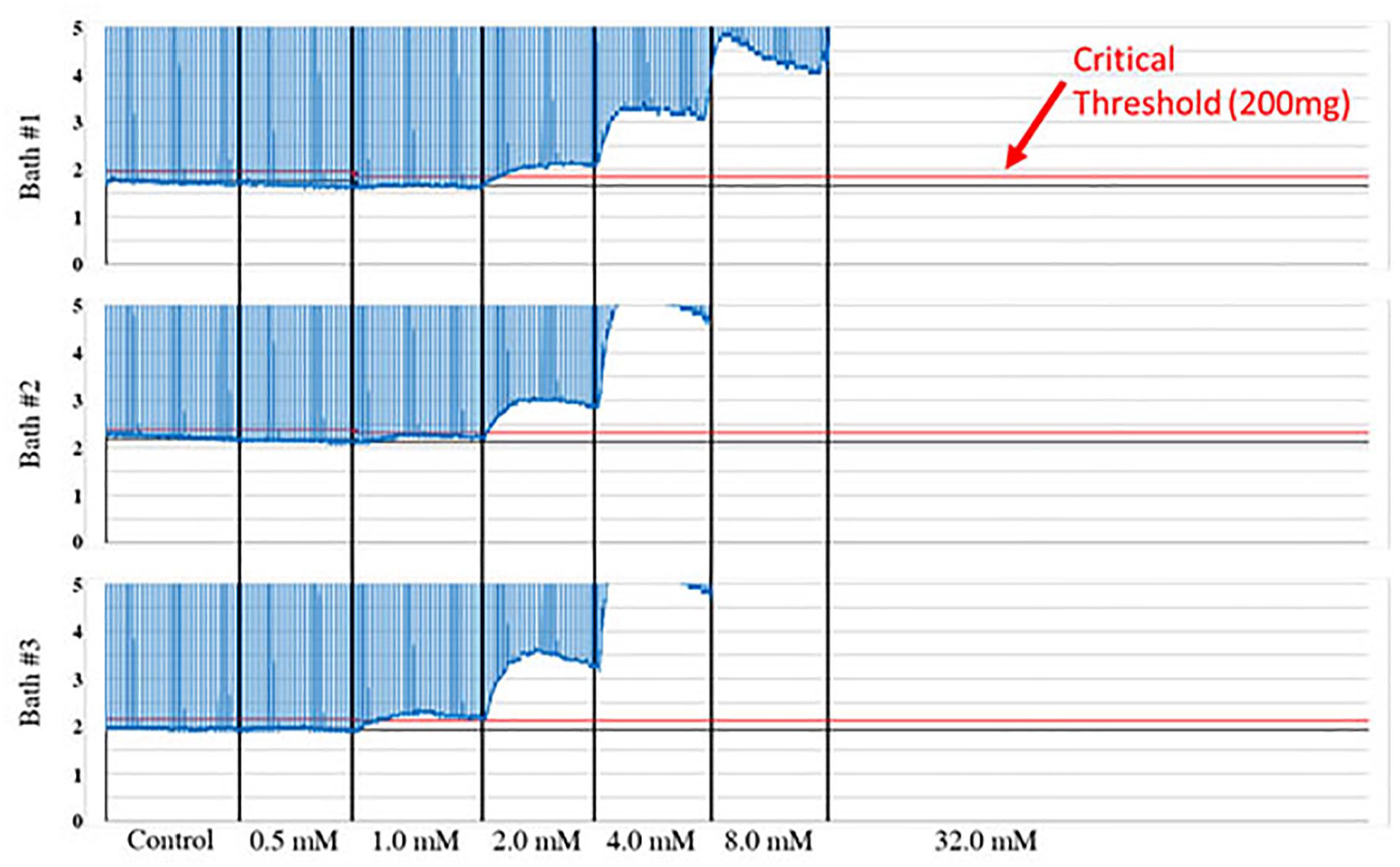
Positive caffeine contracture test performed on three muscle bundles. Sustained drug (caffeine) induced force production (contracture) that exceeds 0.2 g threshold at 2.0 mM caffeine concentration, defined by our laboratory. (A color version of this figure is available in the online journal.)
Experimental setups
Tissue baths for contractile studies can be found or generated in a variety of sizes, chamber designs, electrode, and/or stimulation configurations. Furthermore, the required instrumentation and data acquisition systems can be purchased or developed as needed. Various research groups have designed custom glass-blown tissue baths with locally constructed data acquisition and processing systems adapted to specific laboratory workflows or purchased commercially available systems with proprietary data acquisition software (e.g. ADInstruments, Harvard Apparatus, BIOPAC, Scintica, MDE Research, Radnoti). Despite these variations, the important constant is the ability to provide an in vitro environment capable of maintaining the viability and function of the isolated tissues or organs.
Components of tissue baths in the Visible Heart® Laboratories
The 24 tissue baths utilized within the Visible Heart® Laboratories (VHL) include double-walled chambers that contain a physiological buffer within the inner chamber, the location where the tissue preparation is submerged. Each chamber has a 20-mm-diameter opening at the top for placement of the mounted tissue preparation within the 10-cm-long cylindrical chamber. The second connected cylindrical side chamber, with similar dimensions, is used for gassing and adding reagents. The surrounding water-jacket chamber has an inlet and outlet that allow for water circulation; temperature-controlled water (we add antimicrobials for long-term use) is continuously pumped through the baths to maintain the inner chamber at a desired physiological temperature. The physiological buffer is continuously gassed through an incorporated aerator chamber component. In most studies, carbogen (95% O2 and 5% CO2) is utilized which also maintains a physiological pH. Yet, O2 can be replaced with nitrogen if one wants to induce ischemic conditions. Figure 3 illustrates two tissue bath configurations that we have utilized over the years, with the aerators identified. Minimal gassing is required to sufficiently saturate the buffer solution; however, it serves the dual purpose of mixing the buffer solution (with dye studies, we observed uniform mixing in ~1 s). Note, rigorous gas bubbling can introduce noise into the force recordings, which is more of a concern when the aerator is positioned below the suspended tissue preparation (Figure 3(a)). Again, to alleviate such issues, John Blinks, a former Professor of Pharmacology at the Mayo Medical School, designed the tissue bath shown in Figure 3(b), with the side-chamber aerator sequestered from the main tissue chamber.
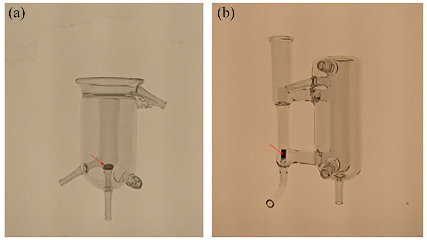
Two different tissue bath designs. The aerator is identified by the arrow. (A color version of this figure is available in the online journal.)
Tissue baths can be readily used to study isotonic or isometric contractions. Isotonic transducers measure the change in length of tissue under a constant load. For isometric contractions, the specimen is held at a constant length, while the force production is measured (this is the common mode of assessment in our laboratory). Yet, normal physiologically functioning muscle typically operates between these two modes – auxotonic contracting muscle shortens against an increasing load. Historically, analog signals were recorded from these transducers with instruments such as the kymograph, which made manual analyses of data quite time-consuming and tedious. Yet, the manual study of data offers the benefit of greater familiarity of responses. With the widespread adoption of computers, data acquisition systems have been implemented to handle the force data, and the digitization of greater volumes of data has enabled the automation of data analyses. Commonly within our laboratory, experiments are performed with multiple tissue baths running in parallel. Our current setup has eight tissue baths connected to each data acquisition system. This allows us to vary protocols or tissue types from the same tissue source simultaneously (e.g. human trabeculae, bronchial rings, and diaphragm bundles from a human organ donor heart-lung bloc specimen). Currently, our laboratory has three tissue bath systems, allowing us to collect data from 24 tissue baths simultaneously. Figure 4 shows one of the data acquisition systems and its connected “Blinks”-style tissue baths. Each 50-mL tissue bath is mounted to a stainless steel support rod that is fixed to a marble slab base for stability. A pair of platinum electrodes, a stationary rod with a hook at the distal end, and a force transducer attached to a micromanipulator are also mounted to the support rod (Figure 5).

Tissue baths connected with data acquisition system. (A color version of this figure is available in the online journal.)

“Blinks” style tissue bath. 6
Experiments can be further automated by programming a stimulator to repeatedly excite the muscle preparations using electrical (field) stimulation with electrodes placed in the tissue bath. This setup results in the generation and collection of large amounts of data and requires the use of computational tools and software to effectively analyze the functional data. To date, we have a database of >10,000 performed contractile responses.
Our laboratory’s implementation of a data acquisition system is illustrated in a block diagram in Figure 6. Grass Technologies Model FT03 force transducers (West Warwick, RI, USA) have been employed in all systems that we have utilized for more than 35 years. These transducers determine the force applied by measuring the strain of a cantilever beam with four bonded strain gauges arranged in a bridge circuit. 27 Due to the small potential voltage changes produced by the force transducer, an amplifier is required. A Grass Technologies P11T amplifier is used to amplify and filter the signals. Our current system allows for both gain control and voltage balancing, thus all force transducer circuits need to be calibrated before each use. We recommend that calibration be completed by applying at least one weight: 0 g (no weight) and 20 g. In our system, each analog signal is digitized (PCle-6363, National Instruments Corp., Austin, TX, USA) and saved at a sampling rate of 1000 Hz using custom-built software (LabVIEW, Austin, TX, USA).

Block diagram of Visible Heart® Laboratories data acquisition system for tissue bath studies. (A color version of this figure is available in the online journal.)
Dissection of skeletal muscle bundles
Tissue specimens for preparing muscle bundles or functional strips, from a whole organ or isolated tissue, must be obtained from: (1) a recent organ donor; (2) waste or biopsy tissue from a patient in the operating room; (3) an anesthetized animal; or (4) an animal recently euthanized. The use of fresh tissue placed immediately into an oxygenated buffer is critical in studying functional contractile responses. Our laboratory fortunately has access to a variety of fresh tissues for study ex vivo, as we routinely perform large animal (medical device) research; tissues are obtained during or following termination of in situ studies via cardioplegia euthanasia. Furthermore, we are grateful for our partnership with LifeSource (Minneapolis, MN, USA) for more than 20 years, as they have provided us the opportunity to study various fresh human cardiothoracic tissues via organ donations not deemed viable for transplant. As noted above, in the case of clinical diagnostic testing for MH susceptibility, fresh tissue is obtained via muscle biopsy under non-triggering anesthetics; remaining viable or waste tissues have also been used for subsequent research.
Numerous reports from other research groups exist in the literature that describes a variety of tissue bath setups using tissues from various species.28–31 For example, the Journal of Visualized Experiments published detailed protocols for the study of smooth32–35 and cardiac muscle. 36 We have been fortunate to study many different tissues in our laboratory.
First, we describe, in some detail, the dissection of muscle bundles from a skeletal muscle biopsy. Maintaining tissue viability requires thoughtful consideration, including the use of a physiological salt solution. We commonly utilize a Krebs–Ringer solution aerated with carbogen gas (95% O2, 5% CO2). Following the initial muscle biopsy, isolated tissue is placed in a specimen container filled with preoxygenated Krebs–Ringer solution and transported to the laboratory for dissection. Upon arrival, the tissue is transferred to a Sylgard-lined dissection dish filled with Krebs–Ringer solution that is continuously bubbled with carbogen (Figure 7(a)), where it is pinned down under slight longitudinal tension. Because blood is no longer being perfused through the vasculature, the muscle fibers rely on diffusion for oxygen delivery. It is important to dissect quickly while treating the tissue preparation with care. Excessive roughness during this process can damage the fibers which will, in turn, impair function. First, it is recommended that any fascia or other connective tissues can be removed from the specimen, and then the muscle bundles are dissected along identifiable fascicles (i.e. bundles of muscle fibers), thus more readily exposing muscle fascicles to the oxygenated Krebs–Ringer solution (Figure 7(b)). Dissection is best performed under a light microscope. Skeletal muscle bundles should be dissected to a thickness of ~2–3 mm in diameter, which allows oxygen to diffuse to the muscle bundle core or innermost muscle fibers. While it is important to dissect along the fiber’s border to minimize damage, muscle fibers are somewhat resistant to minor damage. Iaizzo and Lehmann-Horn 37 demonstrated that muscle bundles from MH susceptible swine that had been cut at the ends despite having a depolarized membrane were still sensitive to caffeine and halothane, and remained reliable for in vitro contracture testing. Later, Lehmann-Horn and Iaizzo 38 showed that long fiber segments transected from human skeletal muscle regained a resting membrane potential and other electrophysiological properties similar to intact fibers, within 2–3 h of dissection. Electron microscopy showed a resealed sarcolemma at the cut location. Yet importantly, this ability to recover was a function of fiber length, with shorter fiber segments repolarizing to a lesser extent (Figure 8). Therefore, fiber segments greater than 2.5 cm should be used when studying skeletal muscle.

A typical skeletal muscle dissection. (a) Muscle biopsy from the vastus of a malignant hyperthermia susceptible swine placed within a dissection dish with Sylgard silicon layer below, allowing for needles to secure the muscle biopsy and dissected bundles. (b) Human vastus specimen being dissected into muscle bundles, while submerged in room-temperature Krebs–Ringer solution gassed with carbogen. (c) Muscle bundles with “suture loops” from silk suture being tied around each end of a prepared bundle. (d) Muscle bundle mounted in a tissue bath.

Resting membrane potential of muscle fiber segments transected at different lengths. 38 (A color version of this figure is available in the online journal.)
Following the preparation of a muscle bundle, 2-0 silk suture is used to tie a loop at each end, for mounting the specimen in the tissue bath (Figure 7(c)). When fiber segments are used, the suture loops must be secured around the muscle bundles themselves, without cutting through the somewhat fragile fibers (this is often the location of a hypercontraction zone). In the case of intact fibers (those with tendons present at each bundle end), suture loops can be tied to the tendons more rigorously. Each prepared muscle bundle is then transferred to the tissue bath where it is hung between a stationary hook and the second hook attached to a force transducer (Figure 7(d)).
Other isolated preparations
In our study of skeletal muscle physiology, we prefer to use muscle bundles with intact fibers, which requires time and practice to develop the skill needed to properly dissect. However, whole muscle preparations obtained from smaller animal species can also be successfully used (e.g. the tibialis anterior or gastrocnemius from mice), minimizing the need for dissection. For these muscle preparations, optimal sustained function in vitro may require lower tissue bath temperatures (see Segal and Faulkner 39 ). In pharmacological assessment studies, determining the impact on cardiothoracic tissue function is an important part of assessing drug safety. Hence, our laboratory and other researchers 40 have utilized an in vitro experimental approach to study different isolated tissue segments (e.g. papillary muscle, ventricle, and atria) and the potential inotropic effects of various compounds. More specifically, our laboratory has utilized cardiac trabeculae isolated from the endocardial surface of the right or left ventricles of large animal models and humans. These preparations are preferred as opposed to cardiac strips, due to the relative alignment of fibers and extracellular matrix; this also results in elicitation of larger contraction forces following electrical stimulation. 41 Yet, it should be noted that the prominence of trabeculae varies among animal species and is very prolific in the human heart. The porcine endocardial surface is relatively smooth in large animal species, increasing the difficulty of dissection. In some cardiac preparations, we have observed spontaneous contractions. Various smooth muscle preparations may also be studied (Figure 9). In contrast to skeletal and cardiac muscle, smooth muscle is much more resilient during the dissection process.

Example smooth muscle preparations. (a) Silk suture is threaded through the lumen of a porcine carotid ring to be tied into loops for mounting on the tissue bath. (b) A swine bladder strip, taken from the lateral wall of the bladder, is mounted on the tissue bath. (c) Human myometrial muscle strip. (d) Segment of swine esophagus being dissected for study. The esophagus is first opened up by a longitudinal cut. The mucosa layer is dissected off and muscle strips are prepared from transverse or longitudinal sections.
Stimulation of contractile tissues
To study muscle function in the isolated tissue bath, a contractile response must be elicited. Typically, this is accomplished by electrical and/or chemical stimulation (e.g. observation of twitch force potentiation in the presence of caffeine or halothane). In the case of a muscle preparation with an intact neuromuscular junction, the attached motor neuron may be stimulated which will then excite the muscle. In addition, smooth muscle can elicit myogenic spontaneous activity, contracting independent of external stimulation and can contract in a coordinated manner due to a syncytial structure. 42
Point stimulation or field stimulation may be used to electrically excite the tissue as well. Excitation of muscle tissue by point stimulation requires placing an electrode (or lead) at a defined point on the tissue sample (muscle bundle). The application of electric current depolarizes the resting membrane potential of muscle cells in this region. In skeletal muscle fibers, this depolarization will propagate down the length of individual fibers. Cardiac muscle fibers and unitary smooth muscle cells possess gap junctions that allow for action potentials to propagate to adjacent cells (cardiac myocytes). However, variable cell-to-cell conduction, decremental propagation, a lack of electrical coupling in multiunit smooth muscle, 43 and electrical isolation between skeletal muscle fibers (cut fibers without neuromuscular junction) make field stimulation more suitable for experimental work. Field stimulation has the potential for more simultaneous and equal stimulation of all cells in the specimen. For a given set of stimulation and length parameters (e.g. voltage and length–tension), field stimulation allows for measurement of the maximum isometric developed tension. We use platinum plate electrodes to provide uniform electric fields when the muscle tissue is suspended equidistance between the two; also, platinum is resistant to corrosion and is a stable metal for electrode use. The relative voltage of the stimulus should be increased until a supramaximal level is reached. This is a point just above the level at which maximal isometric tension is produced and where the muscle is fully activated. We typically utilize a square wave stimulus. A twitch response from skeletal and cardiac muscle can be reproducibly elicited with a 1 ms pulse width. Tetanic responses require a train of pulses. Smooth muscle undergoes tonic and phasic contractions, in vivo, and its contractile state is regulated by autocrine/paracrine/endocrine signaling, autonomic innervation, and mechanical stretch. 44 In the tissue bath, smooth muscle contractions have been best elicited reproducibly with a train of pulses. Again, using platinum plate electrodes 0.9 cm wide, 5 cm long, and spaced 1 cm apart, we have been able to supramaximally stimulate tissues at 10–20 V using the stimulus parameters listed in Table 1. Figure 10 shows representative contractile responses from different smooth muscle tissue preparations studied in our laboratory.
Stimulus parameters.


Examples of contractile responses from different smooth muscle tissues. (a) Myogenic activity observed in swine bladder strips taken from the lateral wall. (b) Contractile response of swine carotid rings, stimulated in 10-min intervals at 50 Hz. (c) Single response of human esophagus to a 50 Hz stimulus. (d) Human myometrial contractility over a 60-min period. Pitocin was administered at the 0-min time point.
Length–tension relationship
When using tissue baths to study isometric contractions of contractile tissues, it is important to optimize the length of the specimen for maximum force production before the application of a treatment. With regard to skeletal muscle, the relationship between active tension and muscle length is shown by the length–tension or Blix curve which was first described by Magnus Blix toward the end of the 19th century. 47 Gordon et al. 48 then showed that regions of the length–tension curve coincided with the sarcomere structure, with optimal actin myosin alignment at the plateau region at the top of the curve. Length–tension curves vary across species due to differences in myofilament lengths. 49 The length–tension curve for frog skeletal muscle is shown in Figure 11. Like skeletal muscle, cardiac muscle is striated with an organized cellular structure; it displays a length–tension behavior similar to skeletal muscle.

Length–tension relationship of frog skeletal muscle (derived from Gordon et al. 48 ). A relationship between force and length was observed in single fibers. (A color version of this figure is available in the online journal.)
Smooth muscle differs from striated muscle both structurally and functionally, as well as biochemically. Compared to skeletal and cardiac muscle cells, smooth muscle cells are quite small in size with a spindle shape that is 50–200 µm long and a diameter that is 2–8 µm wide at the broadest point. 50 They appear unstriated due to the lack of a true sarcomere ultrastructure. Instead, actin thin filaments are dispersed throughout the cell where they are secured to dense bands or dense bodies which are connected to a network of intermediate filaments. 50 One still can identify the length–tension relationship of the isolated smooth muscle preparations, similar to the procedure for striated muscle. 51
Normalization
During tissue dissection, attempts should be made to produce uniform, similar sized specimens from the same biopsy or from multiple ones if the research extends over time. Despite best efforts, specimen sizes can vary due to anatomies, with larger-sized preparations assumed to generate a greater contractile force. It is therefore common practice to normalize the force data to some metric of size or to initial twitch amplitudes. For example, when studying skeletal muscle preparations, we often choose to normalize muscle bundle contractile force to the cross-sectional area (CSA) using the following formula 52
with a muscle density of 1.06 mg/mm3. In cases where we applied a treatment (e.g. pharmaceutical and ablative) in vitro, we normalized the data to the function (average of contraction amplitudes over a period of time) before the treatment. Results are then presented as a percentage of the baseline function. In published data on in vitro contractile studies, the choice of parameter to normalize with varies considerably among investigators. For example, Erdogan et al. 53 recently studied the effectiveness of normalizing rat aortic rings and bladder strips by weight, length, and CSA using correlation analysis and coefficient of variation. However, their results suggested only a minor impact in reducing variability between tissue samples and suggested that the size parameter for normalization be tested for each tissue type.
Basic and translational research in the Visible Heart® Laboratories
Studies of neuromuscular disorders
For more than three decades, our research group has routinely performed in vitro tissue bath investigations of various muscle types. Early work involved studies examining neuromuscular diseases54–56 with a large focus on MH and MH testing.37,57–59 For example, Iaizzo and Lehmann-Horn 54 reported on the electrical activities and forces, measured simultaneously, generated by myotonic muscle bundles obtained from recessive generalized myotonia diseased and non-diseased (pharmacologically induced myotonia and control) patients. This article confirmed that in certain forms of myotonia (i.e. recessive generalized myotonia and myotonia congenita), the elicitation of electrical after-activity was responsible for delayed relaxation. Simultaneous measurements of electrical activities and forces were also used in Lehmann-Horn’s report 55 to study bundles of intact intercostal fibers from a patient with Schwartz–Jampel syndrome. Here, procainamide, a Na+ channel blocker, had a therapeutic effect on the spontaneous myogenic activity and reduced the twitch myotonic after-activity observed, while other agents did not; note this therapy was then used to treat the patient successfully. Subsequently, additional electrophysiology studies were performed using intercostal fibers and latissimus dorsi fiber segments which showed unique characteristics from other forms of myotonia. 55 It was determined that this patient suffered from multiple defects that affected Na+ channel gating, reduced Cl– conductance, and altered myoplasmic [Ca2+].
Periodic paralysis, another neuromuscular disease characterized by sudden attacks of muscle weakness, has also been studied utilizing the in vitro tissue baths. 56 A method for diagnosis in instances of inconclusive provocative testing and genotyping by use of the tissue baths to alter ion concentrations and administer drugs in vitro was presented. Since then, diagnostic advances have meant that genetic testing is first performed when there is suspicion and can identify a causal mutation in 60–70% of cases, and provocative testing is now avoided to prevent full-body paralysis. 60
As mentioned previously, MH is a muscular disorder involving improper Ca2+ regulation within all skeletal muscles which can be induced by certain triggering agents. In one set of in vitro studies, using a Ca2+ indicator, we observed increased myoplasmic [Ca2+] in muscle bundles isolated from MH susceptible patients in response to caffeine-halothane testing. 61 An initial focus of our research into MH involved examining the reliability of the caffeine-halothane contracture test. The sensitivity of different skeletal muscles to halothane and caffeine was evaluated, and it was shown that muscle bundles of cut trapezius, cut intercostal, and intact intercostal fibers produced appropriate results, whereas the extensor digiti II muscles showed reduced sensitivity to caffeine (MH susceptible and normal) and a reduced sensitivity to halothane (MH susceptible only). 37 Therefore, selection of the specific muscle biopsied was critical to the test. In another associated study, Lehmann-Horn and Iaizzo 62 showed that it is possible for patients with myotonia and myotonic dystrophy to produce false positive or equivocal results to contracture testing via electrical after-activity and abnormal myoplasmic [Ca2+], respectively. Hence, the lack of specificity of in vitro contracture testing is the reason we require a histological sample alongside a test specimen in our own clinical testing. In 1991, Iaizzo et al. 58 described in detail the equipment and methodology for performing the contracture test more accurately and reliably, while ensuring the administration of appropriate concentrations of caffeine and halothane. It should be noted that many groups around the world utilize similarly described systems. These aforementioned works contributed to refinement of the clinical testing protocols.
Susceptibility to MH is inherited in an autosomal dominant manner in humans, and it is also seen in swine with a ryanodine receptor mutation. We utilized the swine model for MH susceptibility in multiple studies both in vivo and in vitro to assess diagnoses as well as to elicit the phenotype.37,57,59,61,63–66 For example, in our study reported by Seewald et al., 57 swine that was normal, heterozygous, or homozygous for MH susceptibility was subjected to an in vivo halothane challenge and in vitro contracture testing, with plasma creatine kinase measured and postmortem muscle properties also assessed. Note that this study was performed before our current understanding of the underlying genetics of MH susceptibility and was designed to gain further understanding of MH. Interestingly, swine heterozygous for MH susceptibility responded negatively to the barnyard in vivo halothane challenge but positively to in vitro contracture testing, indicating a decreased penetrance in swine compared to humans as well as a high sensitivity of the contracture test. Further MH research performed within our laboratory investigated in situ and in vitro comparisons of potential triggering agents. The effects of the volatile anesthetics desflurane, isoflurane, and halothane on MH susceptible swine were compared with halothane resulting in faster onset of an MH episode67,68; in each of these studies, MH susceptibility was verified by in vitro contracture testing and differential effects of the triggering agents were noted. In addition, the ability of 4-chloro-m-cresol, a proposed agent for testing, to trigger MH in MH susceptible swine was investigated. 59 We observed no episodes of MH at low doses, but high doses triggered MH events. Tissue baths were used to determine threshold concentrations in normal and susceptible muscle bundles to induce contractures. More recently, we utilized tissue baths to study the impact of propofol, halothane, isoflurane, and sevoflurane on myotonic swine skeletal muscle bundles using a pharmacologically induced model; 9-anthracenecarboxylic acid, a chloride channel blocker, was added to the baths before administering anesthetics. 52 While the volatile anesthetics enhanced myotonic responses, propofol was shown to reverse the drug-induced myotonia at concentrations above 64 µM, suggesting that propofol is the preferred general anesthetic agent for patients with chloride channel myotonia.
Pharmacological pre and postconditioning for ischemia-reperfusion injury
While neuromuscular disorders and their responses to anesthesia have been a major focus of our work with the isolated tissue baths, more recently our projects have shifted to translational research of medical therapies and cardiovascular devices. 69 For example, a long-standing interest has been in pharmacological pre and postconditioning, partially due to a desire to optimize and prolong isolated Visible Heart® preparations and also to establish its clinical value in minimizing ischemia-reperfusion injury. In Seewald et al., 70 1 µM Deltorphin D, a delta opioid agonist, was added to the reperfusion buffer (modified Krebs–Henseleit buffer) used in the reanimation of isolated porcine hearts. It was shown to reduce metrics of reperfusion injury. It should be noted that we first arrived at the concentration of 1 µM Deltorphin D by performing in vitro dose–response studies in the tissue baths. In this case, “ischemia” was modeled by replacing the carbogen gas with nitrogen gas (95% N2 and 5% CO2) before swapping back to carbogen for “reperfusion.” In Hong et al., 71 we used this in vitro protocol to study the preconditioning effect of hibernating woodchuck plasma on porcine muscle bundles using isolated tissue baths. The plasma had a protective effect that was abolished in the presence of naloxone (opioid antagonist), suggesting involvement by opioid receptors. We continue to use this model of ischemia-reperfusion injury to study potential pre and postconditioning agents, with our current focus on omega fatty acids.
Physiological tissue response to devices and ablation modalities
We used isolated tissue baths extensively to study physiological responses to various medical device therapies. For example, in Marshall et al., 41 we looked at how temperature impacts pacing thresholds in vitro. Cardiac trabeculae were dissected from canine, swine, and diseased human hearts and attached to active fixation pacing leads. Strength–duration curves were determined from pacing capture thresholds at pulse widths of 0.02, 0.10, 0.50, and 1.50 ms for each sample and at bath temperatures of 35–42°C in 1° increments. For human trabeculae, capture thresholds were reduced at 42°C, providing insight relative to potential lead heating due to magnetic resonance imaging.
In addition, we used tissue baths to study responses of many different tissues to various ablation modalities72–81 (Table 2). Cardiac catheter ablation has proven to be an effective treatment for certain arrhythmias when the substrate is well defined, such as the pulmonary veins in paroxysmal atrial fibrillation. The requirement of a transmural lesion coupled with complex cardiothoracic anatomy also introduces the possibility of collateral damage to nearby anatomical structures, such as the diaphragm and esophagus, and has been a focus of our recent research. For example, in the report of Singal et al., 78 the physiological response of swine diaphragm to five different ablation modalities (chemical ablation, cryoablation, high-intensity focused ultrasound ablation, microwave ablation, and radiofrequency ablation) at different doses (e.g. ablation duration, temperature, etc.) was evaluated with in vitro tissue baths. In the case of chemical ablation, the substance was injected directly into the muscle, whereas the thermal modalities required temporary removal of the muscle bundles and placement in a custom fixture (filled with oxygenated Krebs buffer and maintained at 37°C) for energy delivery. For radiofrequency ablation, cryoablation, and microwave ablation, a contact force of 0.1 N was applied. Force data (i.e. twitch force and passive tension) were then collected for 3 h to allow for tissue necrosis and/or recovery. Upon completion of functional testing in ablation studies, we performed histological staining 75 or biomechanical testing; in this study, uniaxial tensile testing was conducted until failure. Results showed dose-dependent sustained reductions in twitch force and transient contractures for all ablation modalities. In Singal et al., 79 the impacts of radiofrequency, cryoablation, and microwave ablation on human vastus lateralis muscle bundles were investigated using the tissue bath methodology described above (without histopathology or biomechanical assessment). Another tissue susceptible to collateral damage from cardiac ablation is the esophagus. Atrial-esophageal fistula is a potentially fatal complication occurring in <0.1–0.25% of atrial fibrillation ablation procedures. 82 Thus, we studied the effects of radiofrequency, cryoablation (unpublished work), and electroporation 80 on muscle bundle preparations of both swine and human esophageal tissues. Currently, there is considerable enthusiasm about the use of irreversible electroporation in cardiac ablation because it is a non-thermal modality that may cause minimal collateral damage. 83 The tissue baths have proven to be a useful technique for assessing cardiac ablations and the susceptibility of different cardiothoracic tissues to collateral damage from different ablation modality procedures.
Types of ablative therapies applied.

Other studies and tissues
For over a decade, our laboratory has operated as a muscle functional assessment core facility at the University of Minnesota, and we have been fortunate to collaborate with other research groups to perform various contractility studies. For example, with Venkatasubramanian et al., 84 we performed functional assessments of vascular smooth muscle cells following exposure to freeze-thaw protocols (to simulate cryoplasty). This was performed by mounting arterial rings on the tissue baths and exposing them to 10–6 M norepinephrine and 10–6 M acetylcholine to induce contractions and endothelial-cell-dependent relaxations, respectively. A similar approach can be used in cryopreservation studies since it is important to assess function, not just viability. With the Azzag et al. 85 research team, functional assessments were performed following cellular therapy in a mouse model of limb-girdle muscular dystrophy 2I. In addition, the tissue baths have been used to model compartment syndrome by gassing the Krebs–Ringer solution with nitrogen gas (95% nitrogen, 5% carbon dioxide) to create a hypoxic environment, over a 4-h period. Furthermore, studies have assessed the application of adipokines to isolated human myometrial samples from obese and non-obese women (tissues obtained with IRB approvals from post-Caesarian sections; unpublished work). We also have studied contractile responses in isolated bladder strip experiments (e.g. following pacing and ablations).
Discussion
In this minireview, we discussed some historical uses of the isolated tissue bath for clinical diagnoses and translational research, as well as aspects of experimental implementation including its use in diagnostic testing for susceptibility to MH. We further reviewed important experimental considerations, such as utilizing supramaximal stimulations, identifying length–tension relationships, and using long fiber segments to allow for recovery of resting membrane potentials. In addition, we described how our laboratory configured our tissue bath setups (e.g. with data acquisition systems) which has allowed for higher-throughput experiments (up to 24 tissue baths for experiments lasting up to 20 h). Finally, we provided an overview of some examples demonstrating how we employed isolated tissue baths in experimental translational research investigations. This included work on neuromuscular disorders (MH), pharmacological pre and postconditioning agents, and physiological assessments of different medical therapies. Since its development at the beginning of the 20th century, in vitro tissue bath methodologies remain a valuable experimental tool. We believe this tool will remain relevant for genotype-phenotype assessments, pharmacological and toxicological studies, and medical device testing. In device testing, experiments are typically performed using expensive large animal models. Isolated tissue studies, in concert with in situ work, could significantly reduce the number of preclinical animal trials and be more cost-effective.
Footnotes
Authors’ Contributions
WJU drafted the manuscript. PAI contributed to manuscript preparation and review.
Declaration of conflicting interests
The author(s) declared no potential conflicts of interest with respect to the research, authorship, and/or publication of this article.
Funding
The author(s) received no financial support for the research, authorship, and/or publication of this article.
